THE VENOUS DRAINAGE
A complicated system of venous sinuses within the cranial cavity and vertebral canal is connected at intervals to the exposed regional veins. The cranial sinuses enclosed within the dura mater are divided into dorsal and ventral systems between which there is only limited communication (Figure 8-67).
The dorsal system collects blood from the dorsal parts of the brain and the diploe of the bones of the cranial vault. It includes a dorsal sagittal sinus within the falx cerebri. The dorsal sagittal sinus receives numerous tributary veins directly from the cerebral hemispheres, and it is joined toward its caudal end by the straight sinus, which runs within the ventral part of the falx and collects blood from a major vein draining deeper parts of the brain. The dorsal sinus splits (in a variable manner) into bilateral transverse sinuses within the tentorium cerebelli; each later divides—one branch leaving the skull through a foramen, the other connecting with the ventral system.The ventral or basilar system drains the ventral part of the brain (and other cranial contents and walls) and also receives a major inflow from a vein that enters the cranial cavity from the orbit after draining much of the face, including the nasal cavity. The rostral part of the longitudinal trunk of the ventral system, the cavernous sinus (Figure 8-67/6), is connected with its fellow both before and behind the hypophysis. It divides cau- dally into the basilar sinus, which continues through the foramen magnum as the main component of the internal vertebral plexus, and a branch that receives a connection from the dorsal system before emerging through a ventral foramen to contribute to the maxillary vein.
The flow of blood into the cranial cavity from the face is noteworthy for two reasons. First, it provides a potential pathway for the spread of infection from the face to the cranial contents.
Secondly, it provides for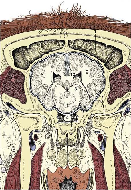
Figure 8-67 Position of the brain in relation to the roof of the bovine skull. Some features of the meninges are also shown. 1, Frontal sinus; 2, cerebral cortex; 3, diencephalon; 4, hypophysis; 5, sella turcica; 6, cavernous sinus; 7, dura mater; 8, arachnoid; 9, pia mater; 10, falx cerebri with dorsal sagittal sinus; 11, temporomandibular joint.
cooling of the arterial supply to the hypothalamus, the part of the brain responsive to and concerned with the regulation of body temperature. The cooling is due to the passage of the internal carotid artery (or rete substitute) through the cavernous sinus, where bathing by venous blood at a somewhat lower temperature (because it is drained from the nose and superficial structures of the head) promotes heat exchange. (An additional mechanism for protecting the brain from damaging hyperthermia is provided by the course of the common carotid artery, which is extensively related to the trachea at no great depth below the skin. These relationships promote heat loss, especially because any physical exertion that tends to raise body temperature also increases the flow of air within the upper respiratory tract.)
The vertebral venous plexus is probably more important clinically. It runs the whole length of the vertebral column and drains blood from the vertebrae, the adjacent musculature, and the structures within the vertebral canal. It gives rise to segmental veins that leave the canal through the intervertebral foramina to join the principal venous channels of the neck and trunk: the vertebral, cranial caval, azygous, and caudal caval veins (see Figure 7-43/18). The major part of the plexus consists of paired channels within the epidural space ventral to the cord. They are composed of crescentic segments that extend between successive intervertebral foramina (see Figure 26-5). The enlarged midpart of each segment swings toward and is generally joined to its neighbor over the middle of the vertebra, which produces a ladderlike pattern of vessels.
The connections with segmental veins through the intervertebral foramina form a plexus around the emerging spinal nerves, protecting them from injury.The veins composing the plexus are thin walled and, being without valves, may pass blood in either direction. They are capacious and adjust in size to compensate for variations in venous return to the heart induced by the intrathoracic pressure changes that accompany respiration. Since the system provides alternative channels to the major systemic veins, it may mitigate the effects of jugular obstruction (when the neck is compressed) or caudal caval obstruction (when pressure within the abdomen is raised). The intermittency of flow caused by these several factors facilitates the spread of septic or neoplastic disease to the vertebral column when the lungs would be the expected destination; blood diverted into the vertebral plexus when the flow through other channels is impeded may be temporarily held stagnant, which allows tumor seeds or microorganisms to settle within the tributaries that issue from the bones.
A further point of clinical importance lies in the risk of hemorrhage when epidural or subarachnoid puncture is performed. The risk is greatest at the atlantooc- cipital space, where tributaries of the plexus most often encircle the dural tube.
There are no lymphatics in the central nervous tissue.