» The Vertebral Canal
The vertebral canal is widest within the atlas and tapers rapidly within the sacrum; in between, it is most expanded where it contains the cervical and lumbar enlargements of the spinal cord that give rise to the nerves that form the limb plexuses.
Access to the vertebral canal is frequently necessary to withdraw cerebrospinal fluid from the subarachnoid space or to introduce local anesthetic into the epidural space. Therapeutic agents are also occasionally injected into these spaces. Examination of the skeleton shows that, although entry is theoretically possible through any of the interarcuate spaces, it is easiest at the wider gaps between the atlas and the skull, at the lumbosacral joint, and between the first two caudal vertebrae of the tail (Fig. 26.3). The first intercaudal space is conveniently large, measuring about 2 ? 2 cm. Most other interarcuate spaces measure only a few millimeters in each direction, and because they lie at a considerable depth below the skin, they are not easily located. The cranialmost interlumbar interarcuate spaces are occasionally used to perform epidural injections to obtain local anesthesia of the flank. A slightly oblique approach, from a point of entry a little lateral and caudal to the target space, gives the least risk of having the needle impinge on bone.The cord reaches to the first sacral vertebra in adult cattle and considerably farther in young calves, perhaps into the caudal half of the sacrum. It may occupy almost the whole sacrum in the small ruminant species.
The spinal cord is divided into 8 cervical, 13 thoracic, 6 lumbar, 5 sacral, and (usually) 5 caudal segments. The 8 cervical segments are accommodated within the 7 cervical vertebrae, while each of the thoracic and cranial lumbar segments shows an almost exact correspondence with the vertebrae of the same designation. The cranial shift of the more caudal part of the cord leaves the canal within the last lumbar vertebra occupied by the 5 short sacral segments (Fig. 26.4).
The subarachnoid space extends well into the sacrum, and its dimensions are sufficiently generous to make subarachnoid puncture a relatively simple procedure at the lumbosacral level (Fig. 26.3/4).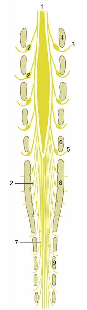
FIG. 26.4 The relationship to the vertebrae of the caudal end of the spinal cord and its branches (schematic dorsal view). Note the position of the spinal ganglia (2). The schema indicates the situation in adult cattle. The cord extends to the second or even third sacral vertebra in the newborn calf and in adult sheep and goats. 1, Spinal cord; 2, spinal ganglia; 3, second lumbar spinal nerve; 4, section of arch of second lumbar vertebra; 5, sixth lumbar nerve; 6, section of arch of sixth lumbar vertebra; 7, cauda equina; 8, section of sacrum; 9, section of arch of second caudal vertebra.
The internal vertebral plexus of the vertebral column (Fig. 26.5/1) presents two features of potential interest. The first involves the possibility of the plexus conveying blood diverted from the caudal vena cava when this is narrowed or obstructed by ruminal tympany; compression of the vena cava may be direct or exerted indirectly by a shearing displacement of the liver against the diaphragm (Fig. 26.6). The second significant feature involves the risk of hemorrhage in the performance of subarachnoid or epidural puncture.
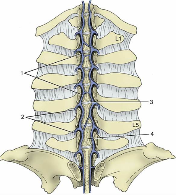
FIG. 26.5 Dorsal view of the venous drainage in the bovine vertebral canal. The internal vertebral plexus, with its internal connections and its lateral segmental branches, has been exposed. 1, Internal vertebral plexus; 2, intervertebral veins; 3, intervertebral disk; 4, vertebral body; L1, first lumbar vertebra; L5, fifth lumbar vertebra.