» The Vessels of the Tail
The median artery and vein of the tail require brief notice. The artery, which continues the median sacral, is ventral to the vein for most of the length of the tail and is commonly used for pulse taking, usually about 18 cm from the root of the tail.
The vessels lie side by side in the proximal part of the tail (Cd2 or Cd3), where both artery and vein are available for obtaining blood, although this site is an unwise choice because of the inevitable fecal contamination (Fig. 26.7B). At this level both vessels lie against the ventral aspect of the caudal vertebrae, where they are protected by the hemal processes (Fig. 26.7A), arches on the first few vertebrae (see Fig. 2.12E/9). The vessels are thus accessible only at intervertebral levels. It is usual to dock the tail of lambs.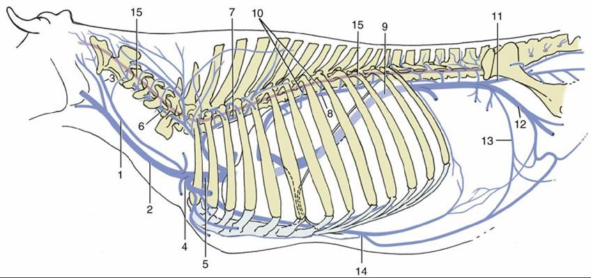
FIG. 26.6 The connections of the major veins with the vertebral plexus-azygous system. Note specifically the connections between the internal vertebral plexus (15) and the intercostal veins (10) and between the plexus and the branches of the vertebral vein (6). 1, Internal jugular vein (v.); 2, external jugular v.; 3, occipital v.; 4, axillary v.; 5, cranial vena cava; 6, vertebral v.; 7, supreme intercostal v.; 8, left azygous v.; 9, caudal vena cava; 10, intercostal veins; 11, internal iliac v.; 12, external iliac v.; 13, deep circumflex iliac v.; 14, cranial epigastric v.; 15, internal vertebral plexus (red).
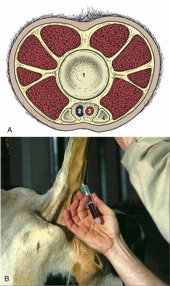
FIG. 26.7 (A) Transverse section of the bovine tail between Cd3 and Cd4. 1, Intervertebral disk; 2,
median caudal vein; 3, median caudal artery; 4, hemal process. (B) Collection of blood from a median
caudal vessel.
Comprehension Check
Review the anatomic boundaries of the flank, and determine the reasons for its suitability for surgical access to the abdomen.
* This description refers to cattle of European origin. The pronounced hump in cattle of the Zebu (Bos indicus) line (and their crosses) is mainly due to enlargement of the rhomboideus muscles.