» The Vertebral Column
The vertebral column comprises 7 cervical, 18 thoracic, 6 lumbar, 5 sacral, and about 20 caudal vertebrae. Variations in number are not uncommon, with the most frequent being the reduction of the lumbar vertebrae to 5, especially in the Arabian.
The impression of shortness in the loins in other breeds is more often due to a marked caudal inclination of the last ribs.The vertebral column inclines ventrally below the withers to reach its lowest point at the cervicothoracic junction, although the external elevation creates a contrary impression. It then changes direction abruptly, and as it ascends toward the poll, it shifts closer to the dorsal contour (Fig. 19.1).
The cervical vertebrae are individually long. Those behind the axis have rudimentary spinous processes, large divided transverse processes, and broad articular surfaces. The thoracic vertebrae are unremarkable apart from the great length of the spinous processes that form the basis of the withers. Independent centers of ossification develop for the summits of the first 12 or so spinous processes, and these may not fuse until comparatively late (10 or more years), if at all. The lumbar vertebrae have long horizontal transverse processes; synovial joints sometimes develop between those of the fourth and fifth bones and are constant between the fifth and sixth bones and between the sixth bone and the wings of the sacrum. In saddle horses exostoses sometimes develop on the summits of the thoracic spinous processes (mostly 14th-17th), bringing these into painful contact with their neighbors ("kissing spines") and resulting in minor local deflections of the vertebral axis.
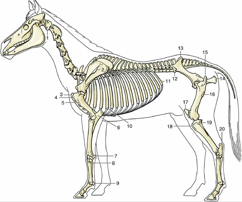
FIG. 19.1 The equine skeleton. The features labeled are among those normally palpable. 1, Wing of atlas; 2, tuber of scapula; 3, manubrium; 4, greater tubercle; 5, deltoid tuberosity; 6, olecranon; 7, accessory carpal bone; 8, proximal end (base) of lateral splint bone; 9, proximal sesamoid bone; 10, sixth rib; 11, last (18th) rib; 12, coxal tuber; 13, sacral tuber; 14, ischial tuber; 15, greater trochanter; 16, third trochanter; 17, patella; 18, tibial tuberosity; 19, head of fibula; 20, calcanean tuber.
The intervertebral disks are relatively thin, collectively accounting for only 10% to 11% of the length of the vertebral column. Each consists of a peripheral anulus fibrosus and a central nucleus pulposus, but the boundary between these parts is less distinct than in many species. Age changes include dehydration and fragmentation of the outer fibrous part but rarely calcification of the center. The disks most severely affected tend to be those of the neck and that between the last lumbar vertebra and the sacrum, which are the regions where movement is greatest. The clinical importance of these changes is not clear.
The nuchal ligament, which divides the dorsal cervical muscles into right and left groups, is massively developed and supports much of the burden of the head without interfering with the ability to lower the neck when grazing (Fig. 19.3). It consists of two clearly defined parts, each paired. The dorsal (funicular) part is a thick cord extending between the highest spines of the withers and the external occipital protuberance of the skull. It is flattened at its cranial attachment, becomes rounded shortly behind this, and flattens again as it nears the withers, where it forms a broad flange extending almost to the scapular cartilage. It is continued behind the withers as the narrower supraspinous ligament. The second (laminar) part forms a fenestrated sheet closely applied to its fellow. It fills the space between the funicular part and the cervical vertebrae and consists of bundles of elastic fibers that run cranioventrally from the funicular part and the spines of T2 and T3 to attach to C2 to C7. Synovial bursae are interposed between the funicular part and certain bony prominences to minimize pressure. One, the cranial nuchal bursa, is constantly present above the dorsal arch of the atlas; a second, the caudal nuchal bursa, is sometimes found above the spine of the axis; and a third, the supraspinous bursa, is constantly present over the most prominent processes of the withers (Fig.
19.3/2, 2', and 2"). Infections of the first and third bursae, leading to conditions known as "poll evil" and "fistulous withers," respectively, were formerly frequent and required extensive surgery for their eradication.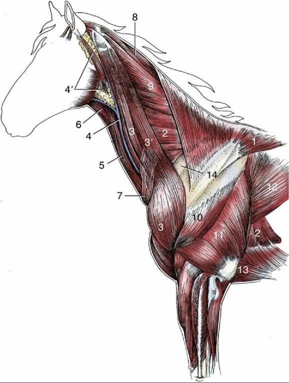
FIG. 19.2 Superficial dissection of the neck and shoulder region. 1, Trapezius; 2, serratus ventralis; 3, brachiocephalicus; 3', omotransversarius; 4, external jugular vein; 4', parotid gland; 5, sternocephalicus; 6, omohyoideus; 7, cutaneous colli; 8, rhomboideus cervicis; 9, splenius; 10, deltoideus; 11, triceps; 12, latissimus dorsi; 13, pectoralis ascendens; 14, subclavius.
The complicated arrangement of the powerful epaxial muscles of the back and neck conforms, but only in a general way, to the account given in Chapter 2 (pp. 43-44). The many features of difference are fortunately not of clinical importance, and illustration of their arrangement in transverse sections of the neck and back will suffice for a description (see Fig. 18.38). One specific feature of the associated deep fascia does, however, require notice. In the horse this thoracolumbar fascia possesses, opposite the scapula, an additional superficial lamina of importance. This, the dorsoscapular ligament (Fig. 19.3/5 and 5'), has an origin, in common with the deeper layers, from the supraspinous ligament over the highest spines of the withers. In its ventral passage it is applied to the deep surface of the rhomboideus and gradually transforms from a purely fibrous to a largely elastic nature. It detaches a number of side branches that insert on the deep face of the scapula, alternating with divisions of the serratus ventralis muscle. The arrangement provides an elastic mechanism that helps absorb shock when the foot strikes the ground, limiting the dorsal shift of the scapula that would otherwise occur.
As always, the cervical part of the vertebral column is so mobile and flexible that the mouth may reach the flank or the pasture. The latter movement is not always so easy for draft animals, which have relatively short necks that lead them to adopt a spreading posture of the forelimbs; they may lean forward when grazing. Only small movements are permitted to the back and loins except at the very mobile lumbosacral joint.