The Vestibular System Provides Sensory Information for Reflexes Involving Spinal Motor Neurons, the Cerebellum, and Extraocular Muscles of the Eye
As noted earlier, vestibular hair cells synapse on sensory neurons whose axons form part of the eighth cranial (vestibulocochlear) nerve and that carry action potentials to the medulla.
Almost all these axons synapse in the vestibular nuclear complex, a bilateral group of four distinct nuclei occupying a substantial portion of the medulla beneath the fourth ventricle. From here, second-order neurons (those on which cranial nerve VIH axons synapse) project to three important areas of the nervous system. Some of the neurons of the vestibular nuclear complex receive significant input from the utricle and saccule (the otolith organs), and their axons in turn form the lateral vestibulospinal tract. This tract provides excitatory facilitation to gamma (γ) and alpha (α) motor neurons of antigravity muscles of the trunk and limbs in response to linear acceleration/deceleration or static tilt of the head (see Chapter I O).Other neurons of the vestibular nuclear complex receive significant sensory input from the cristae ampullaris of the semicircular ducts, and their axons in turn form a pathway that projects to cranial nerve nuclei that control eye movements. This pathway, called the medial longitudinal fasciculus (MLF), produces compensatory eye movements in response to rotary acceleration/deceleration of the head. The vestibular nuclear complex also sends projections to, and receives projections from, the cerebellum, especially the flocculonodular lobe. Fhrough these reciprocal connections, the cerebellum can fine-tune the coordination of postural and visuomotor reflexes that are controlled by the vestibular system. Finally,
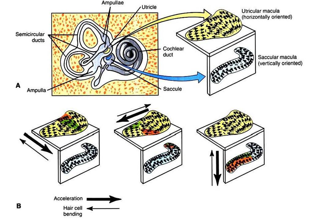
FIGURE 11-7 A, Macula of the utricle is horizontally oriented, and macula of the saccule is vertically oriented.
Small arrows in a macula represent the approximate orientation of the hair cells in that region, with respect to their cilia. For a given hair cell, the arrow tip represents the position of the largest cilium, and the arrow tail represents the shortest cilium. B, Acceleration in a given direction (large thick arrows) results in bending of hair cell cilia in the opposite direction (large thin arrows) caused by otolith drag. Hair cells whose cilia are bent directly toward the largest cilium (green regions) will be depolarized the most and will produce the greatest increase in action potential frequency in their associated sensory neurons. Conversely, hair cells whose cilia are bent directly away from the largest cilium (red regions) will be hyperpolarized the most and will produce the greatest decrease in action potential frequency in their associated sensory neurons. Hair cells whose cilia are bent along other axes will be less significantly affected. (Portions modified from FuchsAF: Peripheral motor control: the vestibular system. In Patton HD, Fuchs AF, Hille B, et al, editors: Textbook of physiology, ed 21, Philadelphia, 1989, Saunders.)some of the projections leaving the vestibular nuclear complex participate in neural circuits leading to cerebral cortex, resulting in conscious vestibular sensations.