The yellowish white adrenal glands (see also pp. 207-208) (Fig. 14.28/7 and 7') of the dog are dorsoventrally flattened, about 2 to 3 cm long and 1 cm wide.
Each occupies the retroperitoneal space medial to the kidney, cranial to the renal vessels, and dorsolateral to the aorta (the left gland) or the caudal vena cava (the right one).
The capsule of the right adrenal gland may be continuous with the tunica externa of the vena cava. The right adrenal gland is located ventral to the transverse process of the last thoracic vertebra, with its cranial two thirds covered by the caudate process of the liver. The left adrenal gland, which has a somewhat dorsoventrally flattened oval cranial portion and a cylindrical caudal projection, is positioned ventral to the transverse process of the second lumbar vertebra, just caudal to the origin of the cranial mesenteric artery and adjacent to the origin of the phrenicoabdominal artery. This paired artery courses on the dorsal surfaces of both left and right glands. The ventral surfaces are crossed and indented by the phrenicoabdominal veins; on the left, this surface is also related to the pancreas.The glands are diffusely supplied by branches from adjacent vessels: the aorta and the renal, phrenicoabdominal, lumbar, and cranial mesenteric arteries. The right and left adrenal veins enter the vena cava directly and the left renal vein, respectively.
The nerve supply is derived from a dense network on the dorsal surface of the glands that appears continuous with the nearby celiac and mesenteric plexuses. The fibers that actually enter the glands are preganglionic and are provided by the splanchnic nerves that enter the abdominal cavity close by.
In cats the adrenal glands are shorter and similar to oval disks. The adrenal glands of older cats are occasionally calcified and if so are visible on radiographs. The topography is the same in both species.
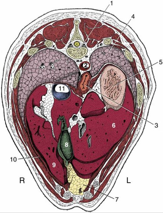
FIG. 14.26 Transverse section of the canine trunk at the level of the 11th thoracic vertebra. 1, 11th thoracic vertebra; 2, aorta; 3, esophagus; 4, left lung; 5, fundus of stomach; 6, left lateral lobe of liver; 7,
fat-filled falciform ligament; 8, gallbladder; 9, right medial lobe of liver; 10, diaphragm; 11, caudal vena cava; L, left; R, right.