» The Pancreas
The slender pancreas (see also pp. 129-131) consists of two limbs or lobes that diverge from the vicinity of the pylorus. The left lobe is directed caudomedially and crosses the median plane behind the stomach to end against the left kidney (see Fig.
3.56/5). It divides the branches of the celiac artery from those of the cranial mesenteric artery and is enclosed within the deep leaf of the greater omentum where the latter passes dorsal to the transverse colon. The dorsal surface of the left lobe is crossed by the portal vein where the lobe makes contact with the hilus of the liver to the right of the median plane.The longer right lobe is directed caudodorsally and follows the dorsal surface of the descending duodenum within the mesoduodenum. It is related dorsally to the visceral surface of the liver and, behind it, to the ventral surface of the kidney (Fig. 14.27/9). The lobe lies lateral to the ascending colon and dorsal to the small intestine.
Two secretory ducts open into the duodenum where the two lobes diverge. The smaller and inconstant pancreatic duct joins the bile duct just before the latter opens on the major duodenal papilla, 3 to 6 cm distal to the pylorus. The accessory pancreatic duct, the main channel, opens on the minor duodenal papilla 3 to 5 cm farther down the gut. Both papillae can be detected with the unaided eye. The duct systems of the two lobes communicate internally. In the cat the main duct is the pancreatic duct. In a minority of cats (around 20%) an accessory duct can also be found that, as in dogs, opens onto the minor duodenal papilla, some 2 cm distal to the major papilla.
The major part of the pancreas is supplied by two of the three branches of the celiac artery. Only the caudal part of the right limb of the pancreas receives blood from the cranial mesenteric artery. The left lobe is entered by branches of the splenic artery whereas branches from the hepatic artery supply the body of the pancreas (gastroduodenal artery) and the cranial half of the right lobe (cranial pancreaticoduodenal artery).
Duodenal branches are given off from cranial pancreaticoduodenal artery and course through the pancreatic tissue to supply the gut itself. Anastomoses between these various vessels occur within the gland. Lymphatics are abundant and drain into the duodenal lymph node, if present, or into the mesenteric lymph nodes.Pancreatic Tumor: One of the most encountered problems in the pancreas of the dog is the presence of an insulin-producing tumor, an insulinoma. Thorough inspection for metastases must be performed in the liver, the duodenum, the mesentery, and the hepatic, splenic, gastric, duodenal, and cranial mesenteric lymph nodes. Resection of the part of the pancreas is difficult because it shares blood supply with the duodenum and the spleen. Removal of the spleen is indicated when the splenic artery cannot be preserved.
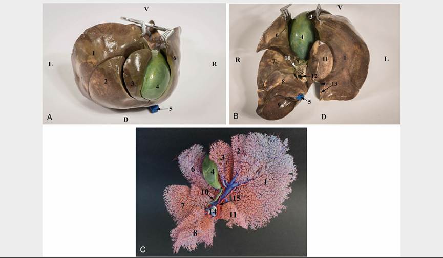
FIG. 14.25 Liver of the dog, diaphragmatic surface (A), visceral surface (B), a corrosion cast (C): 1. Left lateral lobe; 2. Left medial lobe; 3. Quadrate lobe; 4. Gallblader; 5. Caudal vena cava, intrathoracal part; 6. Right medial lobe; 7. Right lateral lobe; 8. Caudate process of the caudate lobe; 9. Renal impression at the caudate process; 10. Bile duct; 11. Papillary process of the caudate lobe; 12. Portal vein (on the fixed specimen); 13. Esophageal impression; 14. Portal vein (on the corrosion cast); 15. Hepatic artery