Topography
The brain and spinal cord are contained within a continuous space provided by the cranial cavity of the skull and the vertebral canal, which is formed by successive bony rings and connecting ligaments and disks of the vertebral column.
The cranial cavity lies directly behind the nasal cavities. It is smaller than is commonly supposed, the form and extent of the cranial cavity not easily being predicted from the external appearance of the head and skull because the paranasal sinuses, horns, muscular ridges, and other projections of the skull, as well as the temporal muscles, all contribute significantly to the conformation of this part of the head. The closest agreement between the external contours and the cavity within the cranium is found in the newborn of all species; among adults, this agreement is best retained in cats and in dogs of brachycephalic breeds. Fortunately, the exact location of the brain is rarely of practical significance except in humane slaughter techniques mentioned in later chapters. It is probably sufficient to know that the caudal limit of the cavity extends to the caudal wall of the skull — thickened by the frontal sinus in cattle—but that the rostral limit shows considerable variation; in dogs and cats, the rostral limit of the cranial cavity is associated with the caudal margin of the zygomatic processes of the frontal bones and in horses and cattle with the rostral level of these processes. In pigs and small ruminants, the rostral limit of the cranial cavity extends to the middle of the orbit.
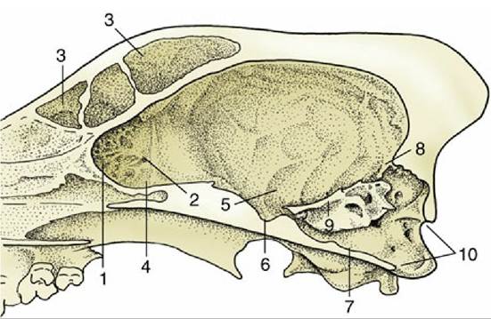
FIG. 8.54 Sagittal section of the cranium of the dog. 1, Cribriform plate; 2, ethmoid foramen; 3, frontal sinus; 4, rostral fossa; 5, middle fossa; 6, hypophysial fossa; 7, caudal fossa; 8, tentorium cerebelli osseum; 9, petrosal crest; 10, foramen magnum.
The interior of the cranial cavity shows a fairly close correspondence with the contours of the brain, although significant intracranial space is required for the meninges and intermeningeal spaces that surround the brain and for the capacious intracranial venous sinuses. Although the roof (calvaria) of the cavity remains largely undivided, the base is divided into three fossae; these need not be described in detail because the main features are depicted in Fig. 8.54. The rostral fossa is formed by the sphenoid and ethmoid bones and extends to the level of the optic canals, the passages of exit of the optic nerves. The rostral fossa contains the olfactory bulbs embedded within recesses of the cribriform plate (Fig. 8.54/1) and the rostral parts of the cerebral hemispheres. The middle fossa extends from the optic canals to the sharp petrosal crests (Fig. 8.54/9) that project inward from the petrous temporal bones of the lateral walls. The floor of the middle fossa is formed by the sphenoid bone, which carries the median hypophysial fossa (sella turcica) into which the hypophysis fits; it also contains various foramina — the orbital fissure and the round and oval foramina—that were encountered in the previous description of the skull (p. 54). This middle fossa, the widest part of the cranial cavity, contains the temporal and parietal lobes of the cerebral hemispheres. The caudal fossa extends from the caudal limit of the hypophysial fossa to the foramen magnum in the caudal wall. Its principal features are the invaginations from the lateral walls made by the petrous parts of the temporal bones (each perforated by an internal acoustic meatus) and the jugular and hypoglossal foramina in the floor. The caudal fossa lodges the midbrain, pons, and medulla ventrally and the cerebellum dorsally.
The caudal, dorsal, and lateral walls of the entire cranial cavity are smoothly joined together. The most prominent internal feature of the cranial cavity is the tentorium cerebelli osseum (Fig.
8.54/8), a large projection at the junction of the dorsal and caudal walls forming the middle portion of the tentorium cerebelli within the transverse fissure of the brain. The tentorium contains passages for branches of the dorsal intracranial venous sinuses.The vertebral canal is widest within the atlas cranially and tapers rapidly within the sacrum caudally; between the two extremes, it is most expanded where it contains the cervical and lumbar enlargements of the spinal cord, from which arise the nerves that form the forelimb and hindlimb plexuses, respectively (see Fig. 8.15). The topography of the spinal cord is of considerable importance in veterinary practice because injections into the canal are frequently made, particularly injections of local anesthetic, with the intention of blocking specific spinal nerves; in addition, there is sometimes a need to locate central nervous lesions to specific vertebral levels, a procedure made possible by the association of specific sensory and motor deficits with particular spinal segments.
Even with the inclusion of its meningeal wrappings, the spinal cord is considerably smaller than the vertebral canal (Fig. 8.55). It is also considerably shorter in craniocaudal length. This discrepancy is due to the unequal growth between the spinal cord and vertebral column, which begins well before birth and continues after. The relative shift in position (ascensus medullae) results in the more cranial location of spinal cord segments in comparison with their original corresponding vertebrae. This shift is most pronounced in more caudally located segments and explains the position of the tapered end of the spinal cord (conus medullaris) in the lumbar or sacral region of the vertebral column. The level at which the cord ends varies among species (and, in early life, with age); it is within L5 or L6 in the pig, L6 in ruminants, L6 or L7 in the dog, S2 in the horse, and rather variably between L6 and S3 in the cat (Fig. 8.56).
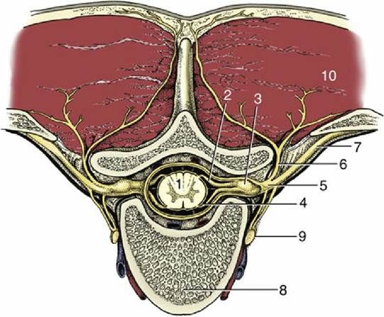
FIG.
8.55 Transection of the vertebral column to show the formation of a spinal nerve. 1, Spinal cord; 2, dorsal root; 3, spinal ganglion; 4, ventral root; 5, spinal nerve; 6, dorsal branch of spinal nerve; 7, ventral branch of spinal nerve; 8, body of vertebra; 9, sympathetic trunk; 10, epaxial muscles.The cranial shift in position of the more caudally located segments also explains the peculiar arrangement of the associated spinal nerves. The spinal nerves associated with the lumbar and sacral spinal segments travel caudally within the vertebral canal to reach their corresponding vertebrae to exit the canal. This collection of caudally directed spinal nerves on each side of the conus medullaris is known as the cauda equina because of its superficial resemblance to a horse's tail (see Fig. 12.9/9).
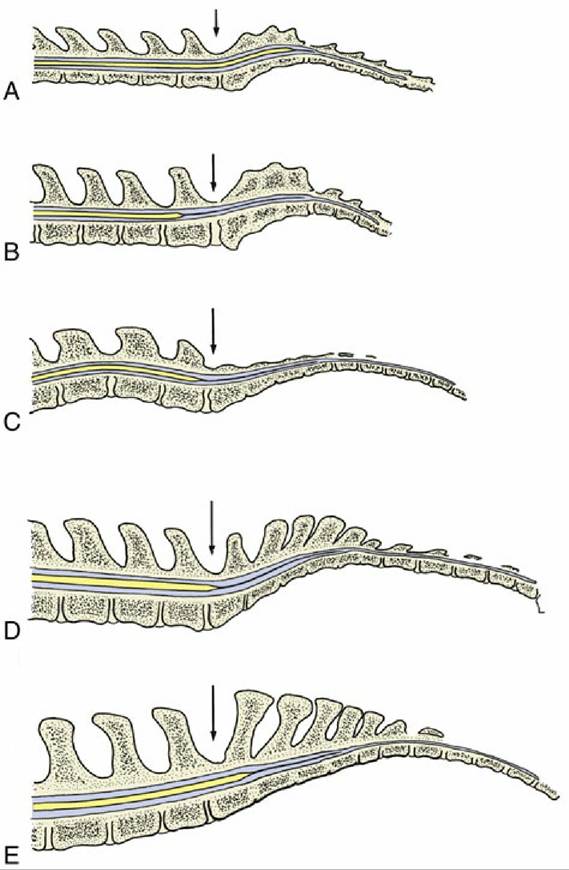
FIG. 8.56 Median section of the vertebral canal and spinal cord of (A) cat, (B) dog, (C) pig, (D) cattle, and (E) horse. The lumbosacral interarcuate space is indicated by an arrow. Notice the difference in caudal extent of the spinal cord in the different species. The thin extension of the spinal cord is the filum terminale that ends on the caudal vertebrae (not shown).