Transport Across Cell Membranes
The plasma membrane and the membranes of intracellular organelles have an important function in determining what enters and leaves the cell or its organelles. Our very life and that of animals depend on this ability to control what enters and leaves the cells.
it is therefore important to understand and appreciate the processes of membrane transport before discussing the functions of the animal’s organs and systems. Transport into and out of cells may occur by simple and facilitated diffusion, osmosis, active transport, endocytosis, or exocytosis. (Endocy- tosis and exocytosis are described at the beginning of this chapter.)Simple and Facilitated Diffusion
Diffusion is a passive mechanism. it is simply the distribution of a substance in a solvent medium, usually water, so that it becomes equally concentrated throughout the medium. Diffusion occurs because all molecules and ions have kinetic energy. They collide with each other and bounce away, becoming so dispersed in the solvent that an equal concentration appears throughout. in solutions, diffusion proceeds from a region of greater concentration of particles to a region of lower concentration, so the net diffusion is said to occur down a concentration gradient.
Only a few substances, such as oxygen, carbon dioxide, and alcohol, are membrane soluble, that is, capable of diffusing freely through the lipid bilayer of plasma membranes. Such molecules must be lipid soluble. Certain drugs, such as barbiturates, a class of anesthetics, are membrane soluble. If a substance cannot diffuse freely through the lipid bilayer of the cell membrane, its ability to diffuse in or out of a cell depends on some other means of crossing the membrane.
one way lipid-insoluble substances cross the cell membrane is via a transmembrane protein or proteins that form a channel, or passageway, through the membrane (Fig.
2-8). If a channel that permits the passage of a given molecule is present, the membrane is said to be permeable to the molecule. The degree of permeability of an individual channel may also be subject to regulation by factors such as the electrical potential across the membrane. Channels whose permeability varies with the electrical potential across the membrane are said to be electrically gated or voltage gated. A change in the conformation or configuration of the membrane protein is responsible for changes in the permeability of the channel (Fig. 2-11).Ions (atoms or radicals having a positive or negative charge) cannot diffuse freely through the lipid bilayer of the plasma membrane. Thus, a channel that is permeable to a given ion must be present for that ion to diffuse through cell membranes. Most channels are permeable only to a single specific ion or a small number of specific ions. This characteristic is important from a clinical standpoint, as some drugs are relatively specific for a given type of channel. With the use of these agents, the movement of a specific ion across cell membranes can be regulated. For example, the inward movement of calcium into cells of the heart can be regulated with such drugs, and this is beneficial in certain types of cardiac arrhythmias (abnormalities in the electrical activity of the heart).
The rate and direction of passage of a charged ion through a channel depends on two factors that may act synergistically. That is, both may
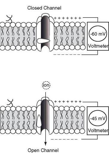
Figure 2-11. Electrically-gated channel opens and closes with changes in the electrical potential across the cell membrane.
have the same effect on the rate and direction of movement. Or they may act antagonistically, each having opposite effects on rate and direction of movement. One factor is the concentration gradient between the two sides of the membrane for the particular ion. Because of diffusion, ions have net movement through permeable channels from areas of higher concentration to areas of lower concentration.
The second factor is any electrical gradient generated by concentration differences among other charged ions on the two sides of the membrane. in all animal cells, the concentrations of charged ions on the two sides of the cell membrane are normally such that the inside of the cell is negative to the outside (Fig. 2-11). The net negative charge on the inside of the cell inhibits the inward diffusion of negatively charged ions (anions), while it promotes the inward diffusion of positively charged ions (cations). The term electrochemical gradient is used to refer to the combined effects of the concentration gradient and electrical gradient on the diffusion rate of an individual ion.Facilitated diffusion is the same as simple or free diffusion in that it operates passively down the concentration or electrochemical gradient. However, facilitated diffusion requires a carrier system in the membrane to assist the crossing. The carrier system is a transmembrane protein that binds the diffusing molecule or molecules on one side of the membrane and then transfers them to the other side, where the transported molecules are released (Fig. 2-12). The movement or transport across the membrane probably entails a change in the shape of the protein, but it does not require any direct use of ATP for energy as in active transport.
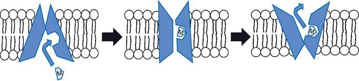
Figure 2-12. Facilitated diffusion of solute (labeled M) across a cell membrane by the action of a transmembrane carrier protein.
Sugars, especially glucose, depend on facilitated diffusion to enter cells by joining with carrier proteins upon reaching the lipid bilayer of the membrane. The glucose carrier complex transports glucose down the glucose concentration gradient to the inside of the cell membrane. Here the carrier releases the glucose to enter the cell. The carrier remains in the membrane and reconfigures itself so that it is available for more transport.
other substances besides glucose, such as amino acids, also depend on facilitated diffusion to cross cell membranes.The rate at which facilitated diffusion occurs also depends on the number of carrier proteins available in the membrane. in the case of glucose, the speed of entry into many cells, such as skeletal muscle, is greatly increased by the hormone insulin, which the pancreas secretes. insulin facilities the entry of glucose into skeletal muscle, in part by increasing the number of carrier proteins in the cell membrane of these cells.
Osmosis
osmosis is movement of water across membranes. Like many solutes, water does not diffuse freely through the lipid bilayer of cell membranes but rather must diffuse through water channels formed by transmembrane proteins. These proteins are aquaporins. If the intracellular fluid within a cell has a higher concentration of undiffusible solutes than the interstitial fluid bathing the cell, water will move into the cell from the interstitial fluid until the concentrations are the same on both sides of the membrane. As the water moves in, the volume in the cell increases. The driving force moving water from the solution on the side of the lower solute concentration to the side with the higher solute concentration is osmotic pressure.
The osmotic pressure of an aqueous solution can be measured by using a U-tube in which the two sides are separated by a semipermeable membrane, which is permeable only to water (Fig. 2-13). The solution is placed in one side of the U-tube, and distilled water is placed in the other. The force of osmosis moves water through the membrane from the side containing distilled water to the side containing the solution. This movement continues until the hydrostatic pressure generated by the increased height of the fluid column on the solution side is equal to the osmotic force (Fig. 2-13). The units of osmotic pressure may be given in centimeters of water (height of water column), or converted to millimeters of mercury (mm Hg; height of a column of mercury creating an equivalent amount of hydrostatic pressure).
osmotic pressure is an important mechanism in maintaining cellular volume by determining whether water will enter or leave cells. If the concentration of solutions on each side of a membrane is the same, as seen with cells in blood, the bathing fluid is said to be isotonic (isosmotic) in relation to the cells. This means that the osmotic pressure is the same on both sides of the membrane. A 0.9% solution of sodium chloride is considered to be isotonic with mammalian red blood cells and for this reason is called a normal or physiologic saline solution. Normal saline can be used to moisten exposed tissues, such as open wounds, without damaging the cells.
If the bathing fluid has a lower osmotic pressure than the cells, it is said to be hypotonic, and water tends to cross the membrane and enter the cells. In the case of red blood cells in hypotonic plasma, the water entering the cells can swell and finally burst them, a condition called hemolysis.
Red blood cells in hypertonic plasma (more concentrated than the cell cytoplasm) lose water to the plasma and become wrinkled. The wrinkling of red cells is called crenation. In relation to mammalian cells, a solution less concentrated than 0.9% sodium chloride is said to be hypotonic; one more concentrated than 0.9% sodium chloride is said to be hypertonic; and of course a 0.9% sodium chloride solution is isotonic.
The osmotic pressure of a solution is determined by the number of solute particles: the more solute particles in a volume of fluid, the greater the osmotic pressure. The number of
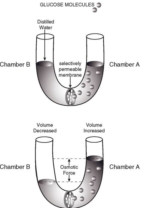
Figure 2-13. Osmosis and osmotic pressure. A membrane impermeable to solute particles prevents their diffusion from chamber A into chamber B. Distilled water from chamber B migrates into chamber A until the difference in the height of the water columns equals the osmotic pressure of the solution in chamber A.
particles is determined by the molar concentration of the solution and by the number of ions formed if the solute is an electrolyte.
For example, glucose is not an electrolyte, having one particle per molecule, but sodium chloride is an electrolyte, giving two particles (Na+ and Cl-) per molecule when placed in solution. A 1-molar solution of sodium chloride has twice the osmotic pressure of a 1-molar solution of glucose, because the sodium chloride solution has twice the number of particles in solution.These concepts of osmosis and osmotic pressure become important when intravenous fluids are administered to animals for problems such as dehydration, anorexia, milk fever, and diarrhea. Furthermore, they are important principles in normal functions of animals, such as the flow of blood and lymph, the excretion of wastes in the urine by the kidneys, and the digestion and absorption of food, as is discussed in subsequent chapters.
Active Transport
some molecules and ions can move across cell membranes (either in or out of cells) against concentration or electrical gradients. The term against, as used here, means that the particles are moving in the direction opposite that of diffusion. This movement across the cell membrane consumes energy produced by the cell and is called active transport.
The best-recognized example of a primary active transport system is the sodiumpotassium (Na-K) pump. This pump is actually
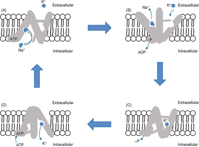
Figure 2-14. A schematic representation of the hypothetical operation of the Na+-K+ pump, Na+-K+-ATPase. The Na+ icon represents three sodium ions and the K+ icon represents two potassium ions.
a membrane protein that is also an enzyme. The protein reversibly binds three Na+ and two K+ ions. The enzymatic activity of this protein permits it to hydrolyze ATP to gain energy. By a poorly understood mechanism, the gain of energy causes the protein to change its shape so that the Na+ and K+ are moved to the other side of the membrane. There the ions are released, and the protein returns to its original shape. These movements are summarized in Fig. 2-14. The Na-K pump, or Na-K-ATPase, is a component of the membrane of all cells, and it is always arranged in the membrane so that Na+ moves out of the cell and K+ moves into the cell. The continuous operation of this transport system is a major factor in keeping the intracellular concentration of Na+ relatively low in all cells, while the intracellular concentration of K+ is relatively high in all cells.
Secondary active transport also requires a membrane protein carrier and cellular energy, but the carrier proteins are not ATPases (enzymes that can use ATP directly). The uptake of glucose from the lumen of the intestine and the lumen of renal tubules by epithelial cells is an example of secondary active transport. In both types of epithelial cells, a membrane protein that acts as a carrier is found in the portion of cell membrane facing the lumen. This protein is capable of binding Na+ and glucose simultaneously when both are in the lumen. After both Na+ and glucose are bound, the protein changes its shape so that both are moved to the opposite side of the membrane. There they are released into the interior of the cell. This transport can move glucose against its concentration gradient because of the potential energy of the concentration gradient for Na+. Recall that the low intracellular Na+ concentration in all cells is maintained by the continuous operation of the Na-K-ATPase. Thus, ATP is used directly in maintenance of the low intracellular concentration of Na+, and this energy is used indirectly or secondarily to transport glucose.
An important characteristic of primary and secondary active transport systems is their degree of specificity. In most cases, a given transport protein transports only specific ions or molecules. For example, the Na-K pump transports only Na+ and K+. other electrolytes are not transported by this system.