TYPES OF ANIMAL EYES
Animal eyes fall under one of two types, based on morphology. These are simple and complex eyes (Erclik et al., 2009) (Figure 9.1).
9.2.1 Simple Eyes
Simple eyes consist of individual or small clusters of photoreceptors, typically accompanied by pigment cells and/ or lens-forming cells that allow the organism to differentiate between light and dark environments, including the source direction.
Organisms cannot use the eyes to create images (Arendt, 2003). Simple eyes comprise a large array of photoreceptors, pigment cells, and lens cells and can further be classified into two mutually exclusive classes called single-chambered and compound chambered (Treisman, 2004). Still on mechanism employed in capturing light rays, single-chambered eyes can be seen in pinhole eyes of the cephalopod Nautilus, which consist of a pigmented pit that relies on shadows to form an image, one that contains a lens to focus light (vertebrates and squids), or a mirror to reflect it (scallops) (Land, 2005).Compound eyes found in insects and crustaceans employ a fundamentally different design. They are made up of ommatidial facets, each of which can be termed a ‘mini-eye’ containing a group of photoreceptors and, in most cases, a lens (Erclik et al., 2009).
9.2.1.1 Parts of the Eye
9.2.1.1.1 Cornea
The cornea is a highly transparent layer that forms the anterior aspect of the outer coat of the eye. It is continuous with the opaque sclera, which is the other component of the outer coat. The transitional zone, where the cornea becomes sclera, is known as the limbus or corneoscleral junction (Downie et al., 2021).
The opaque sclera covers most of the surface of the globe and shows a varying thickness, ranging from 0.3 mm immediately posterior to the insertion of the rectus muscles, up to 1.0 mm near the optic nerve (Dua & Augusto, 2000).
It has been reported that the sclera is composed of collagen bundles that are approximately 64 nm in length, and branched fibrils that are randomly arranged and vary in diameter from 20 to 30 nm (Meek & Carlo, 2015).9.2.1.1.2 Conjunctiva
The conjunctiva is a thin, translucent mucous membrane, consisting of a superficial epithelium overlying loose connective tissue called the lamina propria or stroma (Takahashi et al., 2013). Embryologically, the conjunctiva develops from the ocular surface ectoderm (Shumway et al., 2018). It covers the anterior region of the eyeball, extending from the limbus to the eyelid fornices and then back to the mucocutaneous junction. Major roles of the conjunctiva include immune surveillance (Gipson & Mary, 2022) and production of tear constituents (Doughty, 2012)
9.2.1.1.3 Lacrimal System
The lacrimal system comprises three major divisions. The first is the secretory component, consisting of the exocrine tubuloalveolar serous lacrimal gland and two types of accessory lacrimal glands (Krause and Wolfring glands), which contribute the aqueous components of the tear film (Takahashi et al., 2013). The second component consists of the tear meniscus and blinking. The third component is the system that mediates the drainage of these secretions (Murube, 2012).
Tear film is a protective fluid layer that contains lipids, proteins, and electrolytes, which overlies the surface of the eye (Giudice et al., 2021). The ocular surface and tear
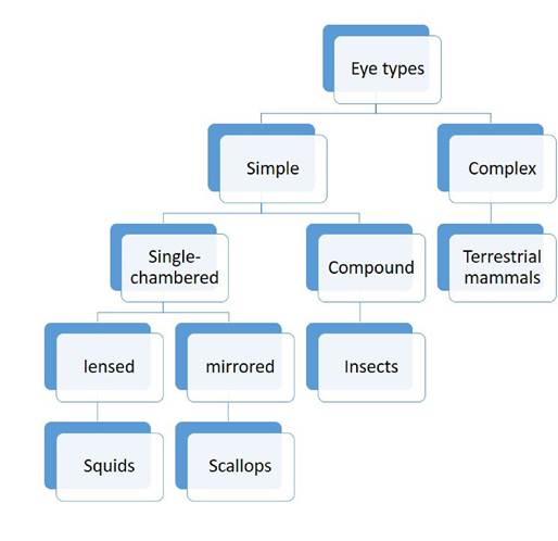
FIGURE 9.1 Schematic hierarchy showing the various eye types with an example of an organism in every case.
film are intimately related; a loss of homeostasis in either component can trigger a vicious circle of dry eye disease, a self-perpetuating cycle of tear instability, tear hyperosmolarity, ocular surface inflammation, and epithelial damage (McKee et al., 2014).
9.3