VASCULARIZATION AND INNERVATION OF THE VESSEL WALL
Like other tissues, blood vessel walls require nutrition. Diffusion from the lumen is sufficient to supply the needs of smaller vessels but requires supplementation by an intramural circulation in those of larger size.
The supplying arteries (vasa vasorum) most often arise at some distance from the stretch of wall they feed, frequently coming from collateral branches. They penetrate the adventitia from outside and ramify within this layer and the adjoining part of the media (Figure 7-31/1). They do not penetrate beyond the middle of the media in arteries, probably because capillaries in the inner part of the wall would be closed by the radial pressure generated by the bloodstream within the lumen. The tunica intima is never vascularized unless diseased.Arteries and veins receive both a motor and a sensory innervation. The vasomotor nerves to the arteries are particularly important because they control the diameters of the lumina and hence the peripheral resistance. Most are vasoconstrictor fibers of sympathetic origin. Some pass directly to the great arteries from sympathetic plexuses within the mediastinum, but most first travel within local nerve trunks from which they later emerge to enmesh the peripheral arteries. The afferent supply is concerned in local and general vascular reflexes; some fibers mediate the sensation of pain perceived from arterial lesions.
In addition, certain specific sites are much more richly supplied with nerves whose endings respond to pressure or chemical stimuli. These baroreceptor and chemoreceptor concentrations, of great importance in the regulation of the circulation, are confined to arteries originating in the pharyngeal (branchial) arches: the
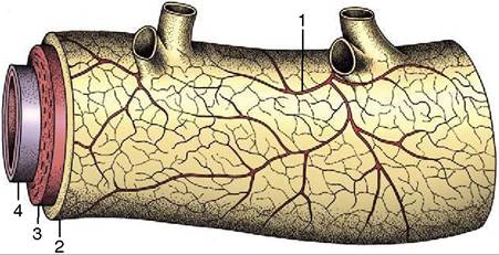
Figure 7-31 Vasa vasorum in the wall of a large artery. 1, Vasa vasorum; 2, tunica adventitia; 3, tunica media; 4, tunica interna.
internal carotid arteries, the aortic arch, the right subclavian artery, and the pulmonary trunk. The best- known examples of each type, the carotid sinus and carotid body (glomus caroticum), are found in close association at the origin of the internal carotid artery (Figure 7-32).
The carotid sinus may be recognized in the cadaver as a slightly expanded and especially distensible stretch at the origin of the internal carotid. Its receptors are stimulated by pressure changes that alter the mechanical tension in its wall. The carotid body is a neighboring nodule (sometimes palpable) that is composed of a richly vascularized mass of epithelioid cells. The chemoreceptors respond to changes in oxygen and carbon dioxide tension and hydrogen ion concentration in the perfusing blood. The afferent fibers from both receptor types travel in the carotid sinus branch (known to physiologists as the nerve of Hering) of the glossopharyngeal nerve to project on centers within the brainstem.
The less familiar receptor areas in the other arteries named are similar but less important. Specific differences exist, and in some animals they appear to decline in importance with the attainment of maturity.