VISCERAL EFFERENT PATHWAYS
Unlike the afferent component, the efferent component of the visceral nervous system is arranged in two divisions, sympathetic and parasympathetic, distinguished by morphology, pharmacology, and physiology.
The final conducting pathway of both divisions, unlike that of the somatic system, includes two motor neurons in succession: the first has its perikaryon within the central nervous system, and the second is stationed within a peripheral ganglion (Figure 8-53). The two are most frequently distinguished as preganglionic and postganglionic neurons and together are equivalent to the lower motor neuron of the somatic system.The preganglionic neurons of the sympathetic division are located within the lateral (visceral efferent) column of the spinal cord between the first thoracic and middle lumbar segments (with some interspecific variation) (see Figure 8-74). The postganglionic neurons are found in paravertebral ganglia of the sympathetic chain or subvertebral ganglia on the aorta; both groups are relatively close to the cord.
The parasympathetic preganglionic neurons are restricted to the nuclei of origin of the oculomotor, facial, glossopharyngeal, and vagus nerves within the brainstem and the lateral columns of certain sacral segments of the cord (see Figure 8-73). The postganglionic neurons are stationed within small ganglia in close proximity to or actually incorporated within the walls of the organs they supply.
The transmitter substance at the last sympathetic relay is norepinephrine and that of the parasympathetic division is acetylcholine; both are collocated with a host of neuropeptides. The two divisions therefore react differently to autonomic stimulant and depressant drugs.
The two systems have broadly similar distributions and are frequently described as antagonist: one inhibits
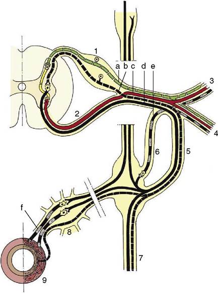
Figure 8-53 Comparison of the organization of the visceral (black) and the somatic (red) nervous system at the thoracolumbar level of the spinal cord. Afferent fibers are indicated by interrupted lines, efferent fibers by solid lines.
The postganglionic sympathetic fibers are indicated by alternating black and stippled lines. 1, Dorsal root ganglion; 2, ventral root; 3, dorsal branch of spinal nerve; 4, ventral branch of spinal nerve; 5, 6, white (preganglionic) and gray (postganglionic) communicating branches, often fused; 7, sympathetic trunk with ganglia; 8, prevertebral ganglion; 9, gut. a, Somatic afferent fibers; b, visceral afferent fibers; c, somatic efferent fibers; d, visceral efferent fibers (preganglionic sympathetic); e, postganglionic sympathetic (to peripheral structures); f, postganglionic sympathetic (to abdominal organs).while the other stimulates a particular activity. This rule is less absolute than was once supposed, and their roles are better regarded as collaborative. The more diffuse anatomy of the peripheral sympathetic nerves (which are described later) and the use of norepinephrine as a transmitter indicate the more general effects produced by sympathetic activity, in contrast to those of parasympathetic activity, which are often local, effecting single specific functions.
The central control is exerted by neurons within the hypothalamus; those that influence the sympathetic division are generally caudal to those controlling the parasympathetic division. The pathways from both sets follow various routes, of which some are direct and others are via multisynaptic chains within the reticular formation.