Zygomatic Gland
The ventral buccal glands comprise a few small, solitary units located in the submucosa, rostral to the masseter muscle, medial to the ventral part of the buccinator, and lateral to the mandible.
The dorsal buccal glands are consolidated in a mass generally known as the zygomatic gland (see Figs. 11.10A, 11.27/28, and 11.36/2). This structure is a large mixed gland located in the ventral part of the orbit, covered by the zygomatic arch, and related medially to the maxillary artery and nerve and medial pterygoid muscle and dorsally to the periorbita. Its swelling, when diseased, may cause protrusion of the eyeball (exophthalmos) or bulging of the oral mucosa near the last upper cheek tooth, where the duct opens into the vestibule. Facial trauma may cause leakage of saliva, and the resulting zygomatic mucocele may produce exophthalmos.
The main duct of the zygomatic gland (Fig. 11.15C) opens on a small papilla lateral to the caudal part of the upper first molar tooth. A small ridge connects the main zygomatic and parotid gland duct openings. Usually there are one to four small accessory ducts opening caudal to the main one. These openings are usually obvious and easily cannulated.
id="Picutre 536" class="lazyload" data-src="/files/uch_group31/uch_pgroup304/uch_uch7239/image/image536.jpg">
FIG. 11.28 Ventrodorsal radiograph of the canine head. Note the position and size of the brain case. 1, Nasal septum; 2, mandible; 3, temporomandibular joint.
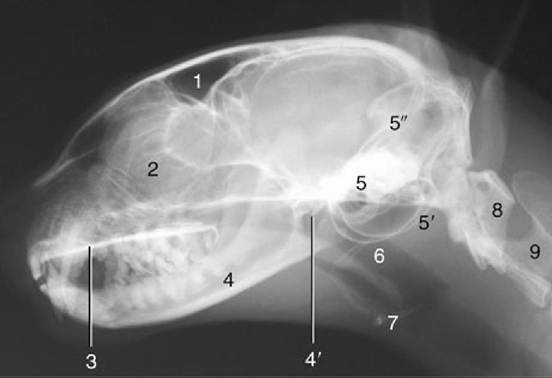
FIG. 11.29 Radiograph of the feline head. 1, Frontal sinus; 2, cribriform plate and ethmoidal conchae; 3, hard palate; 4, mandible; 4', temporomandibular joint; 5, petrous temporal bone; 5', tympanic bullae; 5", tentorium cerebelli; 6, nasopharynx; 7, basihyoid; 8, atlas; 9, axis.