Parotid Gland
The parotid gland (see Fig. 11.6) is roughly triangular, relatively thin, and molded around the proximal portion of the auricular cartilage, against which it can be rolled on palpation.
It occupies a depression formed by the masseter muscle, the wing of the atlas, and the auricular cartilage. Ventral to the cartilage, it is related medially to the facial nerve and maxillary vein and more rostrally to the parotid lymph node and temporomandibular joint. The parotid duct leaves the cranial aspect of the gland and continues over the lateral aspect of the masseter muscle between the buccal branches of the facial nerve. The duct opens into the vestibule at a small parotid papilla opposite the caudal part of the upper fourth premolar tooth, approximately 5 mm from the margin of the gum. The duct makes a right-angle bend just before opening at the papilla; one can make cannulation of the duct easier by grasping the mucosa just caudal to the opening and pulling it rostrally to straighten the bend.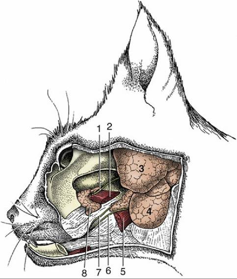
FIG. 11.27 Deep dissection of the feline head to expose the zygomatic salivary gland (8). 1, Parotid duct, cut; 2, medial pterygoid muscle; 3, parotid gland; 4, mandibular gland; 5, digastricus muscle; 6, mandibular duct; 7, sublingual duct emerging from the rostral end of the monostomatic sublingual salivary gland.
More on the topic Parotid Gland:
-
Veterinarian -