Actinobacillosis (Woody Tongue, Wooden Tongue)
Bradford P. Smith
Definition and Etiology
Actinobacillus Iignieresii, a Gram-negative rod, is a normal inhabitant of the rumen and mouth of cattle and sheep and probably goats.
When the organism enters the soft tissues through a lesion, actinobacillosis results in a granulomatous abscessation. Ruminants of all ages can be affected, and the disease appears to have a worldwide distribution. The classic site of infection is the bovine tongue; because the condition causes a very hard, diffuse nodular swelling, it has been given the name “woody tongue” or “wooden tongue.” The prevalence of woody tongue in bovines at slaughter is 0.7% to 3.6%.1 Atypical actinobacillosis lesions of cattle can occur in the lips, nose, or lymph nodes of the head or neck or at other sites. Although the lesions ordinarily occur sporadically, herd outbreaks with morbidity rates of up to 73% have been reported.1,2 Sheep are most commonly affected by hard swellings of the lips, often with fistulous tracts. Actinobacillosis has also been reported to cause tongue lesions in sheep and horses. These lesions appear to be rare in goats.Clinical Signs and Differential Diagnosis
Actinobacillosis lesions usually involve soft tissues. When the tongue is affected, the major clinical signs are inability to prehend food normally, excessive salivation, and sometimes visible enlargement of the tongue, which protrudes from the mouth. The submandibular area often is enlarged and firm. On palpation the tongue is firm to very hard, painful, and nodular (Fig. 32.83). Nodular lesions are often slightly ulcerated. The base of the tongue is most frequently affected, but the shaft may also be involved. An ulceration filled with plant awns or stems often is seen in the sulcus lingualis at the junction of the base and shaft of the tongue. Because cattle use the tongue to prehend food, anorexia results when the tongue is painful and inflexible.
Actinobacillosis must be differentiated from dental disease, oral foreign bodies, pharyngeal trauma, and other diseases that cause oral pain.Atypical lesions in cattle involve sites other than the tongue.3 Lymph nodes of the head and cranial cervical area are most frequently affected, but where any abrasion is present, granulomas or abscesses may develop, followed by licking or contact with pus draining from a lesion on another animal. Because plant awns and stems create entry sites for the organism in the mouth, most lesions are in the head. Granulomas in the nose and eyelids and needle puncture wounds over the left jugular vein have been reported. These granulomas may be confused with tumors, polyps, or cysts. Granulomas have been reported in the esophagus, pharynx, palate, flank, internal iliac lymph nodes, and testes; multiple subcutaneous lesions with regional lymph node involvement also have been reported. Three cases involving cutaneous lesions of the facial tissues were described as extensive swollen plaques under alopecic areas. Most granulomatous abscesses in a herd outbreak of actinobacillosis involved the tongue, muzzle, and lips and the submandibular, parotid, and cranial cervical areas. Generalized involvement or granuloma formation in internal organs may also occur.
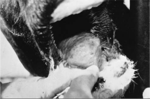
FIG. 32.83 Firm, enlarged bovine tongue typical of woody tongue caused by Actinobacillus lignieresii. Partly ulcerated areas of mucosa overlie hard nodules.
Lesions in sheep typically involve the lips and face or parotid and submaxillary regions. The nasal cavity and internal organs are occasionally involved. Soft tissues of the head may be infected through fight wounds. Lesions of the lips must be differentiated from those of contagious ecthyma, and granulomatous abscesses in other sites must be differentiated from caseous lymphadenitis lesions. Lesions of the tongue in sheep, essentially identical to those found in cattle, have been reported as a cause of green staining of the lips and “cud-dropping” in sheep.
Clinical Pathology
Diagnosis of actinobacillosis requires biopsy and culture of the lesion. The pus usually is not malodorous. Pus crushed between two glass slides shows “sulfur granules,” “clublike rosettes,” or “club colonies.” Similar colonies may be found in actinomycosis and some staphylococcal infections. A. Iignieresii organisms are small, Gram-negative rods. Definitive diagnosis relies on culture. No reliable serologic test is available for actinobacillosis, and the hematologic and biochemical findings may be normal or typical of a chronic infection.
Pathophysiology
A. Iignieresii is a normal inhabitant of the mouth in ruminants and can be found in many plant awns. When mucosal lesions occur as a result of plant awns (e.g., foxtails), thistles, or particularly stemmy coarse feed, actinobacillosis may occur. Many affected cattle have a small ulcer in the sulcus lingualis at the junction of the base and shaft of the tongue. Plant fibers are sometimes found in the granulomatous lesions of actinobacillosis. Once inoculated into tissues, the organism may cause a local lesion, lesions in draining lymph nodes, or both. Lesions elsewhere on the body may be contaminated by saliva or by pus from other draining lesions or directly by plant awns on which A. Iignieresii resides.
Epidemiology
Most cases of actinobacillosis are sporadic. Herd outbreaks may be associated with abrasive feedstuffs and crowded conditions in which the organism is spread rapidly to wounds on other animals by way of saliva. In one herd outbreak, 73% of a group of heifers (4 to 24 months of age) were affected 1 month after feeding of a coarse, stemmy haylage had begun.2 Atypical lesions often are associated with a previous wound at the site, such as a nose lead wound, multiple needle punctures, or a head butting wound. Outbreaks of actinobacillosis in wounds of the head, neck, body, and limbs have been reported.
Necropsy and Biopsy Findings
Actinobacillosis lesions typically are firm, pale, gritty, granulomatous abscesses.
On gross examination, they appear similar to exuberant granulation tissue and connective tissue, often appearing to have a yellowish granular (1- to 3-mm) surface. Masses contain multifocal necrotic foci, often filled with nonodorous, thick, yellow-white pus. Histological examination reveals that the lesion is a granulomatous abscess. An outer capsular region of connective and granulation tissue surrounds an area of leukocytes and rosettes (“club colonies”). Mononuclear cells, plasma cells, and eosinophils predominate. Many neutrophils are seen at the center of the lesion. Multinucleated giant cells or plant fibers, or both, may be present.Treatment and Prognosis
Treatment usually is successful, and the condition has an excellent prognosis when only the tongue is involved. The prognosis may be only slightly less optimistic when internal organs and atypical sites are involved. Sodium iodide (70 mg/ kg given IV as a 10% to 20% solution) is the treatment of choice. Intravenous treatment is given once and repeated at least one more time 7 to 10 days later. In refractory cases, intravenous therapy may be repeated more often (2- to 3-day intervals). In severe cases, daily organic iodides can be administered orally at a rate of 60 mg/kg/day4 in addition to the intravenous iodide. If signs of iodinism develop (excessive tearing, coughing, inappetence, diarrhea, or dandruff), iodine administration should be halted; the adverse signs normally disappear shortly thereafter.
The onset of therapeutic benefit of sodium iodide is remarkably rapid. Within 48 hours after treatment, the tongue is flexible enough to allow the animal to eat. Although the mode by which iodides exert their beneficial therapeutic effect in actinobacillosis is not well understood, they probably exert some antiinflammatory effect on the granulomatous inflammation. Iodides have little in vitro bacteriostatic or bactericidal effect at the concentrations given,5 but the onset of action is very rapid.
They probably act in some other way than by direct antimicrobial effect. The old belief that iodides cause abortion at the recommended dose has been cast into doubt by the author's clinical experience and by reports of others,6 who gave 1.5 to 2 times the recommended intravenous dose without inducing abortion. Nevertheless, there are anecdotal reports of the association, and when products are labeled with a contraindication to their use in pregnant cattle, due caution should be exercised. Iodides should be given slowly and with caution to horses because of the possibility of severe generalized adverse reactions to intravenous sodium iodide.Most strains of A. Iignieresii are sensitive in vitro to a number of antimicrobial drugs, including ceftiofur, ampicillin, penicillin, florfenicol, sulfa drugs, certain approved aminoglycosides, and tetracyclines. Each isolate should be tested for antimicrobial sensitivity. Therapy with an antimicrobial drug to which the isolate is sensitive is recommended in severe, generalized, or refractory cases of actinobacillosis. Therapy should also include iodides.
Surgical debulking of lesions, particularly if they interfere with airflow, is also possible for atypical cases in which a large granulomatous mass is present and the mass has proved refractory to medical therapy. Hemostasis may be a problem after surgical debulking.
Prevention and Control
Prevention relies mainly on avoidance of coarse, stemmy, scabrous feeds and pastures full of hard, penetrating plant awns (e.g., foxtails) or thistles. If an outbreak occurs, immediate change to a softer feed is advised, and affected animals should be treated individually. Rapid resolution of the outbreak can be expected once these steps have been taken. If atypical lesions occur, a cause of skin wounds in the area should be sought and resolved.