Dental and Periodontal Diseases
Sylvain Nichols • Guy St. Jean
Dental Attrition and Erosion
Rapid wearing of teeth is most common in grazing sheep older than 5 years of age. Many sheep and cattle grazing foragedeficient or sandy pastures and arid ranges in Africa, Australia, New Zealand, and the southwestern United States display accelerated tooth wear.7,11 In Rhodesia, the incisors of Hereford cattle had an increased rate of wear because of softer enamel, in comparison with those of indigenous cattle.12
Examination of an animal with dental attrition reveals worn incisor or molar teeth (or both); often only short stumps are seen.
The teeth also may be loose, fractured, or missing. Dental attrition from excessive wear must be differentiated from periodontal disease that causes tooth loss.7,13,14 The pathogenesis of excessive tooth wear relates to tooth hardness and diet quality. Ingestion and mastication of soil and sand with forage abrades and wears the incisor and molar teeth. In New Zealand, dental attrition in sheep was attributed to acids and enzymes in the herbage consumed, which acted on the tooth dentine.11 A calcium deficiency or calcium/phosphorus imbalance, causing softness of the tooth enamel and dentine, may accelerate the rate of wear.In the United States feeding fermented sweet potato cannery waste to cattle resulted in substantial increases in incisor erosion.8,9 Cattle producers feeding sweet potato cannery waste noticed poor growth rate, inadequate calf development, low pregnancy rates in heifers, and worn, mottled, discolored incisors.8,9 Sweet potato cannery waste is highly acidic (pH, 3.2) and causes calcium loss and tooth erosion. Deciduous teeth are etched more rapidly, which places young cattle at higher risk for severe tooth wear and dental infection. In addition, the distance between the original enamel surface and the pulp chamber is less in deciduous teeth than in permanent teeth, which is, again, more significant in younger cattle.8,9 Mixing 10% broiler litter with sweet potato cannery waste raised the pH to 4 and thus provided a palatable high-quality feed and prevented the severe dental problems associated with sweet potato cannery waste alone.8 It is important to allow a withdrawal time of 21 days before slaughter of cattle fed broiler litter to avoid the presence, in the meat, of pharmaceutical residues used in the poultry industry.
A syndrome involving excessive wear of deciduous incisor teeth, maleruption of permanent incisors, and an increased prevalence of dentigerous cysts in sheep has been reported in New Zealand.15 Excessive wear of the incisor teeth of cattle was also recorded on the same farms. Ingestion of soil during winter because of the inclement weather, overgrazing of pastures, and the presence of low copper levels in the blood of affected animals were the main causes for the syndrome.15 Dental attrition is prevented by providing supplemental feed to avoid overgrazing of pastures. The addition of 1% ground limestone to the feed in calcium-deficient areas is recommended.16
Periodontal Disease
Periodontal disease involves the supporting tissue that surrounds the teeth.17,18 Periodontal disease of sheep is endemic in parts of New Zealand. A periodontal disease kfiown as cara inchada, or “swollen face,” has caused losses of cattle in Brazil.19,20 Periodontal disease is characterized by protruding and loose incisors.7,13 With time, incisors, premolars, and molars may be missing. Periodontal disease seems to be more prevalent in sheep than in cattle. However, an abattoir investigation in West Scotland revealed periodontal diseases in 12% of the heads evaluated. The disease was located mainly on masticatory teeth and was more frequent on beef cattle heads.21 Periodontal disease results in pain during mastication, which leads to poor rumination and reduction in food digestibility.
Periodontal disease has been associated with bacterial plaque-induced gingivitis.17,20,22,23 The acute gingivitis is replaced progressively by a chronic inflammation in the gingival sulcus. The periodontal ligament is destroyed by plaque-forming oral microorganisms and host enzymes. At this stage, periodontal pockets are formed, which cause loosening of teeth. This process may take months or years. Over time, the infection extends to the apical area.
The gum margin begins to recede over the lesion, food accumulates within the pocket, and the entire alveolus becomes infected. At that point, the tooth becomes a sequestrum. The alveolar pyorrhea causes a periostitis of the external surface of the alveolar process, and swelling is observed. Once a tooth is lost from an affected alveolus, granulation tissue fills the alveolus and the alveolar bone undergoes remodeling.Bacteria such as Truperellapyogenes, Bacteroides spp., Actinomyces spp., and spirochetes; metabolic or immune disorders in the host; and mechanical or chemical agents have been implicated in the 7222425 pathogenesis of periodontal diseases in sheep.7,22,24,25 Bacterial invasion in the gingival pockets has been associated with a defect of host immune competence.26 Hypomagnesemia has been a common finding in sheep with periodontal disease, but this may be a secondary condition.25 Gingival trauma may have a causal role.27 Dissolution of the enamel at the attachment of the gingival epithelium by organic acids from microorganisms of the soil may be a predisposing factor.28 The serum values for calcium, albumin, ALP, and citrate were lower for sheep with periodontal disease than for sheep not affected by dental disease.29
A postmortem examination of Scottish-Hill sheep revealed that 60% of 478 aged sheep had either loose or missing teeth. Gingival pockets were present in 87% of the population and were correlated with tooth looseness.30 In the United States, a 25% rate of mortality caused by dental disease in a herd of 300 ewes was reported.31 The clinical signs were depression, anorexia, and emaciation.31 Necropsy revealed dental disease of the mandibular teeth with plaque, plant fibers in periodontal pockets, and osteomyelitis of the mandibular bone. An initial trauma to the gingiva from cheatgrass awns in the hay field was implicated.31
A particular type of periodontal disease in cattle was reported in Brazil.19,20 This condition involves an inflammatory process of the periodontium of calves and older Zebu cattle that results in alveolar periostitis of the maxilla or, less often, the mandible.19,20 Examination reveals deep periodontal pockets and loss of or loosened teeth.
Affected animals suffer from malnutrition, diarrhea, and loss of condition, and many die. Bacteroides and Actinomyces organisms often are found in the lesion and are suspected of causing the disease.19 This disorder is seen only in certain areas of Brazil, and the cattle improve if moved to unaffected areas. In one trial, the development of periodontal disease in calves was avoided by administration of the antimicrobial drug spiramycin.20The assumption that incisor condition is a good indicator of future productivity is not well founded. Little scientific evidence exists to support the culling of sheep affected by periodontal disease. Three farms with a high prevalence of periodontal disease were selected, and the body score and weight of affected sheep were compared with those of sheep with no signs of periodontal disease. A significant association between periodontal disease and body condition or weight was identified on only one farm. It was concluded that periodontal disease in sheep may impair productivity on some farms but not others.32,33
Treating periodontal disease on a flock basis often proves impractical. Dental treatment, drug therapy, and management change have been tried in sheep. One form of dental treatment, tooth grinding, consists of trimming the incisors to the level of the lower dental pad.13 Two trials of the productivity effects of tooth grinding were conducted; neither showed any positive benefit from the procedure.32 An attempt to influence the development of periodontal disease in sheep by long-term treatment with tetracycline and metronidazole was also found ineffective.23 In commercial sheep, surgical treatments could improve conditions such as periapical and gingival abscesses, but these treatments are not economically feasible.
Sheep can live without incisors if they do not have to graze too closely. Supplementary feeding should provide a net gain to the owner. When premolar and molar teeth are involved, this approach is unlikely to be effective because of the inability to chew and ruminate efficiently.
Dentigerous Cysts
Dentigerous cysts have been described in ruminants, particularly 1533
sheep.15,33 These are odontogenic cysts of unknown cause that manifest as localized, bony swellings. Radiographs demonstrate the cystic nature of the swelling and reveal one or more teeth in the cyst.15,33 For valuable animals, treatment is surgical.
Developmental Anomalies and Retention of Deciduous Teeth
Developmental anomalies have been reported in cattle.6 Occasionally, tooth buds fail to develop. The fourth pair of permanent incisors is the set most often absent. The mandibular premolar on one or both sides also occasionally fails to form. The permanent incisor teeth have been observed to be rotated up to 180 degrees in the alveolus. Rotation of a first permanent mandibular premolar was observed as well; the rotation was 90 degrees, and no precise cause was identified. Retention of a deciduous premolar is common in 12- to 18-month-old cattle. This results in difficulty in masticating food and excessive salivation. The treatment consists of removing the deciduous premolar with a molar-extracting forceps.
Overgrown or Loose Molar Teeth
Overgrown molar teeth are found most commonly in older ruminants. The opposite tooth is often missing. Food accumulates between the affected tooth and the cheek, causing obvious swelling of the face. Interference with mastication is often noted. The offending tooth should be rasped regularly or removed. A power tool to grind cheek teeth works well. Premolar or molar teeth can become loose in advanced cases of actinomycosis affecting the jaw. It may be difficult to determine whether the loose tooth is a cause or result of the bony abnormalities. One cow with maxillary lymphoma and loose teeth has been reported.34
Broken Teeth
Broken incisors are not common because of the loose alveolar attachment of these teeth. Broken premolars and molars are more common because these teeth are more solidly attached in their alveoli; breakage usually results from attempts to masticate hard objects.
Most fractures usually involve only a portion of the tooth, do not involve the root, and are asymptomatic. A sagittal tooth fracture that includes the root causes pain and may result in reduced feed intake and loss of condition. (Treatment is described later in the Tooth Root Abscess section.)Dental Caries
Dental caries, or decay, creates areas of decalcification of the tooth.4 Dental caries is rare in ruminants,4 but it sometimes occurs in both deciduous and permanent teeth and in both dentin and enamel.6 Interference with prehension is rare. On oral examination, an orange or black pigment is seen in the defective enamel or exposed dentin; these areas should be probed with a fine dental pick or needle. Frequently, the caries fills with food material until it reaches the pulp cavity, which creates a tooth root abscess (Fig. 32.78). At that time, the productivity of the animal decreases, and a swelling on the mandibular or maxillary bone, or purulent discharge secondary to sinusitis appears. A head scan or oblique radiograph is useful for confirming the diagnosis (Fig. 32.79). The infected tooth should be removed. Because the infection is located at the apex of one or multiple roots, the periodontal ligament holds the tooth firmly in the alveolar socket, which makes oral extraction difficult. A surgical repulsion is often necessary. (See the Tooth Root Abscess section.)
Osteodystrophia Fibrosis
Osteodystrophia fibrosa is caused by resorption of calcium from bone and its replacement by connective tissue.35 It is most common in goats. Osteodystrophia fibrosa can result from calcium, phosphorus, or vitamin D deficiencies or from
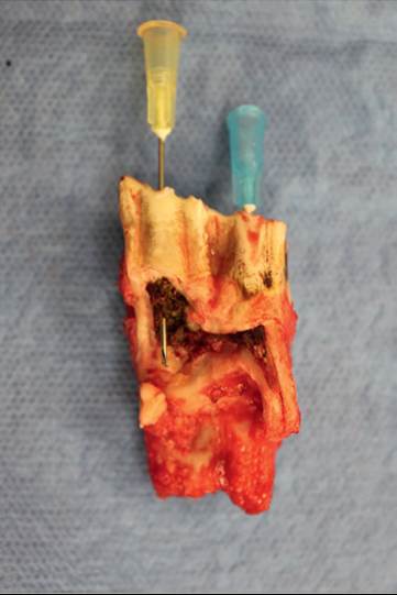
FIG. 32.78 Photograph of a third mandibular molar extracted surgically (the roots are missing). Two hypodermic needles are placed through infundibular caries. The pulp cavity is filled by food material that caused a root abscess. (Courtesy Caroline Constant, Universite de Montreal.
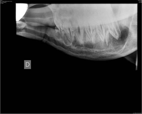
FIG. 32.79 A right lateral oblique radiograph of the mandible of a 3-year-old Holstein cow shows a radiolucency center over the caudal root of the third molar. Lysis of the apex of the root is present. (Courtesy Universite de Montreal.)
hyperparathyroidism. Usually a growing animal with osteodystrophia fibrosa has a bilateral, soft, nonpainful, and swelling of the maxilla or mandible or both jaws.35 The diagnosis is based on radiographic evidence of poorly mineralized bone and inward rotation of the premolar and molar teeth. The treatment consists of supplementing the ration with adequate mineral levels while maintaining a 2 : 1 ratio of calcium to phosphorus.
Tooth Root Abscess
A tooth root abscess can be a complication of periodontal disease or dental caries. It can be diagnosed in aged cattle but seems more frequent in younger cattle. Periodontal disease at the time of permanent tooth eruption could explain the presence of this condition in young cattle. Early on, the repercussions on the health of the animal are minimal. Chewing behavior might change, and growth or milk production could be reduced. With time, a swelling appears around the infected tooth. Eventually, a draining tract becomes evident. If the molars or the caudal root of the fourth premolar is infected on the maxillary arcade, unilateral nasal discharge associated with halitosis appears because the roots of those teeth are located in the maxillary sinus. With time, the mandible or the maxilla becomes distorted from the severe periosteal reaction and the bony proliferation caused by the chronic infection. In some cases, the external appearance of the jaw resembles cases of osteomyelitis caused by Actinomyces bovis (actinomycosis). The oral examination may reveal periodontal disease (exposed roots), a fractured tooth, or caries. A rigid endoscope is useful for evaluating the caudal cheek teeth. The final diagnosis is confirmed by a head scan or oblique radiographs. Medical therapy fails to resolve the infection because the tooth has become a sequestrum; therefore the tooth should be removed. Oral extraction is possible in chronic cases or in cases secondary to periodontal disease. With the animal under sedation or general anesthesia, a mouth gag is used to open the jaw. The affected tooth is located, and the gingiva is elevated on the lingual and buccal side of the tooth. If the tooth is not already mobile, a molar spreader can be used to loosen the periodontal ligament. Molar-extracting forceps are placed on the crown, and the tooth is moved slowly from left to right until the periodontal ligament is completely broken. Finally, the tooth is removed by means of a fulcrum effect. The procedure is, in the author's opinion, difficult when caudal cheek teeth are involved. The limited mobility of the temporomandibular joint reduces the working space in the back of the oral cavity in cattle.
Dental repulsion is done, preferably with the animal under general anesthesia, with the affected tooth uppermost. Mandibular teeth are removed through a ventral or a lateral approach. With the ventral approach, a Galt trephine is used to remove the bone covering the roots of the infected tooth. Then a straight dental punch is placed over the roots before the tooth is hammered out. With the lateral approach, the lateral mandibular bone plate lateral to the infected tooth is removed with a pneumatic burr or with a bone rongeur and curette. The roots are freed from the bone, and a curved dental punch is used to hammer the infected tooth out. In the author's experience, the lateral approach works best to remove mandibular molars. The surgical approach is more challenging, and care must be taken to preserve the facial artery and vein that travel just over the third molar.
Maxillary premolars are removed through a dorsal approach by trephining the maxillary bone above the infected tooth. To remove maxillary molars, a maxillary bone flap is made above the maxillary sinus. Straight or curved dental punches are used to hammer the tooth out. It is important to seat the punch perfectly on the tooth to prevent the punch from slipping and fracturing the hard palate or lacerating the palatal artery. Intraoperative imaging is essential to identify the proper position of the dental punch and to ensure that no tooth fragments are left behind.
The alveolus is packed with dental wax, gauze, or dental acrylic, such as Optosil (Heraeus Kulzer GmbH, Hanau, Germany).8 On the mandible, the ventral aspect of the incision can be left open for drainage. If sinusitis is present at time of surgery, a Foley catheter is left in the sinus postoperatively to allow daily flushing. Antibiotics are administered for 1 to 2 weeks, according to culture antimicrobial sensitivity results. The dental plug is removed 3 to 4 weeks after surgery. Sedation and the use of a mouth gag may be necessary to remove the dental plug safely. The prognosis after oral or surgical extraction is good.
Dental Fluorosis
Dental fluorosis is associated with continuous ingestion of small but toxic amounts of fluorine in the diet or drinking water. Most reported occurrences of fluorosis are in cattle. Sheep are less susceptible. Developing teeth are very sensitive to the ingestion of excess fluorides. Fluorides inhibit the action of ameloblasts and odontoblasts during tooth formation, which results in failure of the tooth to incorporate minerals. Fluorosis causes striations or patches (yellow, brown, green, or black) in enamel. Some erosion of the permanent tooth can occur. Cattles are susceptible to dental fluorosis during enamel matrix formation from approximately 6 months to 3 years of age. Severe dental fluorosis results in dental wear, inability to graze properly, and difficulty with mastication and food intake because of tooth pain. In addition to clinical signs, the fluorine concentration in the blood and urine can be analyzed to confirm the diagnosis. Normal cattle have blood levels of up to 0.2 mg/ dL of blood and 2 to 6 mg/kg in urine. Cattle with fluorine intakes sufficient to cause intoxication may have bloods levels of 0.6 mg/dL and urine levels of 16 to 68 mg/kg. Treatment usually entails removing the animals from the source of fluorine.
Rostral Mandibular Masses
Vascular gingival hamartomas are defined as disorganized and excessive proliferations of vascular tissue that are present at birth (Fig. 32.80).36,37 They are considered a developmental anomaly rather than a true neoplasm because of their limited growth. En bloc excision combined with rostral mandibulectomy is curative.38
Odontogenic tumors are not common in cattle. They occur generally on the rostral mandible but can be found more caudally on the mandible and maxillary. 39 Ameloblastic tumors are the most frequent odontogenic tumor.40 Depending on the tissue involved in the mass, they are called ameloblastic fibroma (dental epithelium), fibrodentinoma (dental epithelium and small island of dentine), orfibroodontoma (dental epithelium, dentine and enamel). They are benign tumors with continuous growth and are found in younger cattle. A mass of variable
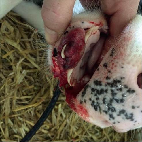
FIG. 32.80 Photograph of a vascular gingival hamartoma in a 3-day-old calf. (Courtesy Sylvain Nichols, Universite de Montreal.)
size covered with gingival mucosa with distorted deciduous and permanent incisors is present (Fig. 32.81). The mass has no effect on the health of the animal. Eventually, ulcerations appears from rubbing while eating. Rostral intraoral radiographs help determine the extent of the tumor in the mandible (Fig. 32.82). En bloc excision is curative. To obtain clean margins, a rostral mandibulectomy is necessary.41
Odontoma is a benign tumor composed of normal dental tissue that grows in an irregular way. It affects permanent teeth before eruption and is therefore found in younger cattle. It does not appear to be common in cattle, however.
Other masses affecting the rostral mandible are fibrosarcomas and osteosarcomas. Those malignant tumors occur in older cattle. Rostral mandibulectomy, if performed early, may be curative.
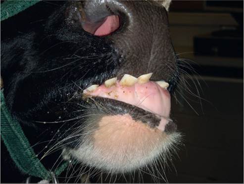
FIG. 32.81 Photograph of an ameloblastic fibroma in a 4-year-old
Holstein cow. (Courtesy Sylvain Nichols, Universite de Montreal.)
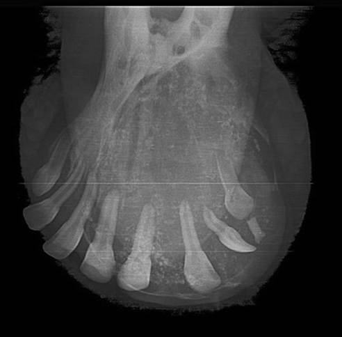
FIG. 32.82 An intraoral radiograph of the cow in Fig. 32.6. The tumor has changed the normal anatomy of the rostral mandible. (Courtesy Universite de Montreal.)
ROSTRAL MANDIBULECTOMY. This procedure is best performed with the animal under general anesthesia in sternal recumbency. With a mouth gag in place, the gingiva is incised at the predetermined site of the mandibulectomy. A periosteal elevator is used to retract the gingiva attached to the periosteum rostrally and caudally. An oscillating saw or a Gigli wire is used to amputate the rostral mandible. The saved gingival mucosa is sutured to cover the bone. Bleeding is to be expected during the dissection of the gingival mucosa and from the mandibulectomy site. Bone wax helps control bleeding from the mandible. A pressure bandage may be needed postoperatively. It incorporates the lower lip and the remaining rostral mandible.
The surgery site heals rapidly without complications. Because cattle graze mainly with their tongue, no repercussions have been observed with regard to food intake after rostral mandibulectomy.
Salivary Gland Diseases
Sylvain Nichols • Guy St. Jean
The parotid, mandibular, and sublingual glands are the three largest salivary glands in ruminants.1 In cattle, the mandibular gland is larger than the parotid gland. The parotid gland is located behind the caudal border of the masseter, from the zygomatic arch to the most ventral aspect of the mandible’s vertical ramus. Its duct enters the gland at its most ventral location. It travels along the facial vein to enter the oral cavity abaxial to the second maxillary molar. The mandibular gland lies medial to the caudal portion of the mandible from the basihyoid bone to the paracondylar process. The duct exits the mandibular gland axially and travels rostrally between the deep mylohyoideus muscle and the sublingual gland. It enters the oral cavity rostral to the frenulum of the tongue. The sublingual gland has two parts: the monostomatic and the polystomatic portions. The monostomatic portion is 10 cm long, and its main opening is near the entrance of the mandibular gland’s duct. The polystomatic portion has multiple microscopic ducts along the base of the tongue.2
A dairy cow in lactation produces 100 to 200 L of saliva in 24 hours. Saliva is secreted continuously, but the rate of secretion is increased by feeding, rumination, and the presence of coarse feed in the rumen.1 The saliva provides a fluid medium for transport of ingesta during deglutition and regurgitation. It also maintains adequate phosphate for bacterial digestion of cellulose in the rumen and contains bicarbonate, which acts as a buffer to maintain ruminal pH above 5.5. Ruminant saliva contains approximately 90 to 140 mmol/L of bicarbonate. The normal pH is around 8.2.3
Excessive Salivation
Excessive salivation (ptyalism) is a sign of many pathologic conditions. The volume of saliva may be normal, but if it is not swallowed, salivation can appear excessive. As a precaution against exposure to rabies, gloves should be worn to examine the mouth of any animal that is salivating excessively. Excessive salivation may be seen with dental disease, stomatitis, foreign objects in the mouth or pharynx, or esophageal obstruction. It also has been seen with ruminal disorders, in cows that have eaten spoiled silage, and with impaction of the abomasum. Ptyalism may be a clinical sign in rabies, pseudorabies, meningoencephalitis, listeriosis, tetanus, and slaframine toxicity.1,4 Mercury, iodine, lead, copper, and arsenic also can stimulate secretion by the salivary glands. Treatment depends on the underlying causes.
Salivary Duct Atresia or Agenesia
This condition is diagnosed in younger animals. A soft swelling is palpable along the normal location of the duct. Sialography may be performed to highlight the obstruction. If possible, creating an opening in the oral cavity allows the saliva to drain in the mouth. This procedure is performed with the animal under general anesthesia. A 1.5-cm opening is created by suturing the oral mucosa to the salivary duct mucosa.5 This procedure is difficult to perform when the obstruction is located distally from the oral opening. Other therapeutic options include chemical debridement or surgical removal of the affected gland and duct (see Sialocele section, as follows).
Sialocele
A sialocele develops when saliva escapes from a duct or salivary gland and enters the surrounding tissue. The saliva contains enzymes that irritate the tissue. The accumulation of saliva is surrounded by inflamed tissue, which gives the lesion a cystic appearance. A soft, fluctuant swelling usually is seen. Pain may be observed during mastication. Trauma or a foreign body is usually the cause of a sialocele. The diagnosis is based on the history, palpation findings, ultrasound evaluation, fluid aspiration and cytologic evaluation, and sialography. Needle aspiration may yield yellow mucoid saliva. Two treatments have been described.6,7 The first involves removal of both the involved glands and duct.6 Surgical extirpation of the mandibular salivary gland in the caudal area of the mandibular spaces has been performed successfully in cows, sheep, goats, and buffaloes.1 Exposure of the tissue by opening the capsule of the gland facilitates the process of extirpation and prevents trauma of the surrounding nerves and blood vessels.6 In the second treatment option, the sialocele is opened, drained, and chemically debrided with a caustic agent such as iodine or copper sulfate.7 Post-debridement analgesia is mandatory after chemical resection.
Sialoadenitis
Inflammation of the salivary glands usually is caused by an ascending infection from a traumatized duct, by penetrating wounds or plant awns, or by a hematogenous embolization. Diffuse swelling of the salivary glands may be observed, and the swelling may be hot and painful on palpation. Abscess formation is a common complication. The pain may interfere with mastication and swallowing. Treatment consists of systemic antibiotics, antiinflammatory drugs, and analgesic drugs. Abscesses should be drained when they localize. Salivary fistulas can be sequelae to salivary abscesses. Surgical extirpation is also possible. However, because of the inflammation surrounding the gland, the dissection is more challenging, and the odds of traumatizing neurovascular structures are increased.5
Parotid Gland Carcinomas
In three cows at slaughter that had parotid gland carcinomas, the neoplastic cells appeared to originate from ductal and acinar epithelium.8 Ocular squamous cell carcinoma may spread locally to the parotid or mandibular lymph nodes.9