Actinomycosis (Lumpy Jaw)
Bradford P. Smith
Definition and Etiology
Actinomycosis is caused by A. bovis, a Gram-positive, nonencapsulated, branching, filamentous bacterium that is a normal inhabitant of the ruminant mouth.
The disease occurs mainly in cattle, but on rare occasions it affects sheep or goats, and one report of the disorder in a horse involving Actinomyces viscosus and Actinomyces odontolyticus has been published.1 The bacterium enters the tissues and bone through oral abrasions, openings, and punctures associated with dental or gingival disease, hard plant awns (e.g., foxtails), thorns, stickers, or dry, coarse, stemmy feeds. Lesions may begin as cavities (caries) in the dentin or dental pulp.2 They are sporadic and occur mainly in the mandible and less commonly in the maxilla. Because of the preponderance of mandibular lesions, with the development of periosteal new bone and fibrosis, the disease is commonly known as “lumpy jaw.” Lesions occasionally occur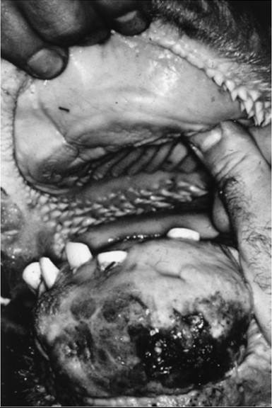
FIG. 32.84 Hard swelling on the distal mandible of a cow infected by Actinomyces bovis. Loss of teeth and bone destruction, along with fibrosis and callus formation, are seen in this advanced case. The oral mucosa is secondarily ulcerated by trauma.
in soft tissues of the head, esophagus, forestomachs, and trachea. A. bovis occasionally causes granulomatous abscesses in other soft tissues. Most of the early reports of esophageal groove and forestomach involvement incriminate actinobacillosis rather than actinomycosis.
Clinical Signs and Differential Diagnosis
Bovine actinomycosis typically causes a hard, immovable, painless, bony mass on the mandible (Fig. 32.84). The lesion is most common on the horizontal ramus. Initially it is nondraining (has no fistulous tracts), but fistulous tracts may develop and involve tooth roots as the condition progresses (Figs.
32.85 and 32.86). When teeth become involved, the animal shows evidence of pain during chewing, and weight loss may result. A careful examination of the mouth is necessary to detect loose teeth, plant awns, or severe gingivitis and to rule out a pathologic fracture. If a fistula is present, the tract should be flushed with organic iodine and contrast radiographs obtained to determine whether it communicates with the mouth. The differential diagnosis includes tooth root abscess, fracture, tumors, and osteomyelitis caused by other organisms. If a mandibular swelling continues to enlarge despite therapy, radiographs should be examined for evidence of a fracture or sequestrum. Atypical actinomycosis with lesions in soft tissue causes a variety of clinical signs, depending on the location.Clinical Pathology and Diagnosis
Hematologic and clinical chemistry findings may be normal or may reflect a chronic infection. Radiographs of the lesion are helpful in determining dental involvement or a pathologic fracture. The radiographic lesion consists of multiple central radiolucent areas of osteomyelitis surrounded by periosteal new bone and fibrous tissue. If a fistulous tract is present, a contrast study performed while the tract is flushed may help
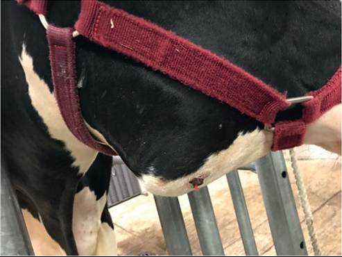
FIG. 32.85 Hard swelling and draining tract on the mandible of a cow, typical of Actinomyces bovis lesion.
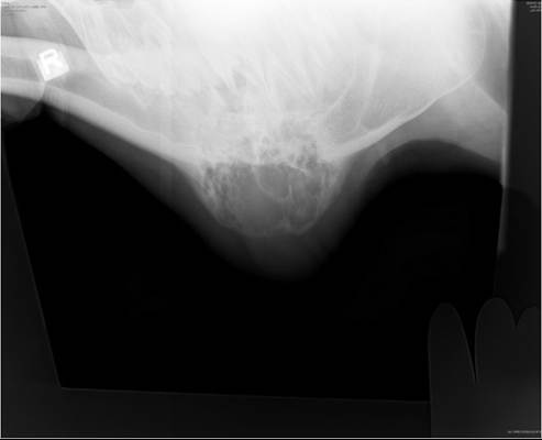
FIG. 32.86 Radiograph of lesion in the cow in Fig. 32.86, showing cavitating central bone loss and peripheral bone density, typical of Actinomyces bovis lesion.
determine the extent of the fistula. Before flushing, material from the core of the lesion should be aspirated or examined in biopsy. A Gram stain and culture of pus should be performed. The organism is Gram-positive, filamentous, and branching. “Sulfur granules” similar to those described for actinobacillosis may be seen.
Many authors report that they were unsuccessful at culturing the organism.Pathophysiology and Epidemiology
A. bovis appears to enter the bone through mucous membrane punctures caused by foreign bodies, plant awns, or coarse, stemmy feeds or through a diseased tooth or areas of gingivitis that allow oral bacteria access to the bone. Cases usually are sporadic.
Necropsy and Biopsy Findings
Actinomycosis causes a granulomatous abscess. Scattered through the mass of tissue are basophilic clumps of bacteria surrounded by eosinophilic clublike projections. The bacteria appear as long, filamentous, branching rods. Surrounding them is evidence of cellular reaction, composed of neutrophils, epithelioid cells, macrophages, and occasional multinucleated giant cells. In the outer fibrous tissue are plasma cells.
Treatment and Prognosis
Treatment of actinomycotic bone lesions usually results in arrest of the lesion, but seldom does the size of the hard mass regress significantly. The prognosis for arrest of the lesion with vigorous treatment is good. If the mass does not have any fistulous tracts and no affected teeth are loose, medical therapy alone may be sufficient. If the mass has fistulous tracts, it should also be vigorously curetted and flushed with povidone-iodine or other organic iodine. The lesion has a rich blood supply, and curettage can result in severe hemorrhage. If the cavity is large, it may be necessary to flush and pack with iodine-soaked gauze daily for several days and then less frequently as healing progresses. Three cases of actinomycosis were treated successfully with a repeated regimen of curettage followed by cryotherapy with liquid nitrogen poured into the lesion. The authors repeated the treatment on days 2, 9, and 16.3 If there is an open fistula into the mouth (as judged by vigorous flushing) or if teeth are involved, the affected tooth or teeth should be carefully removed or repaired endodontically.2 Care must be taken to prevent mandibular fracture, and the animal must be sedated or anesthetized to allow for proper intraoral manipulation.
The empty alveolus should be carefully packed with gauze or a dental acrylic to which a wire or umbilical tape is attached and pulled through the fistula. The wire is tied externally to a small gauze roll to keep the alveolar packing firmly in place. The wire should be untied daily and the tract flushed thoroughly with povidone-iodine until the wound is completely granulated. Once granulation begins, it may not be necessary to check and flush the lesion more than once a week. The alveolus will take several weeks to close, pushing the gauze or acrylic out as it does so.Medical treatment of actinomycosis involves the use of sodium iodide and penicillin or another antimicrobial drug to which the organism is sensitive. Sodium iodide is given IV at a dose of 70 mg/kg as a 10% to 20% solution. It can be given every 7 to 10 days or more often until signs of iodinism occur (i.e., lacrimation, cough, inappetence, diarrhea, and dandruff). If repeated intravenous treatments are difficult, oral organic iodides (total powder) can be given at the rate of 60 mg/ kg/day for 3 weeks. As with actinobacillosis, the beneficial therapeutic effects of iodides appear to arise from their ability to reduce granulomatous inflammation rather than from direct antimicrobial effects. Iodides do not appear to cause abortion and can be safely given to pregnant cows,4 although care should be used because there are anecdotal reports to the contrary, and products are labeled with a contraindication for pregnant cattle.
Isoniazid (10 mg/kg/day given PO for 1 month) is effective at arresting actinomycosis of the mandible in cattle.5 It is inexpensive and readily consumed in a small amount of grain. A prolonged withdrawal period before slaughter for human consumption is required. The drug appears to be nontoxic at this dosage, but it may cause abortion and should not be used in pregnant cattle. The Food Animal Residue Avoidance Databank (FARAD; www.farad.org/vetgram/) should be checked before isoniazid is used.
Penicillin (22,000 U/kg IM twice daily) or another antimicrobial drug such as florfenicol or ampicillin can be added to the treatment regimen in cases involving valuable animals or when twice-daily treatment for 7 to 14 days is possible. Streptomycin can be given twice daily, but because of the prolonged persistence of tissue residues, aminoglycosides generally are considered unacceptable in food animals.
Prevention and Control
A. bovis is a normal mouth inhabitant of ruminants; therefore the only possible means of prevention is to avoid feeding coarse, stemmy feeds; feeds with hard, penetrating plant awns; and feeds with other sharp materials. The recommendations in this regard are similar to those for actinobacillosis.