Acute Renal Failure
Thomas J. Divers • Alexandra J. Burton
Acute renal failure (ARF) in the horse is usually due to exposure to nephrotoxins or vasomotor nephropathy (e.g., hypoperfusion or ischemia).
The most common pathologic lesion with ARF is acute tubular necrosis (ATN).Toxic Nephropathies
Aminoglycosides
Administration of aminoglycoside (AG) antibiotics (gentamicin or amikacin) is one of the most common causes of ATN in horses; however, irreversible ARF due to AGs is uncommon. Aminoglycosides exert their main toxic effect by accumulating within renal proximal tubular epithelial cells. After filtration through the glomerulus, the cationic charge of AGs causes them to bind to tubular epithelial cell phospholipid membranes.1 Once within the cell, AGs disrupt a myriad of ultrastructural cellular elements causing cell swelling, death, and sloughing into the renal tubular lumen.1 More recently, AGs have been found to also cause some glomerular damage and additionally, a decrease in renal blood flow, directly medicated by an AG- induced increase in vascular resistance.2 The detailed molecular mechanisms of tubular cell necrosis, glomerular, and vascular effects caused by AGs are beyond the scope of this text, but in-depth reviews are available.1,2
Most cases of AG nephrotoxicity are not due to singleinstance drug overdose. Horses with normal renal function can usually tolerate a single major overdose without detrimental effects. Thus, AG toxicity is almost always the cumulative effect of repeated administration (e.g., in order to induce a gentamicin nephrotoxicosis model in the healthy equine, doses of 20 mg/kg q8h several days are required; the standard equine dose is 6.6 mg/kg IV q24h).3 Because AGs are water soluble and highly distributed in extracellular fluid (ECF), their clinical pharmacokinetics are influenced by variations in patient ECF volume owing to disease state or age, so different doses and dose intervals may be required to attain desired serum/plasma peak and trough concentrations depending on patient specifics.4-10 Gentamicin is the most commonly used AG in horses.
A retrospective study in hospitalized horses showed significant clinical variability in peak gentamicin concentration for a given dose and emphasized the importance for therapeutic drug monitoring (TDM) when using gentamicin to ensure adequate peak plasma concentrations, which is important for AG efficacy.6 However, because renal cellular uptake of gentamicin is saturable, nephrotoxicity is associated with insufficiently low plasma trough (i.e., sampled just before the next IV dose), rather than excessively high peak concentrations.1 The maximal trough concentration permissible to minimize the risk of nephrotoxicity is unknown in horses. In humans, gentamicin trough concentrations greater than 2 pg/mL have been associated with nephrotoxicity.11,12 However, the optimal trough concentration of gentamicin to minimize nephrotoxicity in humans is still controversial, with recommendations ranging from attaining a trough less than 2 pg/mL once in 24 hours to maintaining a concentration of less than 0.5 pg/mL for at least 4 hours.13-15 Recently in human medicine, there have been developments of gentamicin pharmacokinetic models to facilitate opportunistic TDM and to move away from daily trough concentration monitoring.16,17 The rationale behind this is to avoid repeated daily timed venopuncture, especially in neonates and children, and allow AG concentrations to be evaluated within the model at times when blood would be taken anyway for other analyses.16,17 Nephrotoxicity typically develops after several days of AG administration to horses with diarrhea or septicemia that are not adequately hydrated or because of other factors that may exacerbate a decrease in renal perfusion (e.g., concurrent treatment with nonsteroidal antiinflammatory drugs [NSAIDs]).7-10 The shift to once-daily AG dosing in humans and horses, compared with previous dosing of AGs, which was two to three times daily, has become a standard practice that reduces the potential for nephrotoxicity (by ensuring a longer period of the day with sufficiently low serum trough concentrations) but still provides a similar therapeutic response.18-21Prolonged administration of AGs without TDM or serum creatinine concentration monitoring is a common history with AG nephrotoxicity in the horse.
Typically, AGs may be safely administered for longer than 10 days if the patient is adequately hydrated and appropriate serum AG trough and serum (or plasma) creatinine concentrations are maintained. However, with regard to using serum creatinine concentration to monitor AG toxicity, in one instance, experimental induction of gentamicin nephrotoxicity in ponies was reflected by a rather small increase (0.3 mg/dL) in creatinine concentration.22 Thus, TDM and measurement of the trend in urinary γ-glutamyltranspeptidase (uGGT), which always increases rapidly with AG administration (even in the absence of nephrotoxicity), and trend in UGGT:urinary creatinine (uCr) concentrations in at-risk patients will give a much earlier indication of impending nephrotoxicity than serum creatinine concentration alone.23-25 Although it has not been proven, the neonatal equine kidney is likely more susceptible to AG toxicity than the adult kidney. This is probably reflective of the prolonged elimination half-life seen in foals.4,26 Critically ill neonatal foals may be at greater risk for AG nephrotoxicity, although this may simply reflect an increased incidence of septicemia in neonates and longer courses of treatment with AGs.When AGs are administered to high-risk patients (e.g., those with severe dehydration, sepsis or neonates), volume deficits must be continually replaced, and TDM and UGGT:UCr concentrations or, at the bare minimum, serum creatinine concentration monitored frequently.27 In horses receiving appropriate fluid therapy, AG nephrotoxicity rarely develops. Increased urinary sodium (Na+) excretion and fluid diuresis appear to have a protective effect on the kidney. In contrast, hypokalemia (or total body potassium (K+) depletion) and low calcium (Ca2+) intake may predispose horses to aminoglycoside nephrotoxicity by decreasing urine output.25 Supplementation with oral electrolytes (e.g., 1 to 2 oz of NaCl and KCl daily) may be of benefit to horses being treated with AGs by increasing water intake and urine output and by replacing K+ deficits in anorectic horses.
Furosemide should not be administered prophylactically in an attempt to prevent AG nephrotoxicity as it is a potassium-wasting diuretic. In patients with prerenal azotemia that receive AGs, it is important to monitor serum creatinine closely (i.e., to make sure that it is decreasing in the face of AG therapy) and consider prolonging the administration interval, until volume deficits are corrected, or, if possible, select an alternative antimicrobial. However, because nephrotoxicity is a cumulative effect of repeated dosing, delaying administration of the initial dose of an AG in a critical septic patient pending rehydration is often unwarranted.When ARF from AG use develops, it usually manifests as nonoliguric to polyuric renal failure. Outcome is generally favorable, so long as the duration of ARF is not prolonged and other underlying disease processes can be corrected; however, an elevated serum creatinine may take several days (and occasionally weeks) to return to within reference range. Nephrotoxicity should be considered in horses that become inexplicably depressed and inappetent while being treated with AGs or within a few days after AG therapy is discontinued. Renal failure can develop even after the drug is withdrawn, so monitoring renal function for 2 to 4 days after discontinuing AG therapy is advised in high-risk patients. Polyuria may be observed before the onset of depression and anorexia, or, if the patient becomes oliguric, mild stranguria and repeated posturing to urinate may be observed. A tentative diagnosis of nephrotoxicity is based on history of AG use and supportive laboratory data. Abnormal laboratory findings associated with tubular damage that may be detected before onset of azotemia include enzymuria (e.g., GGT) and cylindruria (casts in urine).22,23,27 Although these parameters can be monitored for early detection of tubular injury, enzymuria does not necessarily indicate nephrotoxicity per se, nor does it necessarily indicate if or when AGs should be discontinued or to what degree the interval of administration should be prolonged.
A rising trend in serum creatinine concentration (even if within reference range) and TDM (e.g., gentamicin 24-hour trough >2 μg∕mL) are often more useful indicators of a need to increase concurrent fluid therapy (diuresis), prolong AG dose interval, or discontinue AG therapy.Pigment Nephropathy (Myoglobin and Hemoglobin)
Acute tubular necrosis (ATN) and ARF subsequent to a severe episode of rhabdomyolysis due to myoglobin from muscle necrosis, especially if the associated dehydration is prolonged.28,29 Dehydration and administration of NSAIDs for analgesia may both increase the risk of ARF by decreasing renal blood flow. Pigment nephropathy can also occur secondary to any other cause of muscle necrosis (e.g., muscle trauma, prolonged recumbency). Rhabdomyolysis with ARF has been reported secondary to severe clenbuterol toxicosis in three Quarter Horse racehorses. Observation of grossly discolored urine is not a prerequisite for development of ARF in cases of myoglobin pigment nephropathy. Hemolysis (hemoglobin), which may be secondary to a variety of toxins and primary conditions, appears to be a less common cause of pigment nephropathy than myopathy. Horses with severe hemolysis and those suffering from disseminated intravascular coagulation (DIC) are at greater risk of developing hemoglobinuria and pigment nephropathy. In one study, 41% of 32 horses with red maple toxicosis and hemolysis had evidence of renal insufficiency, but it was not found to be an important risk factor for mortality.31 Hemolysis with associated ARF is also a feature of acute severe cases of equine piroplasmosis caused by either of the erythrocytic protozoa Theileria equi or Babesia caballi3 Pigment nephropathy should be suspected in horses that become anorectic and depressed during the week after an episode of tying up or during a hemolytic crisis. Measuring serum concentrations of creatine kinase (CK) and aspartate aminotransferase (AST) may help identify rhabdomyolysis, if no other etiologies of ARF are suspected.
There is little preformed creatinine in muscle, so rhabdomyolysis alone does not produce an increase in serum creatinine concentration. Even if normal initially, it is also prudent to recheck serum creatinine 2 to 3 days after an episode of rhabdomyolysis.Vitamin K3
Vitamin K3 was a common cause of ATN before its withdrawal from the North American market. Nephrotoxicity can occur rapidly after a single parenteral dose of the menadione sodium bisulfite form of vitamin K3.33,34 Development of ARF was thought to be idiosyncratic and likely involves direct tubular damage via oxidative stress and pigment nephropathy subse- 3334
quent to hemoglobinuria.33,34 Although horses are sensitive to its nephrotoxic effects, oral supplementation of vitamin K homologs with a view to enhancing bone strength has been investigated in horses in Japan, and no nephrotoxicity was reported.35,36
Nonsteroidal Antiinflammatory Drugs
Most horses do not experience appreciable adverse renal effects from NSAIDs as long as they are administered at the correct dose to euvolemic patients. However, NSAIDs may produce ARF in an occasional horse when excessive doses are administered, maximal recommended doses are administered chronically, and/or when dehydration is not corrected promptly or is ongoing.37 A typical situation where this might occur is in a horse that has been on daily NSAIDs for several months/ years and then is administered excessive additional NSAIDs for severe colic. The lesion produced by NSAID toxicity in horses is papillary necrosis in the renal medulla, also termed medullary crest necrosis (Fig. 34.1), which can be manifested by
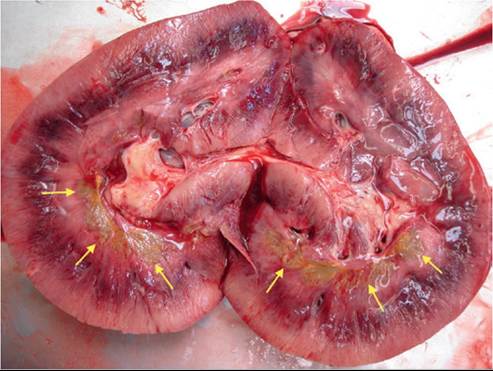
FIG. 34.1 Diffuse renal medullary necrosis (yellow arrows) in a 3-year-old gelding following a 21-day administration of phenylbutazone (2 g [approximately 4.4 mg/kg] q12h PO). This was aggravated by concurrent hypovolemia and hypoproteinemia due to hypoalbuminemia. (Courtesy Montague N. Saulez.)
gross hematuria.37-39 Unless severe, this lesion rarely causes overt clinical signs. One study suggested that ketoprofen was less nephrotoxic in comparison with flunixin and phenylbutazone (PBZ), but this study was short term (12 days) and used an atypically high-frequency dosing regimen of all three drugs, so extrapolation of this finding to inform clinical use is not clear.39 Occasionally, chronic interstitial nephritis (CIN) and nephrolithiasis may occur after prolonged administration (months to years) of NSAIDs at recommended doses.40 The renal medullary rim sign, an ultrasonographic abnormality consisting of a hyperechoic band parallel to the corticomedullary junction, has been demonstrated in NSAID toxicity in adults and foals.41,42 Presence of concurrent gastrointestinal (GI) ulceration (right dorsal colitis, nonglandular gastric and duodenal ulceration) and protein-losing enteropathy (hypoproteinemia) would further support NSAID toxicity. Interestingly, acute interstitial nephritis (rather than papillary necrosis) was the predominant renal histopathologic lesion recorded in clinically normal miniature donkeys administered NSAIDs (ketoprofen, flunixin, or PBZ) for 2 weeks.43 In contrast to horses that may not show increases in serum creatinine while on fluid therapy or until severe renal compromise occurs, donkeys may show more acute increases in serum creatinine with NSAID toxicity.43
The kidney is an important source of prostaglandins (PGs), particularly PGE2, which plays a central role in control of renal blood flow.44 The cyclooxygenase (COX) enzymes COX-1 and COX-2 are required from the production of PG (from arachidonic acid), and both isoforms (COX-1 and COX-2) are expressed by the kidney.44 In humans, broadly speaking, COX-1 is expressed more in the renal medulla and COX-2 in the cortex, but there is considerable variation in expression site dependent on, for example, disease state, hydration, and even age.44 During periods of renal hypoperfusion due to dehydration or hypovolemia, COX-2 expression is upregulated to increase PGE2 and maintain renal blood flow.44 Thus, it is not surprising that NSAIDs, which inhibit the COX enzymes, have increased nephrotoxicity in the dehydrated or hypovolemic patient. Indeed, an early study in horses demonstrated increased renal papillary necrosis when PBZ was administered after water deprivation.45 There are no data concerning differential COX-2 versus COX-1 production in equine renal tissue. Further, there is not enough evidence in horses to conclude whether NSAIDs that more selectively inhibit COX-2 (e.g., meloxicam, firocoxib) are truly less nephrotoxic as opposed to NSAIDs that inhibit COX-1 and COX-2 (e.g., PBZ, flunixin). Indeed, a recent study comparing administration of PBZ with meloxicam to horses that had been administered furosemide or dobutamine found that both NSAIDs similarly reduced urine flow rate.46 Interestingly, NSAIDs and COX-2 selective inhibitors, especially at low to moderate doses, have been shown to be protective in various animal models of acute kidney injury.44
Vitamin D
Although generally uncommon in horses, Vitamin D intoxication may result from ingestion of feed additives or plants (e.g., Cestrum diurnum) containing high amounts of vitamin D metabolites or parenteral administration of vitamin D.47-49 Cholecalciferol (D3) is thought to be more toxic than ergocalciferol (D2) in the horse.48 In general, horses do not need dietary supplementation with vitamin D if they are exposed to adequate sunlight and have access to green forages. The effect of vitamin D supplementation is cumulative, so signs of toxicity may not develop until several weeks after supplementation was started.
Clinical signs of vitamin D intoxication may be referable to mineralization of the musculoskeletal, cardiovascular, and urinary systems.49 Abnormal laboratory findings with vitamin D intoxication include azotemia, isosthenuria, hypochloremia, and increased serum calcium and phosphorus concentrations. Hypercalcemia with hyperphosphatemia is unusual for any other disease in the horse, except for certain neoplasias. A definitive diagnosis of vitamin D toxicosis can be made by measuring serum concentrations of 25-OHD3, 25-OHD2, and 1,25-(OH)2D. Treatment of vitamin D intoxication includes identification and removal of the inciting cause (feed or medication), fluid diuresis, and corticosteroid administration. Provision of feeds low in both calcium and phosphorus may be of benefit in less severely affected horses, but treatment is usually unrewarding once clinical signs due to tissue mineralization have developed.
Heavy Metals
Accidental ingestion of heavy metals may result in ATN and ARF in horses. Mercury, cadmium, zinc, arsenic, and lead are all nephrotoxic but are rare causes of ARF in clinical practice. Mercury has been used experimentally to induce ARF in horses, and there are reports of ARF in horses that have had legs “blistered” or “sweated” with products containing inorganic mercury.50-53 Because inorganic mercury also causes severe damage to GI mucosa, signs of GI irritation (e.g., salivation, oral erosions, colic, hemorrhagic diarrhea) predominate with mercury intoxication. Further evaluation may reveal oliguria. Exposure to excessive amounts of zinc and cadmium can result in nephrocalcinosis and oliguric renal failure, but gait deficits (resulting from osseous effects, particularly in foals) and ill thrift are more likely presenting complaints.53 Laboratory findings with heavy metal intoxication are characteristic for ATN: azotemia, hyposthenuria to isosthenuria or anuria (depending partly on concurrent hydration status), hyponatremia, and hypochloremia. In horses with ARF with concurrent GI disease, as with mercury toxicity, severe hypocalcemia may be present. A tentative diagnosis of mercury intoxication may be made from history of exposure, clinical signs of erosive GI disease, and oliguric renal failure. The diagnosis can be confirmed by measuring increased blood and tissue (kidney and liver) concentrations of the suspected metal. In addition to judicious fluid therapy, treatment of ARF induced by exposure to heavy metals should include parenteral dimercaprol and activated charcoal via nasogastric tube (see Chapter 54, Disorders Caused by Toxicants). Visceral analgesics and/or sedatives are often necessary to control abdominal pain.
Acorn Poisoning
Although less commonly seen in horses than cattle, acorn poisoning can result in equine ATN.54,55 Death in horses is usually due to erosive, hemorrhagic GI disease, changes in vascular permeability, and resulting shock rather than a consequence of ARF. Immature leaves and green acorns are considered more toxic than mature acorns as they have a higher tannin content. Clinical signs may include diarrhea, edema, and body cavity effusion, and laboratory evaluation usually reveals azotemia, isosthenuria to hyposthenuria, hyponatremia, and hypochloremia. Detection of increased urinary excretion of phenols may be useful to confirm the diagnosis. There has recently been a suggestion that the prevalence of acorn poisoning in horses is increasing in the United Kingdom.55
Miscellaneous Drugs and Agents
Several other drugs and agents (oxytetracycline, polymyxin B, amphotericin B, imidocarb dipropionate, ethylene glycol, ochratoxins, pyrrolizidine, cantharidin toxicosis [blister beetle poisoning]; the nematode Halicepbalobus gingivalis; and the apicomplexan parasite Klossiela equi) have been suspected of causing ARF in horses. When high doses of oxytetracycline (usually around 44 mg/ kg IV diluted in saline) are administered to neonatal foals for correction of limb contracture, ARF is a potential complication, especially if the foal is dehydrated or has concurrent sepsis or hypoxic-ischemic encephalopathy.56 Polymyxin B is sometimes used as an adjunct treatment for endotoxemia, and it is prudent to remember that it has nephrotoxic potential. However, studies in adult horses and foals with experimentally induced endotoxemia have demonstrated that the risk of polymyxin B nephrotoxicity is low, especially when it is conjugated with dextran 70.57-59 Amphotericin B also has considerable nephrotoxic potential but is rarely administered systemically to horses. Imidocarb dipropionate, used in treating Babesia equi infections, has been shown to cause reduced renal function after multiple dosing.60 Blister beetle poisoning (cantharidin toxicosis) may cause abdominal pain, shock, hematuria, diaphragmatic flutter, dysuria, and renal dysfunction in horses fed alfalfa hay grown in regions where the beetles are prevalent.61 There are some reports of renal failure, as well as brain involvement, associated with granulomas caused by the nematode Halicepbalobus gingivalis.62'63 The coccidian parasite K equi infects the kidneys of equids and can cause ATN and interstitial nephritis if infection is heavy and/or the patient immunocompromised.64 Many anecdotal reports exist of horses developing renal disease after administration of bisphosphonates for treatment of orthopedic disease, especially when administered frequently and in conjunction with NSAIDs. Renal disease is a reported complication in other species after bisphosphonate administration.65
Vasomotor Nephropathy
Any condition that causes sustained marked hypotension or release of endogenous pressor agents may initiate hemodynami- cally mediated (vasomotor) ARF. Although poorly documented, renal compromise due to a combination of vasomotor and nephrotoxic mechanisms probably occurs more frequently than clinically identified, especially in sick horses receiving drugs with nephrotoxic potential.66 Hemorrhagic shock, severe intravascular volume deficit (e.g., enterocolitis), septic shock, and coagulopathy are important risk factors for vasomotor ARF in horses.66 Another cause may be adverse drug reactions, including those accompanying IV administration of vitamin and mineral products or immunomodulators. The predominant lesion in vasomotor nephropathy is ATN, although diffuse renal cortical or renal medullary necrosis may also occur. Clinical signs of vasomotor ARF are nonspecific and more often referable to the primary disease (e.g., hemorrhage or diarrhea). Additional subtle signs (e.g., more marked depression and anorexia than would be expected with the primary disease), with or without signs of mild colic, should increase suspicion of ARF. Occasionally, horses with severe vasomotor ARF are markedly ataxic and/or manifest neurologic signs similar to hepatic encephalopathy. Oliguria, often seen as a lack of expected urination in response to fluid therapy, is an important early indicator of vasomotor ARF, and production of dilute urine (urine specific gravity [USG] hemolysis that caused distention of glomerular capillary loops with fibrin thrombi and accumulation of large amounts of proteinaceous debris in the Bowman capsule was described in two horses.69 Affected horses presented with oliguric ARF accompanied by hematuria, proteinuria, and intravascular hemolysis; response to treatment was poor.69 The cause of the syndrome in horses is not definitively known. Renal lesions resemble those found with hemolytic-uremic syndrome (HUS) in humans, which is caused by toxins of Escbericbia coli.66 In a case report of HUS with glomerular thrombotic microangiopathy in a postpartum mare, E. coli O103:H2 was isolated from the uterus and gastrointestinal tract.70 Glomerulopathy with ARF caused by Streptococcus mitis has also been documented as a component of toxic shock in a horse.71 Bacterial toxins, coagulopathy, immune complex deposition, vasoactive amines, and hemodynamic alterations may all be contributors to this syndrome in horses. Acute glomerulopathy should be considered in horses with severe ARF that do not have a predisposing primary disease leading to vasomotor ARF and that have not been exposed to nephrotoxins. Gross hematuria, proteinuria, and oliguria would support an acute glomerulopathy, and renal biopsy can be pursued to confirm the lesion.68
Acute Interstitial Nephritis
Acute interstitial nephritis (AIN) is a syndrome of ARF accompanied by rapid elevations in creatinine and clinical signs of uremia. In a recent multicenter study of equine renal biopsies, tubular interstitial disease/interstitial nephritis was the most common histopathologic finding, accounting for 63/142 (44%) of diagnostic biopsies.68 With that said, not all of these cases may have presented as fulminant ARF and interstitial nephritis is the most common finding in horses with chronic renal failure (CRF). Lesions in AIN include interstitial edema with a mild inflammatory infiltrate. Adverse or idiosyncratic drug reactions may be a cause, but the main etiopathogenesis of this disease in horses is unknown. In humans, eosinophilic infiltrates in renal biopsy tissue are supportive of adverse drug reaction. Although there are no published reports of the syndrome in horses, one of the authors (TJD) has examined three horses with apparent AIN. However, experimentally, in miniature donkeys, interstitial nephritis (rather than medullary crest necrosis) was the predominant renal histopathologic lesion recorded following IV administration of NSAIDs twice daily for 2 weeks.43
Leptospirosis
Krista E. EsteH
The bacteria Leptospira spp. is commonly found in the horse. It is predominantly a cause of uveitis and abortion but can also infect the equine kidney, causing renal disease in some instances. Several case reports document L. interrogans as a cause of interstitial nephritis in adults and foals.72-75 Infection with L. interrogans should be considered in equine ARF when an underlying renal or vasomotor disease is not apparent and there has been no exposure to nephrotoxins. The most frequent clinical sign in both experimental and natural infection is fever, but anorexia and depression may also be present, and hematuria has also been observed.74-76 Clinical pathologic abnormalities include azotemia and isosthenuria. Leptospiruria is not a consistent finding with ARF due to leptospirosis, making definitive diagnosis difficult. Diagnosis of Leptospira spp. can be achieved by urine polymerase chain reaction (PCR); administering furosemide and testing the second voided sample may increase sensitivity of this test. Serum titers performed by microscopic agglutination rise quickly after experimental infection.76 Demonstration of a rising titer or a high baseline titer above 1: 6400 indicates active infection. Treatment includes fluid and antimicrobial therapy (see section on Bovine Leptospirosis).
Diagnosis of Acute Renal Failure
ARF should be suspected in patients showing more marked depression and anorexia than would be expected with the perceived primary disease process and in those that fail to produce urine within 6 to 12 hours of initiating fluid therapy. Diagnosis of ARF is confirmed on the basis of history, potential exposure to nephrotoxins, clinical signs, and laboratory findings. Azotemia (elevated serum or plasma blood urea nitrogen [BUN] and creatinine concentrations) is present with clinical ARF, but note that azotemia of purely renal origin only manifests when 65% to 75% of total functioning nephrons are lost. Relative to baselines, the increase in serum creatinine is often several-fold greater (e.g., up to 5 to 15 mg/dL) than BUN (e.g., up to 50 to 100 mg/dL), often resulting in a serum BUN-to-creatinine ratio less than 10 : 1. It is important to remember that serum creatinine concentration may be as high as 8 mg/dL with purely prerenal azotemia (e.g., due to dehydration). A BUN-to-creatinine ratio of greater than 20:1 is suggestive of prerenal azotemia. With ARF, hyponatremia, hypochloremia, and hypocalcemia are usually present. In more severe cases, with oliguric or anuric ARF, hyperkalemia, hyperphosphatemia, hypercalcemia, and metabolic acidosis may also be detected and indicate a guarded prognosis. Urinalysis and USG should be performed on all horses in which ARF is suspected. A low USG (2 : 1 [see Chronic Renal Failure section later]) suggests the presence of glomerular disease. Examination of urine sediment may reveal casts and increased numbers of erythrocytes and leukocytes, as well as a decreased amount of urine crystals. Increased fractional clearances of sodium and phosphorus are also common findings with ARF. However, it is important to remember that administering IV fluids to healthy horses will also result in increased fractional clearances of sodium, chloride, and phosphorus.79 Electrolyte clearances are ideally determined using the initial urine sample voided after admission or a sample collected by catheterization (i.e., before urine is substantially altered by fluid therapy).
The most accurate assessment of renal function is glomerular filtration rate (GFR), which can be measured by performing timed urine collections and measuring inulin and endogenous or exogenous creatinine clearances, or assessing plasma disappearance of several exogenous compounds including radiolabeled substances (see Diagnosis in Chronic Renal Failure section later).80 Computed tomography (CT-GFR) has also been used to determine GFR in small animals and a foal with upper urinary tract disease.81 In a clinical setting, measurement of GFR in cases of ARF is rarely pursued because multiple measurements are required to assess changes in GFR, and prognosis for recovery is more likely related to the duration of decreased GFR rather than to the magnitude of the decrease. Because of the inverse relationship between GFR and creatinine, changes in GFR can be more practically and economically assessed by trend in serum creatinine concentration measured q12-24h. Symmetric dimethylarginine (SDMA) is a renal biomarker specific to kidney function used in small animals, but it has not been investigated in horses.
Renal damage may be further confirmed by performing a renal biopsy.68 However, biopsy is often not indicated in horses with ARF because the diagnosis is usually evident and response to initial therapy will serve as a better prognostic indicator. Immunofluorescent (IF) testing and electron microscopic (EM) examination are routinely performed on human renal biopsy samples to assess specific mechanisms of renal injury and extent of damage to glomerular and tubular basement membranes. If such detailed evaluation of renal biopsy tissue were routinely performed in horses with ARF, then more informed information regarding antemortem etiopathogenesis and prognosis could likely be obtained. At present, renal biopsy is most often indicated to evaluate horses with ARF when exposure to nephrotoxins or another underlying primary disease process is unapparent. Life-threatening hemorrhage is a potential complication of renal biopsy. A multicenter study of 151 horses that underwent renal biopsy found complications in 11.3%, generally associated with hemorrhage or signs of colic, and there was only one fatality.68 Risk factors for complications included biopsy of the left kidney, a diagnosis of neoplasia, and low USG.68 Biopsy of the right kidney with US guidance, usually through the 17th intercostal space, is the preferred procedure.82 Proper instrumentation (automatic or spring-loaded biopsy instruments) and adequate restraint (stocks and sedation) are important considerations. Renal tissue collected should be placed in formalin for histopathologic examination, plain tube and swabbed for culture if indicated, as well as frozen (or placed into additional media specified by the testing laboratory) for immunofluorescent (IF) and electron microscopy. Although biopsy of the right kidney alone is usually adequate for assessment of the disease process affecting both kidneys, samples of the left kidney can also be collected, by guiding the biopsy instrument through the spleen. When collecting a biopsy from the left kidney or when biopsy of a specific area of either kidney is desired, US guidance is vital.
General Principles of Treatment for Acute Renal Failure
Initial therapy should always focus on judicious IV fluid administration to replace volume deficits and to correct electrolyte and acid-base abnormalities. The magnitude of azotemia and serum concentrations of sodium, chloride, potassium, and bicarbonate should be monitored q12-24h. Horses with polyuric ARF require sodium chloride (NaCl) replacement, accomplished by administering IV 0.9% NaCl with or without supplementation of NaCl in grain feedings or as oral pastes. Some horses require an initial dose of hypertonic NaCl (2 mL/kg), but care should be taken not to correct hyponatremia too quickly if it is suspected to have been present for several days. Serum potassium concentration in horses with nonoliguric ARF is often normal, except in cases of postrenal problems (e.g., obstruction or rupture), where hyperkalemia is often present, therapy to lower serum potassium is usually unnecessary. Similarly, it is usually unnecessary to correct the mild hypocalcemia that can accompany ARF in horses.
After correcting volume deficits and electrolyte and acid-base abnormalities, an attempt should be made to determine whether the animal is oliguric or nonoliguric (polyuric); the prognosis for recovery appears to be more favorable with nonoliguric ARF. This often becomes apparent by simple observation: Oliguric horses fail to produce expected amounts of urine in the initial 12 to 24 hours of IV fluid therapy, and the bedding remains dry. Nonoliguric horses repeatedly void moderate volumes of dilute urine during the initial 6 to 12 hours of treatment. Fluid and sodium replacement in horses with oliguric or anuric renal failure must be monitored closely as overzealous fluid administration will result in rapid edema formation, often initially noticed in the conjunctiva. Other manifestations include generalized subcutaneous swelling of dependent areas, progressing to tachypnea and altered mental status if pulmonary and/ or cerebral edema develop. In horses with prerenal azotemia (as opposed to intrinsic ARF), serum creatinine concentration should decrease by at least 30% to 50% within the initial 24 hours of fluid therapy. In contrast, creatinine concentration may remain unchanged or even increase in ARF.
During therapy for ARF, regular assessments of attitude, vital parameters, packed cell volume (PCV), and total plasma protein concentration are important. Monitoring should also include measurement of body weight q12-24h: Patients that are responding to treatment should not gain weight with continued fluid therapy after initial rehydration. An attempt should be made to compare fluid input (IV fluids) and intake (voluntary drinking) with urine output. Although there is no convenient method of collecting all urine voided by ambulatory foals or mares, urine output can be quantified in male horses by placing a urine collection bag, slung around the abdomen. When desired, monitoring urine output in critically ill foals and mares can be accomplished by use of an indwelling Foley catheter and urine collection bag (closed system), but ascending microbial infection is a serious risk.
In horses with severe ARF, especially those with vasomotor nephropathy, systemic blood pressure (BP) should be monitored to confirm that fluid therapy has been adequate to restore BP. Recent studies have demonstrated that indirect BP monitors have good precision and accuracy for measuring mean arterial pressure when corrected for height.83,84 Some horses may remain hypotensive (systolic BP but if it is successful, it may be continued for up to 48 hours if necessary. Dopamine administration should only be performed in a hospital setting where heart rate and BP can be monitored frequently and thus development of tachycardia and hypertension avoided; in the author's (TDJ) experience, the mean BP should not rise above 110 to 120 mm Hg. Dopamine use for selective renal vasodilatory and natriuretic actions has been called into question because most studies in humans have not demonstrated prevention of ARF in high-risk patients or improved outcome in those with established ARF, and in one study dopamine actually worsened renal perfusion in human patients with ARF.88 Further, the drug may precipitate serious cardiovascular and metabolic complications in critically ill patients. As an alternative to dopamine, fenoldopam CRI (0.04 μg/kg/min IV) may be tried in foals. In oliguric patients with a normal CVP but low-normal BP, a dobutamine infusion (1 to 12 μg/kg/min IV, diluted in D5W or 0.9% NaCl to a concentration of 500 pg/ml.) or mannitol (0.5 to 1.0 g/kg IV) can be tried. Dobutamine should not be used concurrently with furosemide. Mannitol should not be used in anuric horses. If these treatments are successful in converting oliguria to polyuria (may require 24 to 72 hours), they should be discontinued, but maintenance of urine production must be monitored closely over the next few days. Fortunately, the majority of horses with ARF resulting from ATN are nonoliguric rather than oliguric, and thus furosemide, mannitol, dobutamine, or dopamine are unnecessary.
When this treatment approach to oliguria remains unsuccessful for more than 72 hours, the prognosis becomes grave. One study of horses with colic or colitis found that horses with persistent azotemia after 72 hours of fluid therapy were three times as likely to die or be euthanized as were horses without persistent azotemia.89 Dialysis therapy may be a further treatment option in selected patients. Hemodialysis has been successfully used to treat an adult horse with myoglobinuric ARF and a neonatal foal with oxytetracycline-induced ARF.90,56 Continuous-flow peritoneal dialysis has been used to successfully treat ARF in an adult horse, and continuous flow (rather than intermittent peritoneal dialysis) helps avoid the problem of omentum blocking the peritoneal tubing.91 Recently, renal replacement therapy (RRT), a blood purification modality, has been investigated in horses and a foal and appears to be a viable option, where finances permit and equipment is avail- able.92,93 Pleural dialysis is another option for which fluid exchange is less problematic but has not been investigated in horses. Hemodialysis or dialysis would likely be most effective in horses with nephrotoxic ARF rather than those with vasomotor ARF.
After volume deficits have been restored and polyuria achieved, patients usually need only continued fluid therapy (0.9% NaCl + 20 mEq/L KCl or another balanced electrolyte solution, at a rate of 40 to 80 mL/kg/day) to promote a continued diuresis and decrease in serum creatinine. Fluid therapy may have to be continued (20 to 40 mL/kg/day) for several days until serum creatinine returns to the normal range or a steady-state value and the horse is eating and drinking adequate amounts. Supplementation with oral electrolytes (1 to 2 ounces of NaCl twice daily) will also promote greater oral fluid intake and diuresis. Potassium supplementation (1 oz KCl PO twice daily) may also be required because diuresis results in kaliuresis. When horses remain anorectic during treatment, the addition of 50 to 100 g dextrose/L fluids can provide immediate calories in the short term. If anorexia persists for several days, caloric intake may have to be provided by nasogastric tube feeding or via total parenteral nutrition (TPN). Within the week after fluid therapy is discontinued, serum creatinine should be measured again to ensure it has not increased. Occasionally, creatinine may not decrease to below
2 to 3 mg/dL despite continued fluid therapy. As long as the horse is eating and drinking well, IV fluids can be discontinued. In some horses, further recovery will be seen as a return of creatinine to normal range within the next couple of months, whereas in other patients a persisting elevation in creatinine indicates permanent loss of renal function. In the author's experience, some normal Quarter Horses have a serum creatinine in the range of 2 to 2.5 mg/dL.