Autoimmune Skin Disorders
Stephen D. White
Pemphigus Foliaceus
■ Definition and Etiology The term pemphigus is derived from the Greek word for “blister” and is used to describe a group of autoimmune Vesiculobullous disorders characterized histologically by intraepidermal acantholysis and immunologically by intercellular deposition of immunoglobulin.
Pemphigus foliaceus is the most common of this group of autoimmune diseases; in large animals it has been reported in horses,1,2 goats,3,4 sheep,5 and a donkey.6 In small animals pemphigus foliaceus has been putatively associated with drugs,7 but this has not been identified in large animals. The factors precipitating the development of pemphigus foliaceus in large animals are unknown. The clinical lesions recognized in horses and goats are primarily scaling and crusting.■ Pathophysiology Pemphigus foliaceus is characterized by the production of autoantibodies. In humans and dogs these are directed against transmembrane proteins (desmoglein-1 in humans and a minority of dogs; desmocollin-1 in most dogs8). The pemphigus autoantibody binds to the transmembrane protein, resulting in release or activation of one or more proteolytic enzymes. These enzymes destroy the attachments between adjoining epidermal cells. The result is acantholysis; the epidermal cells assume a rounded shape and separate from one another, leading to the formation of intraepidermal clefts and vesicles.9
■ Clinical Signs Pemphigus foliaceus is characterized clinically as a generalized exfoliative dermatitis (Fig. 40.1, Color Plate 40.1). Ventral or peripheral limb edema and crusts are the most common clinical signs.1 In one study, no age, breed, or gender predilection was noted, although 80% of affected horses first exhibited signs between September and February.1 In the horse, lesions are usually first noted on the head, limbs, or ventrum.
Initial lesions also may be associated with fever, depression, or rarely urticaria. The disease usually progresses to involve the entire body over days to weeks. The primary lesion is a pustule, but these are fragile and transient lesions. Pustules rupture soon after formation, resulting in erosions, epidermal collarettes (rings of exfoliating superficial epidermis),*Contributions to previous editions by Anne G. Evans are acknowledged. scale, and crust. The lesions may or may not be associated with pruritus or pain.1,2
In the goat, pemphigus foliaceus also presents as a generalized exfoliative dermatitis. In the limited number of cases described, lesions initially were noted on the limbs, perineal region, and ventrum. The lesions consisted of crusting and scaling resulting from rupture of vesicles and bullae. Pruritus and malaise appear to be variable findings.3
■ Diagnosis Diagnosis of pemphigus foliaceus in large animals is typically based on biopsy of lesions submitted for routine histopathology. Characteristic histologic findings include intragranular to subcorneal cleft and vesicle formation associated with acantholysis. Both follicular and surface epithelia are frequently involved. Neutrophils tend to predominate in the inflammatory infiltrate, although eosinophils may also be present.1-3 Because certain strains of Trichophyton species of dermatophytes may also cause acantholysis, any histology suggestive of pemphigus foliaceus must be stained for fungi.10 Direct immunofluorescence or immunohistochemistry as an aid in the diagnosis of pemphigus foliaceus in horses and goats has also been reported but is somewhat limited to research and academic institutions.3,9,11 Indirect immunofluorescence testing (for pemphigus antibodies in serum) is reported to be unreliable for the diagnosis of pemphigus foliaceus in horses, although this may be due to variability in the substrate used.9,12
■ Therapy Treatment is corticosteroids at immunosuppressive doses, such as prednisolone at 1 mg/kg orally (PO) every 24 hours (q24h) or dexamethasone at 0.08 to 0.1 mg/kg PO q24h, then tapering.
Oral prednisolone is preferred to prednisone because some horses are unable to metabolize prednisone into the active metabolite of prednisolone.13 Injectable gold (Solganal [Schering, Kenilworth, N.J.]) was also used successfully, but this product is no longer available. There are anecdotal reports of benefits using another gold salt, aurothiomalate (Myochrysine [Sanofi-Aventis, Bridgewater, NJ]), 1 mg/kg intramuscularly (IM) every 7 days. Gold salts take 1 to 3 months to reach effectiveness, after which dosage frequency can be tapered to every 14 to 30 days. Adverse reactions of gold salts, although rare in the horse, include thrombocytopenia and glomerulonephropathy.There are also reports of azathioprine (1 to 3 mg/kg PO q24-48h) being used for various autoimmune skin diseases in horses.14,15 A potential side effect is thrombocytopenia because horses have low levels of the enzyme thiopurine methyltransferase (TPMT),16 which is responsible for the metabolism of
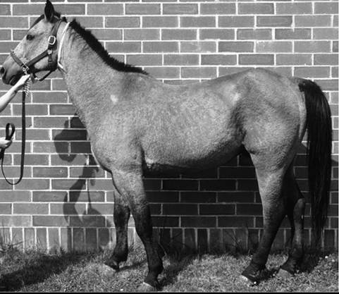
FIG. 40.1 Pemphigus foliaceus in a horse; note the generalized crusts.
azathioprine in other species, including humans. However, the author has administered azathioprine (1 to 3 mg/kg PO once daily for 1 month, then q48h) to eight healthy horses with no deleterious effects.17 Azathioprine is used as a steroid-sparing drug with corticosteroids, eventually decreasing the amount of steroid needed. Approximate cost of daily azathioprine for a 500-kg horse is $300/month.
Goats have also been treated successfully with corticosteroids (dexamethasone, prednisolone) and aurothioglucose.3,4,11 Dosages approximate those for the horse.
■ Prognosis The response to treatment in equine pemphigus foliaceus varies from patient to patient. Many horses require lifelong administration of medication to control the clinical signs; others may be weaned from medication gradually without further relapse.
In one study where follow-up information was available for 13 horses, 4 were euthanized because of complications of the disease or its treatment. The reported cases of caprine pemphigus foliaceus are insufficient in number to establish a reliable prognosis.Pemphigus Vulgaris
Pemphigus vulgaris is a rare autoimmune skin disease anecdotally reported9 in horses and recently definitively diagnosed in a Welsh pony.18 In that report, the diagnosis was confirmed with both direct and indirect immunofluorescence and immunoprecipitation studies, the latter identifying circulating immunoglobulin G (IgG) directed against the epidermal transmembrane protein desmoglein-3, similar to the pathogenesis in humans and dogs. Clinical signs are vesicles and ulcerations seen in mucocutaneous and cutaneous areas. Initial corticosteroid treatment improved the clinical signs, but onset of laminitis necessitated a dose decrease with a recurrence of lesions and development of oral ulcers. Despite successive adjunctive treatment with azathioprine, gold salts, and dapsone, the disease progressed, and the pony was euthanized. Necropsy showed additional lesions of the cardia of the stomach.18
Bullous Pemphigoid
Bullous pemphigoid is an autoimmune vesiculobullous and ulcerative disorder that affects the cutaneous basement membrane zone (BMZ). It has been rarely noted in horses.9,19 Initiating triggers for the disease in the horse are unknown.
The pathophysiology of bullous pemphigoid in horses is assumed to be similar to that described in other mammals. Complement-activating anti-BMZ antibodies bind to a glycoprotein antigen in the lamina lucida of the BMZ. In horses this has been shown to be bullous pemphigoid antigen II (also called collagen XVII). Complement activation results in degranulation of mast cells and chemotaxis of neutrophils and eosinophils. Eosinophils release tissue-destructive enzymes with resultant injury to the BMZ, loss of dermoepidermal adherence, and subsequent blister formation.9
Equine bullous pemphigoid is characterized clinically by painful, crusted, or ulcerative lesions of the skin (face and axillae), mucous membranes, and mucocutaneous junctions.
Bullae are rare.9,18 Ulceration may involve the gastrointestinal (GI) tract. The diagnosis is based on histopathologic and, when available, immunofluorescent findings. Treatment is the same as for pemphigus foliaceus; the few cases reported have not had favorable outcomes, but the author has seen a case that initially responded well to corticosteroids.Alopecia Areata
Alopecia areata is a disease of horses and cattle typified by areas of nonpruritic alopecia.9,20,21 One horse has been described with severe hoof dystrophy on all four legs.22 In one report the median age was 9 years, with an age range of 3 to 15 years. Alopecia was the primary dermatologic abnormality in all horses and most commonly affected the mane, tail, and face, although any part of the body could be affected (Color Plate 40.2).23 Histologically, a lymphocytic infiltrate surrounding the base of the hair follicle is seen, but in long-standing cases this infiltrate may not be present.24 A mare with a T-cell lymphocytic infiltrate causing a mural folliculitis of the isthmus (central section) of the hair follicles was described.24 Antibodies targeting the hair follicle itself have been documented.25 Most horses regrow the hair, but this may take up to 2 to 3 years, and the disease may wax and wane, worsening most commonly in the spring and summer months.23 Corticosteroids may hasten hair regrowth. Mane and tail “dystrophy” of Appaloosas and other breeds may in fact be a form of alopecia areata. Because of the circular nature of the alopecia, this disease is often mistaken for ringworm.