Clinical Presentations
Cats with SNA are usually presented for sneezing and unilateral or bilateral nasal discharge. Intermittent epistaxis occurs in a third of cases and fever is variably present. In severe or very chronic infections, erosion of nasal or frontal bones results in a discharging sinus or facial deformity.
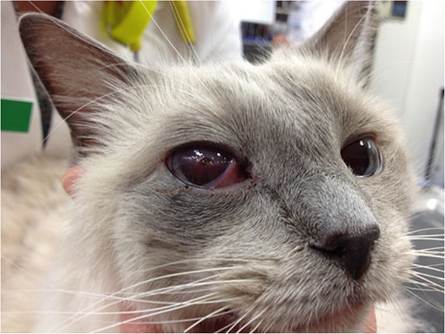
Most cats with SOA are presented for clinical signs associated with a retrobulbar fungal granuloma, unilateral exophthalmos with dorsolateral deviation of the globe, a prolapsed inflamed third eyelid and exposure keratitis (Fig. 15.4) (Wilkinson et al. 1982; Hamilton et al. 2000; McLellan et al. 2006; Barrs et al. 2012; Declercq et al. 2012; Kano et al. 2008, 2013; Barachetti et al. 2009; Giordano et al. 2010; Smith and Hoffman 2010). Resistance to retropulsion of the globe and normal intraocular pressure readings on tonometry enables differentiation of exophthalmos from buphthalmos (abnormal enlargement of the globe) (Smith and Hoffman 2010). Anterior and/or posterior uveitis may be present. Vision loss occurs late in disease due to severe ulcerative keratitis, as well as infiltration of the optic nerve and/or optic
Fig. 15.4 Brachycephalic pure-bred Ragdoll cat with sino-orbital aspergillosis. A retrobulbar fungal granuloma in the right orbit has caused exophthalmos, resulting in prolapse of the third eyelid and exposure keratitis with central corneal ulceration
chiasm (Hamilton et al. 2000; Tomsa et al. 2003; Barachetti et al. 2009; Barrs et al. 2012). In severe chronic infections, exophthalmos may become bilateral (Barachetti et al. 2009; Barrs et al. 2012; Wilkinson et al. 1982). As the ventral floor of the orbit is not encased by bone, the orbital granuloma often invades the oral cavity where it first becomes visible as an ulcerated area of mucosa or a submucosal mass in the pterygopalatine fossa adjacent the last molar tooth. Involvement of maxillary subcutaneous tissues causes facial swelling, although this may be subtle. Nasal signs are absent in 40% of SOA cases at presentation; however the medical history usually reveals sneezing or nasal discharge in the preceding 6 months. Pain on opening the mouth and neurological signs are uncommon at initial presentation. Neurological signs develop in chronic end-stage infection and can include seizures, nystagmus, circling, facial muscle fasciculation and hyperesthesia (Barrs et al. 2012; Giordano et al. 2010; Smith and Hoffman 2010). Similar to SNA, the presence of fever is variable.
15.7