Clostridial Myonecrosis
Steven M. Parish • Stephanie J. Valberg
Various species of clostridial organisms cause acute myonecrosis in many farm animal species. Infections are characterized by a rapid clinical course, fever, systemic toxemia, and high mortality.85-87 Clostridial diseases are infectious but not contagious.
Specific bacteria associated with clostridial myonecrosis include Clostridium chauvoei (Clostridium welchii), Clostridium septicum, Clostridium sordellii, and occasionally Clostridium novyi type B, Clostridium perfringens type A, and Clostridium carnis. Mixed infections involving several agents are common.88 Synonyms for clostridial diseases include blackleg, malignant edema, false blackleg, gas gangrene, and gangrene. Although there may at times be distinct differences among the specific disease syndromes associated with the different clostridial agents, the pathophysiology of these diseases is similar enough to be covered under the general topic of clostridial myonecrosis.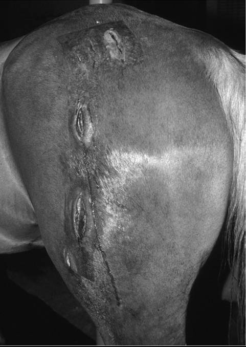
FIG. 42.7 Clostridial myositis in the left gluteal muscle after an injection of flunixin meglumine, which has progressed to involve the biceps femoris muscle. Fenestrations were created for debridement.
■ Clinical Signs Commonly, clostridial myonecrosis is rapidly progressive with the development of tremors, ataxia, dyspnea, recumbency, coma, and death within 12 to 24 hours. As such, many affected animals may be found prostrate or dead.85,86,88 Mortality may approach 100%. Affected animals who are still alive are usually severely depressed, febrile (40° to 41° C∕104° to 106° F), tachypneic, anorexic, and lame. These signs are associated with a rapidly developing muscle infection and toxemia. There is usually only one primary site of infection in an affected animal.
Any skeletal muscle group in the body can be involved, but most infections affect the limb or trunk muscles. Occasionally muscles such as those around the vulva, tongue, and diaphragm can be involved, or the udder in a cow may be the primary site of sepsis. Areas around recent injections are common sites of myonecrosis in the horse.87,89 Initially the skin over the area may be swollen, hot, and discolored; however, as the disease progresses, the skin over the area may become cool and insensitive with progressive sloughing. Crepitus may be detectable, indicating subcutaneous gas production. If a wound is present, malodorous serosanguineous fluid may discharge (Fig. 42.7). Aspiration of the swelling often reveals fluid with similar qualities.Clostridial myonecrosis generally has characteristic pathologic lesions that are absent in most other conditions, making diagnosis relatively straightforward. Differential diagnoses may include other fulminant disease processes in which there is rapid debilitation or death of the animal.
■ Clinical Pathology Clinicopathologic data alone are seldom specific enough to confirm the presence of clostridial myonecrosis. Hematology and serum biochemical analyses usually reflect a generalized state of debilitation and toxemia (e.g., hemoconcentration and a stress/toxic leukogram may be present). Elevations in the activities of serum CK and serum AST usually occur; however, they often do not reflect the toxicity of clostridial myonecrosis. Aspirates from the affected tissues can yield diagnostic information. It is preferable to obtain tissue specimens for direct smear examination and fluorescent antibody testing, and for anaerobic bacterial culture from affected tissues.
■ Pathophysiology Clostridial agents are ubiquitous in the environment and can frequently be cultured from the feces, intestinal tract, and other internal organs of a variety of species.85 Spore-forming characteristics allow these organisms to remain in the environment for long periods, but the exact mechanisms involved in the pathogenesis of clostridial myonecrosis are not fully known.
Development of clostridial myonecrosis following an intramuscular injection or penetrating wound may be the result of direct spore deposition into the tissue in association with penetration. If suitable conditions prevail within the muscle, the spores undergo a conversion into the vegetative, toxinproducing form of the organism. In contrast, the pathogenesis of the disease is more difficult to explain when a wound does not exist. It is postulated that clostridial agents gain access to the body through the alimentary tract and are present in liver and muscle in the dormant spore form.90 Subsequently, when local tissue is devitalized and conditions become appropriate for the spores to germinate, the rapid vegetative process ensues. Muscle trauma associated with injection, transporting, herding, and handling has often been incriminated as creating a suitable environment for the development of clostridial myonecrosis. The proliferation of clostridial agents in devitalized tissues is associated with the release of powerful exotoxins responsible for the local necrotizing myositis and systemic toxemia. Toxins are released by multiplying clostridia; the toxins vary, depending on the clostridial species involved. Necrotizing (lecithinase) and hemolyzing (hemolysin) toxins, as well as neuraminidase, appear to be of greatest importance. The toxins act locally and systemically to create widespread organ dysfunction. The toxins of C. sordellii are the most potent of all the clostridial species, and myonecrosis caused by this organism is fatal.■ Epidemiology Clostridial agents are common in the environment, and susceptible animals are constantly exposed to them. Areas where previous death losses from clostridial disease have occurred may have a higher incidence or risk of disease because of increased environmental contamination. In cattle, clostridial myonecrosis is generally a disease of animals between 4 and 24 months of age. However, C. sordellii is a more common problem in older feedlot cattle in which excessive muscle bruising may occur.
Younger animals are probably protected by colostral immunity and older animals by some degree of acquired immunity. Animals on high planes of nutrition and in excellent body condition are more likely to develop the disease. Infections with C. chauvoei occur most commonly during the warmer seasons, with the highest incidence varying from the spring to fall, depending on when calves reach the most susceptible age group. C. septicum, C. novyi, and C. perfringens type A infections can occur at any time and are usually associated with skin wounds such as injection sites, punctures, and castration wounds. The umbilicus may be a site of invasion. Infections in the genital area can occur, usually in association with a recent dystocia.In sheep and goats, clostridial myonecrosis is most frequently associated with wounds such as those occurring after shearing, docking, and unsanitary surgical procedures. Sheep dipped for parasites after shearing may have an increased risk if the dip becomes contaminated with clostridial spores.
Most reports of clostridial myonecrosis in horses suggest an association with puncture wounds and intramuscular injection sites.87,89 Intramuscular administration of irritating drugs (including antihistamines, anthelmintics, and phenylbutazone) may enhance the susceptibility to clostridial myonecrosis. Horses often present with or have a history of another complaint such as colic, exertional myopathy, or laminitis for which they have received injections of drugs in the preceding 48 hours.91 Previously administered drugs (e.g., phenylbutazone) may mask the fever associated with clostridial myonecrosis, potentially confusing the diagnosis.
■ Necropsy Findings Swelling and autolysis are rapid in animals that have died from clostridial myonecrosis. Bloodstained fluid is often observed discharging from body orifices. Extreme swelling and crepitus may be noted over the affected body area. When acting alone, each of the clostridial agents associated with clostridial myonecrosis produces somewhat different postmortem lesions.
However, it is unwise to assume that in clostridial myonecrosis only a single clostridial agent was involved because mixed infections frequently occur.C. chauvoei infection is characterized by engorgement of the subcutis and adjacent tissues with blood-stained fluids and gas bubbles. Cut tissue from the affected area reveals moist, dark-colored muscle in the periphery of the lesion, with lighter colored, drier muscle with gas bubbles separating the separate bundles of muscle toward the center. Other changes include severe degeneration of parenchymatous tissues caused by the systemic toxemia. The carcass usually has a foul odor similar to that of rancid butter. This odor is a characteristic of most cases of clostridial myonecrosis. The lungs are often congested with edema, and hemorrhage and a fibrinohemorrhagic pleuritis are common. The heart may be friable and show evidence of endocardial hemorrhages, particularly on the right side. The spleen may be normal or enlarged and friable. The liver is usually pale and friable and may be autolytic and porous. Lesions are similar in sheep and cattle, except that there is usually less gas and the muscles are not as dry in affected sheep.
Similar necropsy findings are found with myonecrosis caused by C. septicum and C. novyi type B. C. septicum and C. perfringens generally occur as part of mixed wound infections in which abundant malodorous, serosanguineous fluid is found at the wound site. C. perfringens is common in horses.
Myonecrosis resulting from C. sordellii is most often associated with lesions of the neck or brisket area of cattle. Death is frequently so rapid that subcutaneous gas accumulation is rare. In addition to local myonecrosis, these animals often have massive subendocardial hemorrhages in the left ventricle of the heart and hemorrhage in the trachea, bronchi, and thymus. Extensive perirenal edema and hemorrhagic renal calyces and severe congestion of the lungs are common findings.
■ Treatment Although clostridial myonecrosis is often fatal, aggressive specific therapy combined with supportive care may be successful in individual cases.
A presumptive diagnosis of clostridial disease on the basis of history and clinical signs is usually made before obtaining the results of culture and laboratory determinations such as fluorescent antibody tests. In horses, clostridial myonecrosis resulting from infections with C. perfringens seems to be most amenable to treatment and has the best prognosis for survival, although extensive skin sloughing over the affected area is common.87 Antibiotic therapy, aggressive surgical debridement including fasciotomy, and supportive care are the hallmarks of successful treatment.87 With most clostridial infections, penicillin is the drug of choice. In horses, Na or K penicillin IV or procaine penicillin IM is used at a dosage of 44,000 U/kg IV every 2 to 4 hours until the animal is stable (1 to 5 days). The IV dose is then reduced to four times a day or is replaced by oral metronidazole (15 mg/kg three or four times daily). In ruminants, similar intravenous or intramuscular drug therapy is indicated. In all cases, prolonged antimicrobial therapy may be necessary.Surgical intervention at the affected site by means of debridement or fenestration in an attempt to reduce tissue swelling, aerate the tissues, and remove necrotic tissue is considered imperative for survival in horses (see Fig. 42.7). Incisions are made through the skin and into the affected muscle to establish adequate drainage and hopefully alter the anaerobic conditions. Sufficient fenestrations should be made to establish drainage and aeration over the entire affected area.
Use of specific antitoxins is recommended when possible. However, these are often not available or not used for immediate therapy because the exact species of Clostridium causing the myonecrosis is not known. Cost considerations may also preclude their use. Supportive fluid therapy and use of analgesics and anti-inflammatory agents for control of pain and swelling are recommended. Short-acting corticosteroids such as dexamethasone, prednisolone, or hydrocortisone may be used for initial therapy of systemic and toxic shock, but continued use is contraindicated in the face of overwhelming sepsis. If required, specific therapy should also be directed toward any other underlying problems.
■ Prognosis The prognosis for life in all cases of clostridial myonecrosis is guarded to poor. The disease process is often rapidly fulminant, making treatment unrewarding. However, some animals have survived because of early diagnosis, aggressive therapy, and long-term supportive care. This is particularly true in cases involving C. perfringens in horses. The owner should be aware from the start of treatment that extensive skin sloughing may involve most of a limb and may force euthanasia to become a consideration at a later stage.
■ Prevention Protection against clostridial myonecrosis is based on immunization procedures. Although clostridial agents are ubiquitous in the environment and frequently appear in the body of susceptible animals, rarely does adequate natural protection occur, although some colostral and acquired immunity may at times occur. Infection in unprotected animals usually follows a rapid, degenerative clinical course and terminates before the animal can generate an appropriate protective immune response. At present, only ruminants are commonly vaccinated against the agents responsible for clostridial myonecrosis. Vaccines used include multivalent bacterin toxoids containing antigens against two or more clostridial species, including C. chauvoei, C. septicum, C. novyi, C. sordellii, and C. perfringens. A rational program for protection usually involves vaccinating at an early age to establish immunity. Vaccination age is partly determined by other management factors, including when calves are handled for branding and castration, but 4 to 6 months of age is the usual time of initial vaccination. In areas of heavy exposure it may be necessary to vaccinate at 3 months and again at 4 months. In all clostridial species except C. chauvoei, two doses of vaccine are necessary to establish good protection. The duration of immunity is not long, and booster vaccinations should be administered every 6 to 8 months if protection is to be maintained. In many herds it is only necessary to vaccinate animals younger than 3 years of age (i.e., those animals that are at greatest risk), but in some high-risk herds it is necessary to maintain a vaccination program for the life of the animal.
Animals that die of clostridial diseases should be disposed of by deep burial, burning, or removal from the premises to avoid further contamination of the environment.