Rhabdomyolysis Associated With Streptococcus equi
Stephanie J. Valberg
Acute Rhabdomyolysis
Severe acute generalized rhabdomyolysis has been reported to occur in Quarter Horses younger than 7 years of age.92,93 Affected
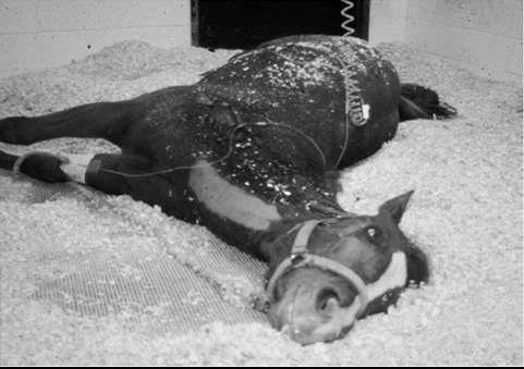
FIG.
42.8 A horse in severe pain unable to rise due to acute rhabdomyolysis concurrent with guttural pouch empyema from Streptococcus equi.horses have evidence of submandibular lymphadenopathy and/ or guttural pouch empyema due to Streptococcus equi. Horses develop a stiff gait, which progresses rapidly to markedly firm, swollen, painful epaxial and gluteal muscles. The majority of reported cases became recumbent and unable to rise. They developed unrelenting pain necessitating euthanasia within 24 to 48 hours of hospitalization (Fig. 42.8). Hematologic abnormalities include mature neutrophilia, hyperfibrinogenemia, and marked elevations in CK (115,000 to 587,000 U/L) and AST activities (600 to 14,500 U/L). Titers to the M protein of S. equi are low in affected horses, unless horses have been recently vaccinated for strangles. Titers to another protein called myosin binding protein have been high in a small number of horses that were tested.92
At postmortem examination, large pale areas of necrotic muscle are evident in hindlimb and lumbar muscles. The histopathologic lesions are characterized by severe acute myonecrosis with a degree of macrophage infiltration. Sub- lumbar muscles often show the most severe and chronic necrosis as indicated by greater macrophage infiltration of myofibers.
One possibile etiology is a toxic shock-like reaction arising from profound nonspecific T-cell stimulation by streptococcal superantigens with the release of high levels of inflammatory cytokines. An alternative explanation for rhabdomyolysis may be a bacteremia with local multiplication and production of exotoxins or proteases within skeletal muscle.
S. equi virulence factors that may account for muscle necrosis include an unidentified cytotoxic protein, several proteases, streptokinase, and streptolysin S.94 Although S. equi has not been cultured in skeletal muscle from horses with rhabdomyolysis, S. equi bacteria have been identified in affected muscle using immu- nofluorescent stains for both Lancefield group C carbohydrate and S. equi M protein.92 There is currently no evidence that the S. equi involved is an atypical genetic strain of S. equi?5 Most recently, a mutation in the gene MYH1 has been identified in association with immune-mediated myositis, and this mutation may also be responsible for cases of S. equi-associated myopathy (see the Immune-Mediated Myositis section).A high mortality rate has been reported in horses receiving intravenous penicillin therapy once clinical signs of strangles and myopathy were well established. It is possible that early recognition of the signs of muscle stiffness in horses with S. equi infections and prompt aggressive treatment may be required for a successful outcome. Although streptococcal species are exquisitely susceptible to β-lactam antibiotics, a mortality rate of 85% has been reported in human group A streptococcal myositis despite penicillin treatment.96 An antimicrobial that inhibits protein synthesis, such as rifampin, combined with intravenous penicillin might enhance survival rates in horses with S. equi rhabdomyolysis. In addition, flushing infected guttural pouches and draining abscessed lymph nodes will diminish the bacterial load. Nonsteroidal antiinflammatory drugs (NSAIDs) and possibly high doses of short-acting corticosteroids may assist in diminishing the inflammatory response. Control of unrelenting pain is a major challenge in horses with severe rhabdomyolysis. Constant rate infusion of lidocaine, detomidine, or ketamine may provide better anxiety and pain relief than periodic injections of tranquilizers. Horses should be placed in a deeply bedded stall and moved from side to side every 4 hours if they are unable to rise. Some horses may benefit from a sling if they will bear weight on their hindlimbs when assisted to stand.