Diseases Manifesting Principally With Forebrain Dysfunction or Multiple-Level Brain Dysfunction
Alphaviruses
Robert J. MacKay
The causative agents of eastern, western, and Venezuelan equine encephalomyelitis (EEE, WEE, and VEE) are New World arboviruses belonging to the genus Alphavirus of the family Togaviridae.1 Ross River virus is an Old World alphavirus in Australia that was implicated in more than 100 cases of neurologic disease in horses during an outbreak of arboviral encephalitis in 2011 and previously was suspected of causing a syndrome of lethargy, muscle soreness, and ataxia in horses in Victoria and South Australia.2,3
EEE, WEE, and VEE affect all common domestic equid species, including horses, mules, and donkeys.
Each of the viruses was first isolated during epizootics in the 1930s.4-6 Alphaviruses are nonsegmented, single-stranded, positive-sense RNA viruses..7 Each of the three equine alphaviral encephalitides is among the 13 equine infectious diseases that must be reported to the World Organization for Animal Health (Office International des Epizooties [OIE]). Data on the occurrence of these diseases in the United States are collected by veterinarians, diagnostic laboratories, and state animal health officials and are reported to the National Animal Health Reporting System.At least three strains of the WEE virus antigenic complex have been identified. Two of these are found in both North and South America, which suggests that circulation of WEE viruses between the New World continents is common.7,8 There is a distinct North American lineage and three South American lineages of the EEE virus antigenic complex.7 South American strains are now classified as a distinct species, Madariaga virus.9 In contrast to the situation with the WEE virus, there is no evidence of movement of EEE virus between North and South America.
The VEE antigenic complex contains six subtypes, eight varieties, and seven species, most of which are maintained continuously in enzootic life cycles in northern South America.7,10 Until the 1990s, when a subtype IE variant caused two outbreaks in Mexico, all equine epizootics had been caused by subtypes IAB and IC.11
■ History
WESTERN EQUINE ENCEPHALOMYELITIS.
What was almost certainly the first recorded outbreak of WEE in horses occurred in the Great Plains of the United States in 1912, when at least 35,000 horses died of the disease.5,12 Epizootics in California and other western states in 1930 to 1932 resulted in the deaths of an estimated 9200 horses and mules (≈50% of those affected).5,13 In 1938, WEE was recognized in every state west of the Mississippi River and affected more than 184,000 horses in that year alone.1 WEE virus was also implicated in outbreaks of fatal equine disease in the early 20th century in Canada and Argentina, and epizootics have since been reported in western Canada, Mexico, Central America, and South America.14 Between 1972 and 1981, an annual average of 267 cases of WEE in horses was reported by the National Veterinary Services Laboratory in Ames, Iowa14,15; in 1993, there were 15 cases in 10 states, and since 2005, no case has been reported in the United States. There have been isolated cases of encephalomyelitis in Florida horses associated with the Highlands J virus, an eastern member of the WEE virus complex with epidemiologic characteristics similar to those of EEE virus.16EASTERN EQUINE ENCEPHALOMYELITIS. An outbreak of what was probably EEE was recorded in 1831 in Massachusetts.17 Approximately 100 horses were affected, and 75 died. Over the subsequent century, there were at least five other outbreaks characteristic of EEE.18 An epizootic in 1933 involved a least 1000 horses in the coastal regions of the mid-Atlantic states.4 Epizootics of EEE in North America have since occurred periodically in the coastal areas of the Atlantic, the southeastern United States, and Texas. In the largest recorded outbreak, in 1947 in southern Louisiana and Texas, 14,344 cases of EEE were recorded and resulted in 11,722 deaths (mortality rate of 82%).19 Focal outbreaks and sporadic cases have also occurred regularly in Michigan and are occasionally reported from other locations in the eastern half of the United States and Canada.
A single case of EEE in a stallion in California was reported in 2002.20 Although the origin of the infection in this horse was not determined with certainty, incompletely activated EEE vaccine was suspected. Despite the availability of effective vaccines since the late 1930s, cases of EEE still occur every year in Florida and other southeastern states. The annual incidence of EEE in horses in the United States since 2003 has ranged from 60 (in 2011) to 712 (in 2003).Epizootics of a mild form of EEE have been widespread in Central and South America, extending southward from Panama to Argentina.18 More recently, outbreaks with high rates of mortality have occurred in Brazil.21 The disease has also caused serious losses in the Caribbean.17
VENEZUELAN EQUINE ENCEPHALITIS. The disease in equids was first recognized and described in Venezuela and Colombia in 1938.6 At intervals of approximately 10 years, there were large subtype IAB or IC epizootics in northern South America that usually involved tens of thousands of horses. Some of these outbreaks may have been caused by incompletely inactivated vaccine. One epizootic in central Colombia during 1962 to 1964 was estimated to have killed 100,000 horses.22 An outbreak of VEE caused by subtype IAB began in Peru in the winter of 1969, jumped to Central America in June 1969, and then entered Mexico in 1970. The outbreak spread north to the U.S. border in the spring of 1971, and the first case of VEE in a U.S. horse was confirmed on June 30.23 By the time of the last case on November 7, 1971, more than 1500 horses had died in the southern counties of Texas. No further epizootic activity was reported until 1992, when a series of epizootics of VEE subtype IC began. Subtype IE has been associated with fatal VEE in Mexico, beginning with small outbreaks in 1993 and 1996.24
Epidemiology
EASTERN EQUINE ENCEPHALOMYELITIS AND WESTERN EQUINE ENCEPHALOMYELITIS.
In North America, the WEE and EEE viruses are maintained between epizootics by low-level cycling between New World passerine birds (i.e., songbirds) and ornithophilic mosquitoes in freshwater, forested swamp habitats.1 Snakes may also be an overwintering host for North American EEE virus.25 Infected mosquitoes can be found throughout the year in Florida and other southeastern states. Under favorable environmental conditions, the viruses periodically spread outward from focal reservoirs to infect the general wild bird population, through which they are spread and amplified by rapid bird-mosquito-bird transmission. The mosquito vectors involved in maintenance and amplification of WEE and EEE viruses are usually Culex tarsalis and Culiseta melanura, respectively. Many avian species, including migratory passerine birds (e.g., starlings and northern cardinals) and wading birds, become infected and develop high-order viremia and high serum titers of EEE virus or WEE virus but usually do not become ill. Among some introduced species, including pigeons, house sparrows, Chukar partridges, and Chinese pheasants, infection with EEE virus may cause high rates of morbidity and mortality.Cases of encephalitis usually begin in susceptible horses 2 to 3 weeks after the virus spreads into birds. Human cases may occur several weeks later. C. tarsalis transfers WEE virus from bird to horse (and human). Although C. melanura may also function in some locations as a bridge vector for EEE virus, mosquitoes of a variety of other species usually transfer the virus from bird to horse.7,18 Viremia in horses is low titer, so mosquitoes feeding on horses with either EEE or WEE virus are unlikely to be infected further, and horses are considered “dead-end” hosts. Both EEE and WEE viruses have been associated with neurologic disease in calves, and EEE virus has caused fatal infections in New World camelids, deer, ratite birds, cats, dogs, mice, foxes, sheep, and pigs.7,26-28
Epizootics of EEE and WEE usually last 1 to 3 months and occur in summer and early fall when warmth and humidity favor breeding, longevity, and mobility of mosquito populations.29 Outbreaks in Massachusetts and Michigan probably occurred during the second consecutive year in which rainfall exceeded the annual mean by more than 20 cm; however, unusually high temperatures did not appear to be a risk factor.29,30 The month in which disease onset peaks varies from June in Florida to August or September in northern and western states.5,26,31,32 In Florida and some other southeastern states, isolated cases of EEE can occur at any time of year.31 Standing surface water for mosquito larval development, bush cover for wild hosts, and the immune status of the various hosts also affect the timing and magnitude of equine epizootics.
Many of these physical factors are significantly affected by the cultivation, clearing, and irrigation of land and by drainage of swamps. The proximity and size of plantations of trees were risk factors for EEE for horses in Florida.33 The equine epizootic usually declines with the onset of cool or dry weather unsuitable for mosquito or bird activity, as well as with the depletion of susceptible equine hosts by death or development of immunity among survivors.
The epidemiologic features of EEE and WEE viruses in South America are poorly understood. Small mammals play a greater role in sylvatic cycles of viral transmission in South America than in North America. Culex (Melanoconion) subspecies and Aedes albifasciatus appear to be involved in transmission of EEE virus and WEE virus, respectively.8,34
VENEZUELAN EQUINE ENCEPHALOMYELITIS. Enzootic VEE viruses are maintained by cycling between mosquitoes and small rodents. The Everglades virus, a subtype II found in the Everglades region of Florida, is of this type, cycling between Culex (Melanoconion) subspecies mosquitoes and wild rodents.10 The epizootic varieties IAB and IC have probably arisen on at least four separate occasions by mutation of enzootic subtype ID strains.11,35 Because of the high-order viremia that develops in infected animals, the defining characteristic of epizootic VEE virus varieties is that equids serve as amplification hosts. Domestic rabbits, goats, dogs, and sheep also suffer fatal disease during VEE outbreaks.10 There does not appear to be any single “epizootic” vector mosquito species; during outbreaks, the virus may be isolated from mosquitoes of many different species.10 Epizootics terminate when susceptible horses are no longer available. The leapfrog-type spread characteristic of VEE is usually attributed to transport of horses incubating the virus. Movement of infected bats may also contribute to discontinuous spread of virus.
In contrast to the situation with WEE and EEE viruses, birds do not have an important role in amplification and spread of epizootic VEE virus. It is not yet clear whether neurovirulent subtype IE virus is amplified in horses. Evidence from experimental infections suggests that viral titers in blood of infected horses may be insufficient to infect feeding mosquitoes.36 The interepizootic reservoir of epizootic varieties, if any, is not known.■ Clinical Findings Clinical signs of infection with each of the viruses are similar, although signs of EEE are typically more severe and progress more rapidly than those of VEE and WEE.4,5,19,20,29,37,38 Infected horses respond in any or even all of the following ways after experimental infections:
1. Inapparent infection with a very-low-grade viremia and fever approximately 2 days after inoculation. This manifestation corresponds to the initial viremia after viral proliferation in regional lymph nodes and probably occurs commonly during outbreaks without progression to subsequent stages.
2. Generalized febrile illness (up to 41.7° C), with anorexia, depression, tachycardia, and diarrhea. This stage is associated with viral proliferation in various body organs after spread from regional lymph nodes.
3. Clinical encephalomyelitis, which is the classic form of the disease.39
Young horses (especially yearlings) are most susceptible: Approximately 90% of 95 horses with EEE admitted to the University of Florida (UF) College of Veterinary Medicine Large Animal Hospital were younger than 5 years of age. The onset of neurologic signs, associated with the second febrile crisis, typically occurs approximately 5 days after infection (range 2 days to 2 weeks), and most deaths occur 2 to 3 days later.38,39 Almost all horses with EEE are febrile at the onset of neurologic signs (mean of 40.3° C in horses admitted to the UF Large Animal Hospital), although the temperature declines thereafter. The onset of severe neurologic signs is peracute to acute, and progression is rapid. Horses may not be found to be affected until they are recumbent and comatose or dead. If they are observed while alive, initial CNS signs are quite variable and referable to diffuse or multifocal forebrain disease; evidence of brainstem and spinal cord involvement quickly becomes obvious as the illness progresses (Fig. 35.7). Obtundation is the most common initial clinical sign, although owners also report suspected colic as part of the initial presentation.
Affected horses often stand apart from pasturemates, have no interest in food, or fail to respond to an owner's call. Further signs of dementia may follow, including head pressing, odontoprisis, hyperesthesia, irritability, aggression (rarely), leaning against a wall or fence, or compulsive walking, often in a circle (especially around the inside of a stall or small paddock). Several observers have noted a “peculiar looseness of the lips” as a characteristic early sign.5,40 Blindness and lack of a menace
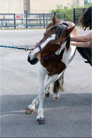
FIG. 35.7 Eastern equine encephalomyelitis in an 18-month-old Paint filly. At presentation, the filly was febrile (39.9° C [103.8° F]), stuporous, and cortically blind and had a marked head tilt and body lean to the left, indicative of asymmetric central vestibular dysfunction. Vaccination history was unknown. With nursing care, this filly completely recovered over several weeks.
response may also be noted at this stage. Signs of cranial nerve disease, including abnormal pupillary light reflexes, head tilt, nystagmus, tight circling, facial and tongue paralysis, and inability to swallow often develop as the disease advances. Abnormal signs usually manifest asymmetrically, at least initially. Ataxia and paresis of the trunk and limbs cause gait to become progressively unsteadier. Tremors occur in the antigravity muscles of the limbs. Once recumbent, affected horses seldom regain their footing and are often noted to make galloping or swimming movements in lateral recumbency.5 Seizures occur in approximately one third of horses with EEE and may happen at any stage. Mucopurulent nasal and ocular discharges and abrasions/lacerations of the face and limbs are the most common nonneurologic findings; the latter occurs secondary to trauma from falling, seizures, or running into objects.
Mortality rates are 75% to 95% for EEE, 19% to 50% for WEE, and 19% to 83% for VEE.5,7,19,34,41 Vaccination within the preceding year was reported to be associated with lower risk of death in horses with EEE.29,31 Death is usually preceded by a period of recumbency during which the horse may be semicomatose and convulsing. Surviving horses gradually recover over a period of weeks. Many horses with WEE apparently recover completely, but it is estimated that two thirds of horses that survive EEE have residual signs of CNS damage, such as central blindness, dullness, and diminished learning capacity or signs of cranial nerve dysfunction such as facial paralysis and head tilt.18 Such horses are often referred to as “dummies.”
■ Laboratory Findings Abnormalities on hemograms and in plasma chemistry panels of horses with alphaviral encephalomyelitis are minor and nonspecific. Horses with EEE may have abnormal WBC counts and high fibrinogen concentration. In 73 horses with EEE admitted to the UF Large Animal Hospital, the following hematologic abnormalities were noted: leukocytosis (56%), neutrophilia (60%), hyperfibrinogenemia (57%), monocytosis (36%), lymphopenia (13%), leukopenia (7%), neutropenia (4%), and lymphocytosis (3%). Mild to moderate elevations in plasma enzyme activities of affected horses either reflect direct organ damage or are secondary to anorexia, fever, stress, recumbency, and seizures. Hyperammonemia is found in some horses with EEE; its significance in horses is unknown but may reflect intestinal or hepatic injury by EEE virus.
The results of CSF analysis for horses with EEE are quite distinctive. More than 95% of horses with EEE admitted to the UF Large Animal Hospital had abnormal CSF cytologic findings. Among patients at the UF Large Animal Hospital, 88% of samples had high protein concentrations (mean of all samples, 139 mg/dL; range, 38 to 526 mg/dL; normal is of choice for use in horses with viral encephalomyelitis because titers of greater than 1 : 400 can distinguish vaccination-induced (IgG only) from viral infection-induced (IgM and IgG) antibody responses.36,42 These assays are available at many state diagnostic laboratories and also at the National Veterinary Services Laboratories (NVSL) in Ames, Iowa. In surviving horses, demonstration of a fourfold or greater rise in hemagglutinationinhibition or plaque-reduction neutralizing titer between acute and convalescent samples provides additional diagnostic support. Positive results of these tests could simply reflect subclinical infection with either virus. Virus can be isolated from fresh or frozen (with low efficiency) cultured brain tissue or mice and can be detected in histologic sections with immunohistochemistry or polymerase chain reaction (PCR) techniques.43
Differential diagnoses include hepatoencephalopathy; gastrointestinal (GI) hyperammonemia; heavy metal or other toxicosis; rabies; other viral encephalomyelitis (West Nile encephalitis, Borna disease, equine herpesvirus myeloen- cephalopathy, Aujeszky disease, louping ill); equine protozoal myeloencephalitis (EPM); trypanosomal (South America), verminous, or bacterial meningoencephalomyelitis; spaceoccupying lesion within the calvaria (e.g., cholesterol granuloma, neoplasia, brain abscess); brain infarction; brain trauma; and leukoencephalomalacia.
■ Treatment Treatment is largely supportive and, in the case of EEE, usually ineffective. Any adult horse with presumptive EEE that is unable to stand should be euthanized. Such horses should not be subjected to slinging. Hyperthermia is known to exacerbate brain injury and must be treated vigorously with cold water or alcohol baths until rectal temperature is less than 102° F. Convulsions can be controlled with diazepam, xylazine, or barbiturates (pentobarbital or phenobarbital).
In addition to receiving excellent general nursing care, all affected horses should be treated for brain edema and inflammation, although only horses with a predominance of forebrain signs are likely to respond. Drugs used for this purpose include dimethyl sulfoxide (DMSO), 1 g/kg as a 10% solution intravenously (IV) or intragastrically; flunixin meglumine (or equivalent nonsteroidal antiinflammatory drug [NSAID]) 1.1 mg/kg q12h for 3 days IV, or orally (PO); dexamethasone (or equivalent corticosteroid), 0.05 to 0.1 mg/ kg daily for 3 days IM or IV and then tapered; and mannitol (0.25 to 1 g/kg as a 20% solution IV) or hypertonic saline (e.g., 7.2% saline 2 mL/kg 4 to 6 times IV during the first day). Anecdotal evidence suggests that a lengthy course of dexamethasone is an important determinant of neurologic recovery in surviving cases. Treatment should be titrated to minimize neurologic signs but generally must be tapered over several weeks. Complete recovery, if it occurs, may take several months.
Treatment with serum from horses hyperimmunized against WEE virus was thought to reduce mortality rates modestly during the outbreak in 1930 to 1932; however, these claims were not tested scientifically.5 Furthermore, administration of large quantities of hyperimmune serum to experimentally infected horses by investigators at Lederle Laboratories was reported to have no beneficial effect.44
Adjuvant antiviral therapy with interferon α or β has salutary effects in some experimental models of alphaviral encephalitis and could be tried in valuable horses.45 A reasonable protocol is described in the section on West Nile encephalitis.
■ Public Health Significance All three viruses infect humans, causing subclinical, febrile, or neurologic disease.7 Since 1964, 640 cases of human WEE have been reported in the United States, with annual average of 27 cases in the period up to 1987. Since 1988, however, only five cases of WEE have been reported. In contrast, the 268 cases of EEE in humans reported in the United States since 1964 have occurred at a fairly steady rate of 6 cases per year. Epidemics of VEE are closely associated with equine epizootics; thus during the 1962 to 1964 epizootic in central Colombia, which killed approximately 100,000 horses, 200,000 humans were infected, of whom 1000 (0.5%) died. The enzootic Everglades subtype II strain, which is nonpathogenic for horses, has caused human encephalitis in Florida. No cases of human disease were recorded during the VEE subtype IE equine epizootics in Mexico in 1993 and 1996, but South American strains of EEE virus have been reported to cause encephalitis in humans. Rates of short-term mortality from EEE, WEE, and VEE in humans are 74%, 4%, and less than 1%, respectively. Most human survivors of EEE suffer long-term neurologic impairment.
■ Postmortem Findings Most organs exhibit congestion, and the brain and spinal cord have a slate-gray discoloration, often with petechial hemorrhage, obvious especially on sections of formalin-fixed tissue.43,46,47 Brain swelling and evidence of occipital subtentorial herniation are common, with brainstem compression. Histologic study reveals evidence of acute to subacute and multifocal to diffuse meningoencephalomyelitis. Gray matter is predominantly involved, with diffuse neuronal degeneration, gliosis, perivascular and neuroparenchymal infiltrates, and meningitis. Neutrophils or, on occasion, eosinophils are prominent in acute EEE, whereas lymphocytes predominate in older lesions.46 Treatment with corticosteroids may suppress the inflammatory cell infiltrates, which then appear mild.
Bronchopneumonia is found in approximately 20% of fatal cases of EEE, probably secondary to pharyngeal paresis and aspiration of particulate material. Up to 10% of horses with EEE have histologic evidence of myocardial necrosis, probably a sign of brain-heart syndrome.47
Because high infection rates in humans have been documented after exposure to aerosols from infected laboratory animals or as a result of laboratory accidents (or possibly exposure to equine carcasses), necropsies of horses with suspected VEE should be performed only by personnel who possess demonstrable immunity in the form of neutralizing antibody.48 All laboratory manipulations must be carried out within certified biological safety cabinets in accordance with physical containment level 3 procedures.
■ Control Active immunization through use of formalinized vaccines began in 1934 and is still the only commercial method of immunization of horses against EEE and WEE viruses. These inactivated vaccines are also used routinely to protect ratites from EEE and WEE and South American camelids from EEE.49 The attenuated tissue culture-origin vaccine, TC-83, derived from a Trinidad VEE virus isolate, which was originally developed to protect military personnel, provides durable immunity against VEE virus and was effective in minimizing the incursion of VEE into the United States in 197123 and in limiting the 1993 and 1996 outbreaks in Mexico. Although TC-83 is no longer licensed for use in horses in the United States, a formalin-inactivated vaccine containing the TC-83 vaccine strain is available and is often included with EEE virus and WEE virus in trivalent products.50 For any of the formalin-inactivated alphavirus vaccines, adult horses should be given an initial two inoculations 4 to 6 weeks apart at least a month before the anticipated risk period or before the onset of vector mosquito activity and then revaccinated at intervals when the risk is likely to be higher. Vaccination should begin in January or February in Florida and in May or June in parts of Canada and the northern United States. Clinical evidence suggests that vaccination against EEE virus or WEE virus provides less than 6 months of protection, and so revaccination at least once during the mosquito season is necessary in areas with warm climates and long mosquito seasons. Vaccinated mares should receive a booster dose 4 to 6 weeks before foaling to provide colostral antibody for newborn foals.
Foals should be given an initial inoculation at 4 to 6 months (if the dam has been vaccinated) or 3 to 4 months (if either the dam is unvaccinated or the vaccination history is unknown) and then a second inoculation 4 to 6 weeks later and a booster at 10 to 12 months of age. The American Association of Equine Practitioners (AAEP) recommends that foals of vaccinated dams in high-risk areas be given a primary series of four doses beginning at 2 to 3 months of age. Two additional inoculations are then given at approximately 4-week intervals and a booster at 10 to 12 months of age, before the onset of mosquito season.
Routine surveillance of the virus pool by serum antibody testing of sentinel chickens and virus detection in trapped mosquitoes by health authorities often reveals early warning of an impending outbreak and allows time for vaccination of susceptible horses. General mosquito control (removal of standing water, spraying by local authorities) will reduce risk of EEE and VEE in horses.29 Because of the role of horses in amplification and transmission of VEE virus, quarantine of infected and exposed horses with area-wide and international restrictions on horse movement is an integral part of the management of VEE epizootics.51