Dysuria, Stranguria, and Incontinence
Dysuria is defined as difficult or painful urination. Stranguria is defined as straining to urinate, with the normal rate of voiding and urine egress being decreased and the effort required to void increased.
Because these signs are often difficult to distinguish from each other and often present concurrently, they are considered together in this section. The most common causes of dysuria and stranguria are urethral obstruction, inflammation of the urethra and/or bladder, and neurologic conditions that prevent normal emptying of the bladder. Adhesions between the bladder and other structures in the abdominal or pelvic cavities can create mechanical interference with bladder emptying, resulting in dysuria and stranguria.The horse voids urine actively and forcefully. Both male and female adult horses may briefly groan and strain slightly during normal urination. This should not be misinterpreted as dysuria or stranguria. In ruminants, urination is more passive, and straining or groaning is not normally observed, although vocalization during urination may signal dysuria. Conditions in which the horse may spuriously appear to have dysuria include lower urinary tract (LUT) disease, abdominal pain, peritonitis, pleuritis, severe musculoskeletal disease, or neurologic disease. With any of these conditions, the horse may attempt to posture and urinate but may not be able to sufficiently increase intraabdominal pressure to allow complete voiding. Owners of horses frequently misinterpret abdominal pain (colic) as “straining to urinate.”
Signs of dysuria and stranguria include treading, repetitive switching or flagging of the tail, pollakiuria (frequent voiding of small amounts of urine), flatulence during voiding, and retention of the urination posture after voiding has ceased. Urine scalding of the perineal region or rear legs may be noted in large animals with dysuria or stranguria.
While straining to void, forceful contractions of the abdominal musculature may be seen. Male horses and ruminants experiencing dysuria or stranguria typically stand with the back slightly extended (mild lordosis), with the front legs held farther ahead of the body and the hind feet positioned farther behind the body than normal (Fig. 10.1).Dysuria or stranguria may be confused with rectal tenesmus (straining to defecate). This is most frequently a dilemma in neonatal foals when trying to distinguish a ruptured bladder from meconium impaction. However, with tenesmus the rear feet of the foal tend to be positioned slightly more anteriorly than with stranguria or dysuria. Stranguria may be severe enough in some cases to induce secondary rectal prolapse; therefore when examining an animal with rectal prolapse, the clinician must establish whether the underlying cause might be urinary tract disease. Conversely, animals with tenesmus may strain to defecate with sufficient force to void small amounts of urine, and the problem may be mistakenly attributed to primary urinary tract disease.
Urinary incontinence is the involuntary voiding of urine. It is most frequently indicative of impaired neuromuscular control of urination. Concurrent fecal incontinence is commonly found if disease or trauma to the sacral segments of the spinal cord is present. Urinary incontinence may also occur with severe cases of LUT trauma and inflammation. In young animals, congenital abnormalities such as ectopic ureter must be considered in the differential diagnosis for urinary incontinence. Causes of dysuria, stranguria, and urinary incontinence are shown in Box 10.1.
Approach to Diagnosis of Dysuria, Stranguria, and Incontinence
Initially, the signalment, dietary and environmental history, onset of signs, duration, progression, and response to treatment should be established. Urethral calculi should be considered a top differential in castrated ruminants on high-grain diets. A history of one or more horses showing clinical signs of spinal cord disease, respiratory disease, fever, stranguria, or urinary incontinence should immediately lead the practitioner to consider equine herpesvirus myeloencephalopathy.
A history of dysuria or stranguria that develops after parturition usually indicates an injury to the LUT; such injuries can increase the female's risk of subsequent urinary tract infection (UTI). For safety's sake, the clinician should consider the potential for rabies as a primary cause before initiating the examination. A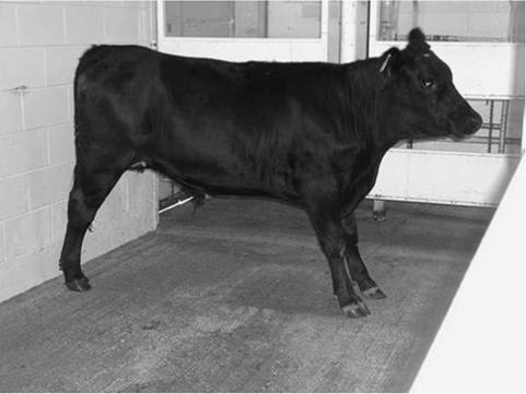
FIG. 10.1 Lordosis and stranguria in a steer with urethral obstruction.
full physical examination should be performed because abnormal urination may be a sign of disease in other body systems, such as those characterized by diffuse muscular weakness.
When possible, the animal should be observed urinating, and a sample of urine should be collected for dipstick urinalysis, measurement of specific gravity, and sediment examination; urine can be collected in a separate, sterile container for culture, if indicated. Urination can be induced in cows by gently rubbing the perineum immediately ventral to the vulva. In male cattle, the examiner may induce urination by placing a finger into the preputial cavity and gently rubbing the preputial mucosa. In ewes, urination can be induced by holding off the nose until the ewe struggles; urination typically occurs at this point. This procedure should not be performed on weak ewes or those in shock or with poor cardiac or respiratory function. In horses, goats, and male sheep, the examiner simply must wait until the animal is ready to void, although urination may be encouraged by placing the animal in a freshly bedded stall. Recumbent animals will often void soon after standing. Horses will often cease urinating midstream if approached quickly; thus, a urine collection cup taped to a long stick or straightened wire coat hanger affords the clinician a better chance of catching urine quickly and safely. In some species, urinary catheterization is possible and sometimes required to obtain a sample. The vulva or penis should be cleaned with dilute chlorhexidine solution, and aseptic technique should be maintained.
In female horses and ruminants the urethral opening is located under the suburethral diverticulum, a mucosal flap on the floor of the vagina. A lightly lubricated stiff urinary catheter can be passed blindly into the urethra by sliding it gently along the ventral floor of the vagina, taking care to stay exactly on the midline. Alternately it can be located by placing a hand in the vagina. In male horses sedation is required to relax and exteriorize the penis, which then should be grasped firmly and extended; then a lubricated flexible catheter can be passed. Acepromazine sedation should be avoided in stallions due to the risk of paraphimosis. Normal equine urine is variably turbid owing to the presence of mucus and calcium-based crystals (Fig. 10.2).The male's preputial hairs and the female's perineal region should be closely inspected for the presence of blood, exudate, or crystalline debris (Fig. 10.3). Sedation and/or epidural anesthesia may be necessary to induce sufficient relaxation of the retractor penis muscles to enable examination of the penis. In prepubescent ruminants the frenulum often prevents complete exteriorization of the penis for examination of the urethral orifice; general anesthesia may be necessary to induce sufficient relaxation. In bulls and steers transrectal massage of the pelvic segment of the urethra may stimulate penile relaxation and visualization. The
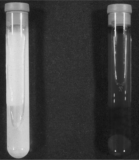
FIG. 10.2 Normal equine urine (left) and urine with severe myoglobinuria (right). Urine containing myoglobin is not always so markedly discolored. (Photo courtesy Paul S. Morley.)
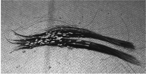
FIG. 10.3 Crystals adherent to the preputial hairs of a steer with urolithiasis. The crystals were analyzed and found to be composed of struvite (magnesium ammonium phosphate).
glans penis and urethral orifice should be carefully examined for masses such as papillomas, evidence of trauma, encircling hair rings, and embedded foreign bodies (e.g., grass awns).
Penile examination is particularly important in the dysuric or stranguric small ruminant because urinary calculi frequently become lodged in the urethral process (see the Urolithiasis in the Ruminant Renal System section in Chapter 34).An accumulation of smegma, composed of mucus and cellular debris, may cause preputial swelling and dysuria in adult male horses. Smegma can usually be found as a hard, waxy mass in the urethral diverticulum. Preputial swelling without urinary dysfunction may be seen with equine metabolic syndrome, and in equine pituitary pars intermedia dysfunction.1,2
In the male the penis and the urethra should be palpated percutaneously from the perineum distally to the sheath. Swelling, pain, abnormal urethral pulsations, and calculi lodged in the urethra may be detected. Urethral calculi are most commonly lodged just below the anus in male horses, and these can occasionally be palpated on the midline of the perineum.
■ BOX 10.1
Causes of Dysuria, Stranguria, and Urinary Incontinence in Horses and Ruminants
Adhesions of the bladder
Balanitis, posthitis, vulvitis, vaginitis
Bracken fern intoxication (R)
Botulism
Cobalt toxicosis (E) Cantharidin (blister beetle) toxicosis (E) Ectopic ureter
Equine dysautonomia (grass sickness) (E) Equine herpesvirus myeloencephalopathy (E) Equine protozoal myeloencephalitis (E) Equine neonatal maladjustment (E) Estrogen-responsive dysuria (E)
Everted bladder Habronemiasis (E) Hemorrhage into the urinary tract
Hendra virus (E, Australia and Southeast Asia)
Laminitis
Lumbosacral spinal cord trauma, inflammation, or neoplasia Lumpy skin disease virus (R, Africa)
Myopathy
Neoplasia of the urinary tract or accessory sex glands Painful conditions of the abdominal organs or abdominal wall Painful conditions of the thoracic organs or thoracic wall Parturition
Penile, vaginal, urethral, or preputial trauma Penile masses or encircling hair rings Pelvic entrapment of the bladder Pelvic, sacral, or vertebral fracture Polyneuritis equi (Cauda equina neuritis) Prolapsed bladder Prolonged recumbency
Rabies
Rectovaginal fistula Ruptured urethra, bladder, or ureter Seminal vesiculitis
Smegma accumulation (E) Sorghum intoxication
Squamous cell carcinoma (penis, prepuce, or vulva) Tetanus
Ulcerative posthitis and vulvovaginitis (pizzle rot) (R) Urachal infection or abscesses
Urinary calculi (urolithiasis) Urinary tract infection
Vaginal prolapse Vertebral or spinal malformation
E, Found only in horses; R, found only in ruminants.
Marked swelling along the prepuce and ventral body wall in a bull or steer with active or recent dysuria or stranguria can indicate urethral rupture.
The vulva, caudal vagina, and urethral orifice should be visualized and palpated in females. Sacrocaudal epidural anesthesia may facilitate examination if painful lesions are present. In females of breeding age the cervix should be visualized or palpated and the uterus evaluated by palpation or ultrasonography because the pollakiuria and apparent dysuria that may occur at the onset of parturition may be the primary complaint of a novice observer. Previous dystocia can result in sufficient soft tissue trauma, laceration, swelling, and pelvic neuropraxia to induce dysuria or stranguria. The ventrum of the tail, perineum, udder, and hindlimbs should be examined for adherent blood or exudate originating from the female's reproductive or urinary tract.
In adult horses and cattle rectal palpation should be performed when dysuria and stranguria are present. Before examination the clinician should take careful note of the tail and anal tone of the animal; reduction of either or both may indicate underlying neurologic or muscular disease. Introduction of the examiner's hand and wrist into the rectum is usually sufficient for palpation of the pelvic segment of the urethra and bladder trigone. The caudal extent of the pelvic cavity should be carefully palpated for masses that might mechanically interfere with voiding. The bladder is typically located on the midline at the level of the pubic brim. Its presence in the caudal pelvic cavity, particularly in the standing animal, may suggest pelvic entrapment of the bladder.
Bladder distention is commonly found in persistently recumbent horses and cattle, and on rectal examination the bladder is often positioned farther caudally than in standing animals. In the horse bladder distention may also be found with abdominal or thoracic pain. Apparently the abdominal pressure necessary to empty the bladder incites sufficient pain of diseased structures to cause reluctance to void. Musculoskeletal and neurologic disease may also result in bladder distention. These other possibilities should be carefully investigated when bladder distention is detected yet no primary disease is found in the urinary tract.
A careful rectal examination of the bladder and the proximal urethra of the horse might allow identification of urethral or cystic calculi. Most cystic calculi in the horse are singular, located in the trigone of the bladder, and are palpable with the examiner's arm inserted to the level of the wrist. If there is a large amount of urine in the bladder, the stone may not be palpable; in such cases, transrectal ultrasound examination may enable visualization of the stone. Detection of calculi in the bladder or urethra should prompt consideration of concurrent nephrolithiasis.
If bladder dysfunction is not caused by structural abnormalities, trauma, or infectious disease, a thorough neurologic examination should be conducted. If neurologic dysfunction is suspected, an attempt should be made to determine whether the primary lesion is affecting the detrusor muscle (lower motor neuron [LMN]) or the urethral sphincter muscles (upper motor neuron [UMN]) of the bladder. This determination is often helpful in localizing the lesion and is important when selecting treatment.
When bladder paralysis is caused by UMN dysfunction, signs of UMn dysfunction may be evident in the rear limbs. The animal frequently postures and strains to urinate but voids only a small amount of urine because the striated urethral muscles are disinhibited from higher centers and their resultant increased tone impedes urine outflow from the bladder. Frequent, small-volume urine egress from the distended bladder occurs when the animal responds to the urge to void or when the bladder undergoes reflex contraction.
With severe disease of the sacral spinal cord or sacral nerve plexus, LMn input to the detrusor muscle is impaired or absent. Urinary incontinence is often the predominant clinical sign (e.g., cauda equina neuritis in horses, spinal cord lymphoma in cattle). The bladder is usually moderately to severely distended, and urine can be expressed easily if pressure is applied to the bladder during rectal examination. With LMN dysfunction, urine may also be voided as the animal walks. Voluntary or involuntary voiding is often incomplete, leading to retention of urine in the bladder. This, in turn, increases the patient's risk of DTI. In horses with bladder paralysis, sabulous accumulations may form a doughy sludge in the ventral bladder, and on rectal examination the clinician may interpret the palpation findings as a bladder tumor.3 Other neurologic signs involving the sacral and coccygeal nerves may be apparent, such as decreased tail and anal tone and atrophy of the gluteal or tailhead musculature. Ataxia or weakness of the rear limbs may or may not be present with bladder paralysis due to LMN dysfunction. Urethral and bladder pressure profiles can be determined to better assess the location of the lesion.4-6
In small ruminants and neonates, transabdominal palpation is useful for evaluation of the urinary tract. In these animals a distended bladder can usually be palpated by simultaneously placing one hand on each side of the caudal ventral abdomen at the level of the pelvic brim and pressing the fingers of each hand toward the abdominal midline. If the bladder has been ruptured, it will be difficult to identify by palpation, but ascites due to uroperitoneum can be detected. Digital rectal palpation of the pelvic segment of the urethra can be performed in neonatal cattle and horses and in small ruminants. The umbilicus should be carefully palpated in neonates with dysuria or stranguria because urachal abscesses and adhesions to the bladder may impair voiding. An infected urachus will occasionally communicate with the bladder lumen, creating concurrent septic cystitis.
Ectopic ureter(s) should be considered in young animals with persistent urinary incontinence; stranguria and dysuria are less common primary complaints. In affected females vaginal urine pooling is often present. Vaginoscopic or cystoscopic examination can be performed, but the opening of the ectopic ureter can be difficult to locate during routine examination. Location of the ectopic ureter(s) often requires intravenous contrast urography or intravenous administration of a dye that is excreted in the urine, such as fluorescein. As for all congenital defects, a careful assessment for defects in other organs should be performed in confirmed cases.
If physical examination and urinalysis do not reveal the source of dysuria, stranguria, or incontinence, transabdominal and/or transrectal ultrasonographic evaluation of the urogenital tract should be performed. Transrectal ultrasound can be performed in geldings to evaluate the accessory sex glands, caudal ureters, and pelvic urethra.7 An endoscope can be used to visualize the vaginal vault or preputial cavity and penis; air insufflation can be used to expand the walls and achieve a clear view of these structures. When advanced retrograde into the urethra, the urethral wall, bladder, and ureteral openings can be visualized. In neonates and small ruminants, plain abdominal radiographs, positive contrast urethrocystography, and intravenous contrast urography are additional options.8
More on the topic Dysuria, Stranguria, and Incontinence:
- Dysuria, Stranguria, and Incontinence
- MANIFESTATIONS OF DISEASE
- Smith Bradford P., Van Metre David C., Pusterla Nicola (eds.). Large Animal Internal Medicine. Part 1. 6th edition. — Elsevier,2020. — 2279 p., 2020