Equine Alimentary System
Samuel L. Jones, Consulting Editor
Examination and Imaging of the Equine Alimentary System
Samuel L. Jones • Michelle H. Barton
A thorough physical examination is compulsory, and tests that provide a minimum database (complete blood cell count [CBC], serum chemistry profiles, and urinalysis) are often indicated in horses with suspected alimentary tract disease.
Once a differential diagnosis is compiled, a number of ancillary diagnostic tests are available to narrow the possibilities. Each diagnostic test or procedure is limited in the type and extent of information that can be obtained, and therefore the clinician should select the complement of procedures that is most likely to provide the information required to make a proper diagnosis and determine the appropriate therapy.■ Rectθl Examination A systematic approach to examining the abdominal and retroperitoneal viscera should be established and applied during each examination to ensure that all pertinent regions and structures are examined. When feasible and if required, the patient should be sedated to allow a more thorough examination. In some cases, W-butylscopolammonium bromide administration or epidural anesthesia is required to obtain adequate access to structures during rectal examination. The principal goal of a rectal examination is to identify changes in size, texture, shape, or location of visceral organs, peritoneum, mesentery, and vasculature, or to detect objects that are normally not present. Rectal examination is often performed with ultrasonographic examination, and information derived from each must be considered in order to draw conclusions. Ultrasonographic examination is described in a later section of this chapter.
In the pelvic region of the normal horse, the urethra and accessory sex glands (male) or the vaginal vault and cervix (female) can be palpated.
The urethra is usually not discernible in the female, but abnormalities such as uroliths may be detected. In the caudal abdominal cavity, the bladder, the uterus in females, and the pelvic flexure and small colon typically should be identified. The pelvic flexure and left ventral and left dorsal portions of the colon are normally located ventrally, on midline, or toward the left side of the abdomen. The small colon, in which formed fecal balls are palpable, courses throughout the caudal abdomen, mostly on the left side. In females the left ovary can be identified in the left dorsal, caudal region of the abdomen. Both ovaries should be palpated in conjunction with palpation of the uterus. The peritoneal surface should be detected along the surface of the abdominal wall and the surfaces of the viscera. It should feel smooth, with no crepitus or irregularities. Farther along the left side of the abdomen, the spleen is palpable as a smooth structure; the caudal border is well-delineated and tapered. The size and location of the spleen are variable because it can extend from the left body wall to the right ventral region of the abdomen. Farther cranially and dorsally, the left kidney can be palpated. The kidney should feel smooth and the renal pelvic fissure should be discernible, although in an overweight horse, extensive perirenal fat may obscure this detail.From the left kidney, toward midline and extending from the abdominal aorta, the cranial mesenteric and ileocecocolic arteries may be palpated. Fremitus detected in these arteries may be associated with arteritis and thrombus formation secondary to Strongylus vulgaris larval migration; however, this association has been very inconsistent. Fremitus is frequently absent when severe arteritis exists, or the arteries may be entirely normal and fremitus felt. Fremitus can often be elicited by compression of the wall of the normal artery, which accelerates flow through the compressed lumen. The mesenteric root of the colon can be palpated ventral to the cranial mesenteric artery.
This should feel like a mildly taut band of tissue extending from the dorsal midline ventrally. Excessive tension, displacement, thickening, or masses within the mesentery are abnormalities. It may be possible to palpate an enterolith, a fecalith, or a gravel impaction in the transverse colon, but this may be beyond the reach of the examiner because the transverse colon is cranial and medial to the left kidney.On the right side of the abdomen, the base and cupola of the cecum can be palpated. The body of the cecum can be followed partially along the medial aspect of the cecum, cranially toward midline. The cecum has a prominent ventral band and
sacculations. Gas, together with ingesta that is soft and mainly of a fluid consistency, can be identified within the cecum. Firm or excessive ingesta suggest an abnormality.
Findings that are different from normal often must be identified as either variations of normal or true abnormalities. Abnormalities of the peritoneal surface are common findings. Crepitus, or a “plastic wrap” texture, is indicative of gas secondary to trauma or infection. An irregular or rough surface may be indicative of fibrin on a visceral surface or neoplasia; with a perforated intestine, ingesta may be adhering to a visceral surface. There are many abnormal presentations of the large colon, most of which are associated with signs of colic. Thickening of the wall of the colon may be appreciated on rectal palpation and is indicative of edema or cellular infiltration of the colon. Abnormal masses palpated in the wall of the colon or in association with the colonic mesentery are indicative of infection, infarction, granulomatous colitis, or neoplasia.
The small intestine is not normally discerned by palpation. On occasion, however, peristaltic contractions may be felt in the small intestine as it courses across midline toward the base of the cecum. In some cases this causes the small bowel to feel firm and tubular. Relaxation of the peristaltic contraction should be discerned in such cases.
Distention of the small intestine is abnormal. In some cases the bowel may feel thickened, which can occur with ileal muscular hypertrophy, edema, or inflammatory disorders of the small bowel.Other abnormal findings that may accompany disorders of the abdominal alimentary system include masses, adhesions, enlarged and thickened mesenteric arteries, and caudal displacement of the spleen (secondary to gastric distention or neoplasia).
■ Paracentesis Abdominal paracentesis is performed routinely in patients with suspected disorders of the abdominal viscera. Cytologic studies are used to quantitate peritoneal fluid; white blood cell (WBC) and red blood cell (RBC) counts; protein, fibrinogen, serum amyloid A, lactate, phosphate, and glucose concentrations; lactate dehydrogenase, creatine kinase (CK), and alkaline phosphatase (ALP) activity; and pH. The results of peritoneal fluid analysis may help establish a specific diagnosis but, of more importance, may help distinguish inflammatory from noninflammatory conditions, identify vascular obstructions or ischemic injury to the intestine, and determine whether surgical intervention is indicated.
■ Endoscopy Two basic types of endoscopic equipment are available: equipment based entirely on a fiberoptic system and equipment based on a video system. Many affordable endoscopes are now available, and endoscopic examination of the proximal alimentary tract in horses is now common. A typical endoscope found in many private practices is a fiberoptic endoscope that has an insertion tube that is 100 to 110 cm in length and 10 to 14.5 mm in outer diameter. The larger-diameter tube can be inserted only through the nasal passages of older yearlings and adults and is not suitable for alimentary endoscopy, whereas a tube 10 mm in diameter can be passed through the turbinates of young foals. An insertion tube length of 110 cm is sufficient to reach the stomach of foals up to 40 days of age. A length of 150 to 180 cm is required for weanlings, and 200 cm is usually required for yearlings and adults.
An insertion tube length of 200 cm is sufficient to reach the stomach of all adults of warm-blooded breeds, although 280 to 300 cm is required to examine the pylorus in adult horses. A 280- to 300-cm-long insertion tube enables duodenoscopy in adult horses.Before a gastroscopic examination, suckling foals up to 20 days of age are not routinely kept from nursing for more than 1 hour. Older foals and mature horses, in contrast, should not have solid feed for 6 to 10 hours so that ingesta from the stomach may be adequately emptied. Longer duration of feed deprivation (12 to 18 hours) is desirable to view the antrum and pylorus of horses. Many foals less than 30 days old do not require sedation for gastroscopy, although sedation with xylazine (0.5 mg/kg) may facilitate the procedure. Sedation is required if the foal is to be placed in a recumbent position so that the entire glandular portion of the stomach can be examined. Older foals and horses must be sedated for gastroscopy. Xylazine (0.5 mg/kg given intravenously [IV]) usually provides adequate sedation. For greater sedation, detomidine (0.005 to 0.05 mg/ kg IV) or a combination of xylazine and butorphanol (0.01 mg/ kg IV) is effective.
After insertion of the endoscope, the stomach is distended with air until the nonglandular and glandular regions of the gastric surface can be observed. Distention with air is tolerated by foals and horses and has been associated only rarely with adverse effects. On occasion, sick neonates with poor intestinal motility have developed distention of the small intestine and experienced discomfort after gastroscopy.
More complete descriptions of techniques of gastroduodenal endoscopy can be found elsewhere.1,2
Endoscopy of the rectum and distal small colon can be performed with most flexible endoscopes in use in equine practice and should be preceded, as much as possible, by evacuation and saline lavage of the rectum and distal small colon. The mucosal surface should appear pink to pale red and should have a smooth, velvety appearance.
Mucosal edema or thickening, hyperemia, irregularities, defects, tears, and intraluminal masses are abnormal findings. Because of the concern for trauma to the rectum and small colon, horses should be adequately sedated and restrained before preparation and examination of the distal alimentary tract.■ Laparoscopy Laparoscopy can offer valuable diagnostic information regarding the abdominal cavity and is only minimally invasive.3,4 It should always be preceded by a thorough physical examination, including abdominal rectal palpation, and by abdominal ultrasonographic examination, with particular attention to the sites for trocar insertion to ascertain the presence of adherent masses or viscera in the area. If abdomi- nocentesis is to be part of the diagnostic workup, it should be performed before laparoscopy because laparoscopy can affect abdominal fluid values. In experimental animals undergoing diagnostic laparoscopy with carbon dioxide insufflation, both the abdominal WBC count and the abdominal total protein increased.5
The indications for laparoscopy include palpable abdominal masses, enlarged viscera, adhesions, acute or chronic colic, weight loss, and the need to obtain visceral biopsy specimens. Contraindications include adherent viscera or masses at the site of laparoscopic trocar insertion, diaphragmatic hernias, or extreme bloating. Horses with acute colic can be safely examined laparoscopically if the trocars and telescopes are inserted carefully.
The basic instruments for laparoscopic examinations include a laparoscopic telescope, laparoscopic cannula and trocar assembly, fiberoptic light source and cable, insufflator, and biopsy and manipulation instruments. Video cameras make visualization easier with less eyestrain but require more powerful light sources. The cost of laparoscopic instrumentation has decreased because laparoscopy has become popular in humans and animals, and the procedure is far more cost effective than in years past.
Horses should fast for 18 to 24 hours before most laparoscopic procedures; water is allowed on an ad libitum basis. Fasting increases intraabdominal visualization and decreases the possibility of penetrating a gas-distended viscus. The animal is restrained in standing stocks if the procedure is to be done while it is standing. Preoperative antibiotics, antiinflammatory drugs, tetanus prophylaxis, and a sedative analgesic combination are administered. It is important to administer the analgesics before abdominal insufflation. The flank areas are prepared for aseptic surgery. Local anesthetic agents are infiltrated subcutaneously (SC) and intramuscularly (IM) in the middle of the paralumbar fossa, slightly above the crus of the internal abdominal oblique muscle, for the insertion of the laparoscopic telescope. It is preferable to begin the laparoscopic procedure
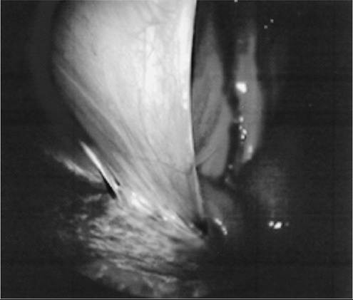
FIG. 32.1 Laparoscopic view of nephrosplenic ligament in a horse.
on the left side of the abdomen to minimize the chance of penetrating the cecal base. Additional ports may be created in the right paralumbar fossa to increase access to intraabdominal and retroperitoneal structures and enable the use of additional instruments. Ports are created with a stab incision, and a laparoscopic cannula and trocar assembly are inserted through the musculature and into the abdominal cavity. The trocar should be oriented toward the opposite coxofemoral joint when it is inserted. The trocar is exchanged for the telescope, and entry into the abdominal cavity is confirmed before insufflation begins. If the abdominal cavity has not been penetrated, a quick thrust with the telescope usually penetrates the peritoneum. Insufflation of the abdomen with CO2 to 8 to 10 mm Hg is usually sufficient for most examinations.
The abdominal cavity is then examined systematically. On the left side of the abdomen, the spleen, left kidney, nephrosplenic ligament (Fig. 32.1), stomach, left side of the liver, diaphragm, and ventral portion of the colon are visualized cranially. A caudal view includes the root of the mesentery, the isolated small intestinal and small and large colon sections, the urogenital tract, the bladder, and the terminal rectum. The procedure is repeated on the right side of the abdomen. The liver, epiploic foramen, right kidney, descending duodenum, cecal base, and large colon are visualized cranially. The urogenital tract, root of mesentery, and intestinal segments are visualized caudally. Technical innovations have enabled extensive examination of intraabdominal structures. For example, it is possible to examine the entire small intestine via laparoscopy.6 Depending on the pathologic condition, diagnostic laparoscopy may provide a diagnosis and allow access to surgically resolve the problem. Liver biopsy and right kidney biopsy samples are taken from the right side of the abdomen; left kidney and spleen biopsy samples are taken from the left side. Mesenteric lymph node biopsy samples are usually obtained via the left flank. Biopsy samples of other masses are taken from the more accessible side. At the end of the procedure, the abdomen is deflated, and the skin is closed with skin sutures only. The skin incision should not be closed until examination of both sides is completed, in order to minimize subcutaneous emphysema.
In some horses with ventral or cranial abdominal masses as determined with ultrasonography, the animal can be anesthetized and placed in dorsal recumbency for better characterization of the mass. Biopsy samples may be readily obtained. In horses with acute colic but without obvious signs indicating the necessity for surgery, laparoscopy can guide the decision to continue medical therapy or proceed to surgery. Strangulated sections of small intestine can be visualized, proximal enteritis can be diagnosed, and edema and vascular compromise to the large colon can be seen. No abnormalities may be detected in some animals with very localized lesions, or lesions may be inaccessible, depending on location.
Laparoscopic complications are similar to those of any other abdominal exploratory procedure. A viscus may be penetrated inadvertently; the left kidney may be perforated if the laparoscope is inserted too far dorsally; and the spleen may be penetrated if the laparoscope is inserted too far ventrally or is not aimed toward the opposite coxofemoral joint. The cecum may be perforated when the laparascope enters from the right side. If the horse has fasted and the laparoscopic trocars are inserted carefully, these problems are minimized. Subcutaneous emphysema occurs commonly but has not caused any clinical problems. The peripheral WBC count increased but stayed within normal limits in experimental animals undergoing laparoscopy.5
■ Transcutaneous Ultrasonography of the Mature Equine Alimentary Tract
MicheUe Henry Barton
TECHNIQUE. One of the most innovative, practical, affordable, and portable diagnostic tools introduced into the field of equine internal medicine since 2000 is transcutaneous ultrasonography. Although several different types of ultrasound transducers exist, adult equine abdomens are most completely imaged with a 2- to 5-MHz curvilinear array transducer, but a high-frequency (6- to 8.5-MHz) linear array transducer can be used to optimize resolution of superficial intraabdominal structures. Ideally, before the procedure, the patient's hair is clipped, the skin cleaned, and coupling gel applied. If clipping the hair is not an option, debris can be removed with grooming aids and the hair soaked in isopropyl alcohol. Most horses tolerate transabdominal ultrasonography without sedation. In patients sedated with α2-adrenergic agonists, small intestinal and cecal motility can be transiently reduced, especially in horses that have fasted, and the luminal diameter of the small intestine may appear more dilated than in unsedated patients.7,8 Likewise, administration of N-butylscopolammonium bromide will immediately reduce duodenal, cecal and left ventral colonic contractions for 30 (cecum and colon) to 120 minutes (duodenum).9
Detailed review of the physics of ultrasonography is beyond the scope of this section; however, several general principles should be kept in mind when transabdominal ultrasonography is performed. First, to ensure that all structures are properly assessed, the approach for covering the entire area to be evaluated must be consistent and systematic: for example, scanning one side at a time, dorsal to ventral, cranial to caudal. Using seven discrete locations in horses being evaluated for acute abdominal pain, the examiner can complete the fast localized abdominal sonography for horses (FLASH) method in less than 15 minutes; this method is extremely helpful in establishing the correct diagnosis.10 The seven locations evaluated in the FLASH method are ventral, gastric, splenorenal, left middle third of the abdomen, duodenal, right middle third, and thoracic. Focused technique can be used to consistently identify the cecum, sacculated large intestine, spleen, liver, and right kidney. The left kidney is not always identified, and successful identification of small intestine may require repeated examination.11
Second, to ensure correct orientation of the anatomy being imaged, the examiner should be mindful of the position of the transducer and its orientation marker on the patient (Fig. 32.2). Because abdominal ultrasonography is often performed with the transducer over an intercostal space, the transducer should be oriented in a slightly oblique transverse plane (i.e., “slicing” across the long axis of the body). Careful attention should be paid to the spatial relationship of the viscera because this may be the only key feature distinguishing normal from abnormal.12 Furthermore, the walls of some sections of the gastrointestinal tract appear strikingly similar and cannot be distinguishable unless information is provided about the probe's exact location on the patient.13
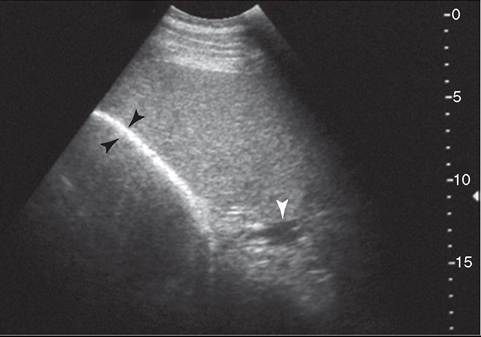
FIG. 32.2 Ultrasound view from the gastric window in the left eleventh intercostal space at the level of the shoulder in a horse. The left side of the image is dorsal. The black arrowheads mark the border between the serosal layer and the mucosal-luminal interface of the stomach wall, which appears as a curved hyperechoic line. The spleen is located in the near field between the stomach wall and the body wall. The white arrow marks the splenic vein. This image was obtained with a 3.5-MHz curvilinear probe set to a depth of 19 cm.
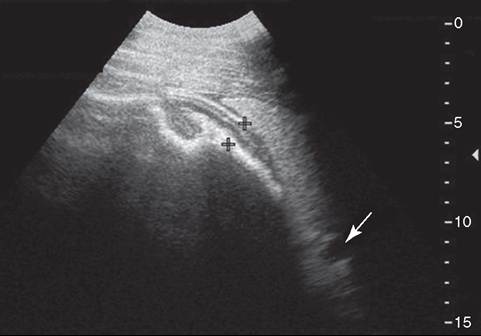
FIG. 32.3 Ultrasound view of the layers of the stomach wall from the gastric window in a horse. The left side of the image is dorsal. The white arrow marks the splenic vein. The electronic calipers (represented by plus signs') are placed at the border between the serosal layer and the mucosal- luminal layer and mark the stomach wall. The five ultrasonographic layers of the wall are visible, and it is abnormally thickened at 1.34 cm. Follow-up gastroscopy revealed that the horse had severe gastric ulceration.
Third, in any ultrasonographic examination, it is important to be aware of the depth of the field of view. Selecting the appropriate frequency for the transducer is the key to producing high-quality images that are suitable for the depth of display. High-frequency probes provide sharp images, but the resolution is compromised as the depth of the viewing field increases.
A final general and helpful principle of ultrasonography is that more sound waves reflect to the transducer if two adjacent interfaces have markedly different acoustic impedances. The more sound that reflects to the transducer, the “whiter” the interface appears on the monitor; these tissue interfaces are called echogenic or hyperechoic. In contrast, less dense tissues reflect less sound, appear darker, and are referred to as anechoic or hypoechoic structures. The acoustic impedance of the soft tissue of the walls of the gastrointestinal tract is several thousand times greater than that of the free gas inside the adjacent lumen.14 Consequently, the image at this soft tissue-to-gas interface appears as a fuzzy hyperechoic border (see Fig. 32.2). Because most of the sound waves at this interface are reflected and the free gas in the lumen has extremely low impedance, sound is neither penetrating nor reflecting from the lumen, and the rest of the lumen is therefore obscure.
Gas within a large luminal viscus is one of the greatest limitations to gastrointestinal ultrasonography in horses, inasmuch as it impedes visualization of deeper structures. While imaging a patient, the ultrasonographer should remember that gas rises to the nondependent portion of the abdomen, and fluid and heavier structures fall to the dependent locations. When a 3.5- to 5-MHz curvilinear transducer is used, the difference between anatomic layers of the intestinal wall in healthy horses is less distinct; the wall typically appears as a single hyperechoic line. Depending on the surrounding tissue and the contents of the lumen, three to five layers of the gastrointestinal wall may be visible when a high-frequency transducer is used; these five layers are the hyperechoic serosa, hypoechoic muscularis layer, hyperechoic submucosa, hypoechoic mucosa, and the hyperechoic mucosal interface with the lumen (Fig. 32.3).15
In the following sections, transabdominal ultrasonography of the gastrointestinal tract is described with a systematic approach of scanning in a cranial-to-caudal direction. For the gastrointestinal tract, the ultrasonographer should be careful to document the following key elements: motility, presence of luminal distention, wall thickness and structure, luminal contents, and viscera position.
ULTRASONOGRAPHY OF THE LEFT SIDE OF
THE ABDOMEN
Stomach. When imaging is started on the left rostral side of the abdomen, the stomach should be located deep to the spleen between the ninth and thirteenth intercostal spaces at approximately the level of the shoulder. In this location, the only part of the stomach that can usually be seen is the wall of the greater curvature, which can be reliably identified as a smooth, curved, hyperechoic line adjacent to the spleen and the splenic vein (see Fig. 32.2).16 The motility of the stomach is sluggish, and the ultrasonographer may get the impression that the greater curvature of the stomach remains stationary.8 The stomach has the thickest wall in the gastrointestinal tract, measuring approximately 7 mm thick from the serosal layer to the mucosal-luminal interface.17 When the stomach is empty, the wall measures up to 1 cm thick, ill-defined loops of small intestine may appear between the stomach and the spleen, gastric rugal folds may be visible, and the gastric wall may be found only below the costochondral junction.8,17
Gastric Impaction and Ulceration. Because only the dorsal portion of the greater curvature is visible and the lumen generally contains gas at this location, transcutaneous ultrasonography of the stomach is not a sensitive tool for diagnosing gastric ulceration, intramural gastric masses, or gastric impaction. However, if the wall of the greater curvature of the stomach extends beyond the fourteenth intercostal space in a horse that has not recently drunk or eaten, gastric distention should be suspected.18 Furthermore, if excessive gastric fluid is present ventrally, a distinct hyperechoic gas-fluid interface may be apparent in the lumen (Fig. 32.4). If the stomach wall appears irregular or thickened, follow-up gastroscopy is indicated for a more definitive diagnosis (see Fig. 32.3).
Spleen and Left Kidney. Although the focus of this section is gastrointestinal ultrasonography, the spleen and left kidney warrant brief review because of their proximity to the gastrointestinal tract. Caudad from the stomach, the spleen should be identifiable immediately adjacent to the body wall, from the left ventral eighth intercostal space to the paralumbar fossa. The size and location of the spleen vary greatly: The
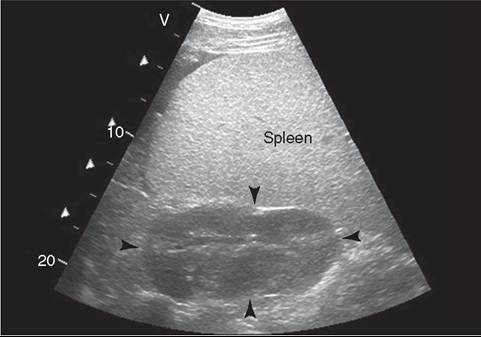
FIG. 32.5 Ultrasound view from the left dorsal fifteenth intercostal space in a horse, showing the normal adjacent orientation of the spleen to the left kidney (black arrowheads). The left side of the image is dorsal. This image was obtained with a 3.5-MHz curvilinear probe set to a depth of 21 cm.
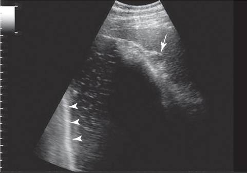
FIG. 32.4 Ultrasound view from the gastric window in a horse. The left side of the image is dorsal. The white arrow marks the splenic vein. The white arrowheads mark the interface between hyperechoic fluid in the dependent portion of the stomach (right) and gas dorsally. This image was obtained with a 3.5-MHz curvilinear probe set to a depth of 23 cm.
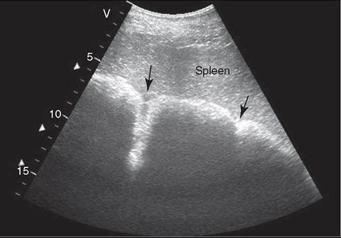
FIG. 32.6 Ultrasound view from the left flank of a horse, showing the spleen adjacent to the left ventral portion of the colon, identifiable by its location and the presence of sacculations (black arrows). The left side of the image is dorsal.
spleen can be left of the midline or extend slightly right of the ventral midline. In the rostral ventral left abdomen of some horses, the most rostral aspect of the spleen can appear either lateral or medial to the liver.12 The spleen's ultrasonographic architecture is usually homogeneous, with vessels that are rarely visible. The echogenicity of the spleen is greater than that of the liver or kidneys.
The left kidney can be found between the fifteenth and seventeenth intercostal spaces and the first to third lumbar vertebrae, medial or deep to the spleen (Fig. 32.5), between the level of the tuber coxae and the tuber ischii.19,20 In rare cases, the left kidney directly apposes the left body wall.12 Gas in the small colon, left portion of the colon, or lung can preclude transabdominal viewing of the left kidney, which is normal, although colonic gas obscuring visualization of the left kidney may be adjunct evidence of dorsal displacement of the left portion of the colon.21 The ultrasonographic dimensions of the left kidney vary according to the location from which the measurements are obtained. The longitudinal axis for cranio- caudal length (i.e., the dorsal plane parallel to the spine) is difficult to measure because of interference by the ribs.20 In Thoroughbred horses, the length of the left kidney varies from 9.6 to 15.6 cm.19 The dorsoventral height in the slightly oblique transverse plane varies from 3.1 to 8.1 cm, and the lateromedial thickness varies from 6.6 to 9.1 cm.19 The corticomedullary junction should be distinct, with the cortex being more echogenic. The adrenal glands are not usually identifiable via transabdominal ultrasonography.12
Left Portion of Colon. Many disorders of the large colon can be palpable by rectal examination, and thus transabdominal ultrasonography is an ancillary tool. Ventromedial to the spleen, the left ventral portion of the colon can be identified by its sacculated wall (Fig. 32.6) and sluggish but visible motility, normally contracting two to six times per minute.8,13,15 Precise measurement of the colonic wall can be difficult because of the indistinct mucosal-luminal interface; in general, however, the wall of the colon should be less than 4 mm thick.13,15,17 The left dorsal portion of the colon is not sacculated and can be dorsal, lateral, medial, or even ventral to the left ventral portion of the colon. Gas in the left ventral portion of the colon often precludes distinct identification of the left dorsal portion of the colon when the latter lies medial or dorsal to the left ventral portion of the colon. Gas in the colon typically generates a hyperechoic wall with an indistinct luminal border and intraluminal acoustic shadowing that precludes identification of the contents and the medial walls.
Colitis. When visualized through ultrasonographic examination of the left portion of the colon, useful diagnostic characteristics include wall thickness and structure, orientation of the colon, and its contents. For example, thickening of colon wall can be caused by inflammation, infiltration by neoplastic cells, hemorrhage, or edema. Inflammation of the colonic wall (i.e., colitis) can result in mural widening, but the mucosal border often appears shredded, undulating, or wavy (Fig. 32.7). These later findings may be particularly helpful in identifying horses in the prodromal phase of colitis, before the onset of diarrhea. The contents of the colon can change from characteristic acoustic shadowing to that of swirling mixed echogenic fluid that may enable visualization of the distal colonic wall. With colitis, colonic motility can be increased or decreased, or it can remain unaffected.
Colonic Volvulus. Colonic edema resulting from a volvulus of the colon also results in uniform hypoechoic mural thickening that is best identified with imaging along the ventral midline behind the xyphoid process. In one study in which colon wall edema was evaluated by transcutaneous ultrasonography through a ventral abdominal window, colon wall thickness of more
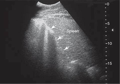
FIG. 32.8 Ultrasound view obtained from the same location as in Fig. 32.5 but in a horse with left dorsal displacement of the colon into the renosplenic space. The left side of the image is dorsal. The gas- distended colon (arrowheads) obscures visualization of the left kidney. This image was obtained with a 3.5-MHz curvilinear probe set to a depth of 20 cm. (Courtesy Science In 3D, Inc., and copyright 2002, University of Georgia Research Foundation, Inc.)
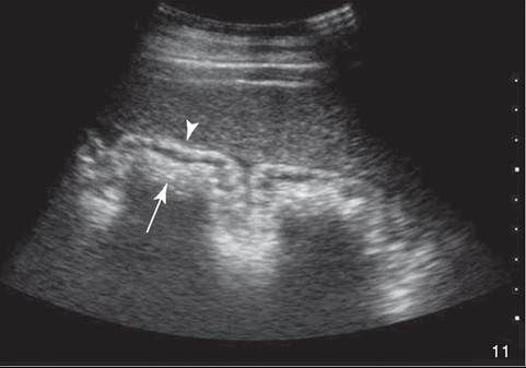
FIG. 32.7 Ultrasound view obtained from the same location as in Fig. 32.6 but from a horse with colitis. The left side of the image is dorsal. The left ventral portion of the colon is identifiable by its location medial to the spleen and by its sacculations. The wall of the colon was thickened to approximately 1 cm with three distinct layers: hyperechoic serosa (white arrowhead), hypoechoic muscularis, and submucosal, hyperechoic mucosal-luminal interface (white arrow). This image was obtained with a 3.5-MHz curvilinear probe set to a depth of 11 cm.
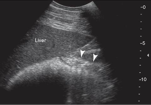
FIG. 32.9 Ultrasound view obtained at the eleventh right intercostal space at approximately the level of the shoulder in a horse. The left side of the image is dorsal. Sand and gravel in the dependent portion of the right dorsal portion of the colon (white arrowheads on serosal surface) created reverberation artifacts in the lumen. The colon wall is thickened. This image was obtained with a 3.5-MHz curvilinear probe set to a depth of 13 cm. (Courtesy Science In 3D, Inc., and copyright 2002, University of Georgia Research Foundation, Inc.)
than 9 mm had a sensitivity of 67% and specificity of 100% for colonic volvulus.22 In addition, in some cases of colonic volvulus, nonsacculated colon tissue replaces sacculated colon tissue in the left ventral abdomen.23 Furthermore, the blood supply to the ascending colon is located in the mesentery along its medial aspect, and thus major blood vessels are normally not visible ultrasonographically on the lateral surface (adjacent to the body wall) of the ascending colon. The ultrasonographic finding of distended vessels with reduced blood flow in colonic mesentery adjacent to the body wall implies that the colon is rotated along its long axis and is consistent with a diagnosis of displaced colon or a colonic volvulus.24 Colonic motility is reduced with a volvulus.
Displacement of Left Portion of Colon. On the left side of the abdomen, dorsal displacement of the left portion of the colon over the nephrosplenic ligament may obscure ultrasonographic visualization of the dorsal aspect of the spleen or left kidney, or the colon may appear lateral to the spleen (Fig. 32.8).21 However, if gas is not present in the entrapped colon, the ultrasonographic diagnosis can be missed. Likewise, gas within the left portion of the colon can obscure the left kidney without entrapment of the colon. When ultrasonographic and other clinical findings support the diagnosis of left dorsal displacement of the colon, serial ultrasonography of the area can be a useful noninvasive method of determining whether the colon has relocated to its ventral position, as evidenced by successful ultrasonographic visualization of the left kidney.
Colonic Impaction. Gas within the colon often precludes accurate ultrasonographic diagnosis of colonic impaction. An impaction should be suspected in the ventral portion of the colon when a hyperechoic border at the mucosal surface casts a strong acoustic shadow and when sacculations are flattened. Sand impactions are best visualized in the ventral abdomen and often compress the mucosal surface, create excessive acoustic shadowing or reverberation artifacts (Fig. 32.9), and significantly reduce visible motility of sacculations.25
Small Colon. The small colon is in the left paralumbar fossa, medial or ventral to the spleen. Because of its small diameter, sacculations, and packed serpentine loops that suspend from the dorsal mesocolon, often only small sections of the surfaces are visible ultrasonographically as short, sharply curving, hyperechoic lines (Fig. 32.10). As with the large colon, the motility of the small colon is slow (no more than three contractions per minute), and luminal surface gas typically prevents visualization of the contents and the distal walls.15 Likewise, the dense contents of fecal balls in the small colon often generate anechoic shadows from the near wall that precludes visualization of the distal wall (see Fig. 32.10).
Impaction in the Small Colon. Impactions or enteroliths obstructing the lumen of the small colon can usually be readily diagnosed by transrectal examination; however, transabdominal ultrasonography may reveal loss of, or enlargement of, sacculations and intense anechoic shadowing (Fig. 32.11).
Small Intestine. The jejunum can be identified in only approximately 10% of fed healthy horses.8 The examiner must carefully watch for peristaltic activity that creates transient expansion of the lumen from movement of ingesta. Because of its medial location, the ileum cannot be identified distinctly by transcutaneous ultrasonography. The jejunum is most consistently found in the left inguinal area, medial to the spleen and the left ventral portion of the colon (Fig. 32.12). The
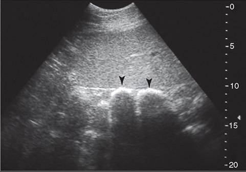
FIG. 32.10 Ultrasound view obtained from the left dorsal paralumbar fossa in a horse. The left side of the image is dorsal. The spleen is in the near field. The serosal surface (black arrowheads) of two adjacent loops of small colon are visible. Note the anechoic shadowing that precludes visualization of the distal small colon wall. This image was obtained with a 3.5-MHz curvilinear probe set to a depth of 21 cm.
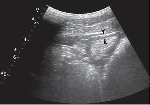
FIG. 32.12 Ultrasound view obtained of the left flank in a healthy, unsedated horse. A single loop of small intestine is visible between the body wall and the left ventral portion of the colon. The serosal to mucosal surfaces are most distinct in the near wall and are marked by the black arrowheads. This image was obtained with a 3.5-MHz curvilinear probe set to a depth of 8 cm.
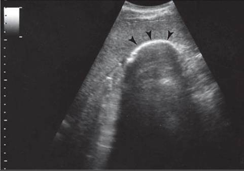
FIG. 32.11 Ultrasound view obtained from the left dorsal paralumbar fossa in a horse. The left side of the image is dorsal. The spleen is in the near field. The serosal surface (black arrowheads) of the small colon is visible and is bulging. Note the dramatic anechoic shadowing that was created by an intraluminal enterolith. This image was obtained with a 3.5-MHz curvilinear probe set to a depth of 21 cm. (Courtesy Science In 3D, Inc., and copyright 2002, University of Georgia Research Foundation, Inc.)
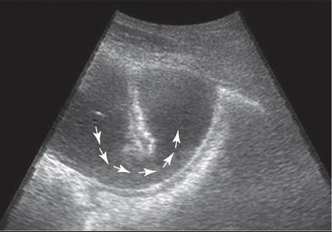
FIG. 32.13 Ultrasound view obtained from the left flank of a horse. A segment of distended small intestine is folding back on itself, creating the appearance of a “U turn” (arrows). This image was obtained with a 3.5-MHz curvilinear probe set to a depth of 10 cm.
small intestine has the most visible motility of any part of the gastrointestinal tract; peristaltic waves produce frequent rhythmic contractions. Fluid luminal contents typically enable accurate measurement of the wall (and motility varies with the duration of strangulation; preobstruction segments may become turgidly circular and completely amotile loops of small intestine, and this development is highly correlated with mechanically obstructive lesions that necessitate surgical intervention (Fig. 32.18).26,27
An important distinguishing feature of strangulated small intestine is the appearance of focally and usually uniformly thickened walls at the site of the strangulation (Fig. 32.19). Thus when regional uniform thickening of the small intestine is identified ultrasonographically, with other evidence of obstruction (hypomotile, hairpin-turn stacks of turgidly round and distended loops of small intestine with dependent ingesta), a strangulating obstruction should be the top consideration. Frequently the strangulated loops are identified in the ventral portions of the abdomen. In one study, the presentation of focal thickening and distention of the small intestine with no motility was 100% sensitive and 100% specific evidence of strangulating lesions of the small intestine.26 If the affected segments are most readily visualized adjacent to the spleen and stomach on the left side of the abdomen (Fig. 32.20) or between the liver and right dorsal portion of the colon on the right side of the abdomen (Fig. 32.21), these respective locations are areas in which gastrosplenic and epiploic foramen incarcerations are readily found.
Intussusception. Intussusceptions of the small intestine are most common in foals and young horses, and affected segments have a characteristic “bull’s-eye” or target appearance in images across the short axis of the small intestine (Fig. 32. 22).30 The intussuscepted intestine tends to fall toward the dependent portion of the abdomen. As in other mechanical obstructive diseases, the proximal small intestine has normal wall thickness but is distended, with reduced motility and hairpin turns, depending on the degree of obstruction.
ULTRASONOGRAPHIC ANATOMY OF THE RIGHT SIDE OF THE ABDOMEN. Right -sided transabdominal ultrasonography is described with a systematic approach of scanning in the direction of cranial to caudal.
Liver and Pancreas. The liver, descending duodenum, and right dorsal portion of the colon have characteristic locations and can be identified in the right rostral abdomen at the level of the shoulder (Fig. 32.23). The liver is located from the sixth to the fourteenth intercostal spaces, between the diaphragm
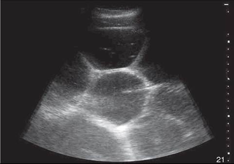
FIG. 32.18 Ultrasound view obtained from the left flank from a horse with a nonstrangulating obstruction that necessitated an exploratory celiotomy for correction. Note the numerous turgidly round and distended loops of small intestine. In real time, these distended loops had no appreciable motility. This image was obtained with a 3.5-MHz curvilinear probe set to a depth of 21 cm.
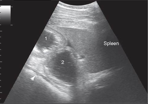
FIG. 32.20 Ultrasound view obtained from the gastric window from a horse with a strangulating gastrosplenic ligament entrapment. The left side of the image is dorsal. Two thickened, distended, and amotile loops of small intestine (labeled 1 and 2) were found between the spleen and the stomach (arrowhead). This image was obtained with a 3.5-MHz probe set to a depth of 17 cm.
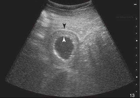
FIG. 32.19 Ultrasound view obtained from the left flank from a horse with a strangulating obstruction. A single distended loop of small intestine exhibits uniform thickening of the wall to approximately 8 mm (between arrowheads'). In real time, this segment had no discernible motility. This image was obtained with a 3.5-MHz curvilinear probe set to a depth of 13 cm.
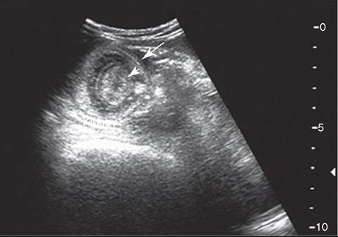
FIG. 32.22 Ultrasound view obtained along the caudal ventral midline in a horse with a jejunojejunal intussusception. The arrowhead marks the inner loop telescoped into the outer loop of jejunum (arrow), which created the classic “bull’s-eye” appearance. This image was obtained with a 3.5-MHz curvilinear probe set to a depth of 10 cm.
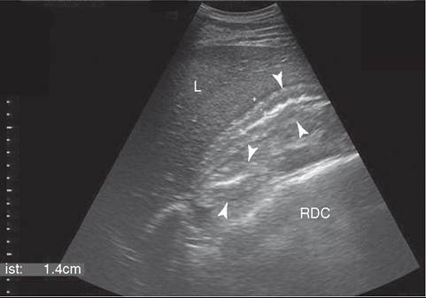
FIG. 32.21 Ultrasound view obtained from the right twelfth intercostal space from a horse with an epiploic foramen entrapment. Arrowheads mark the serosal surfaces of two thickened (14-mm wall) and compressed loops of amotile small intestine between the liver (L) and the right dorsal portion of the colon (RDC). This image was obtained with a 3.5-MHz curvilinear probe set to a depth of 24 cm.
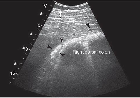
FIG. 32.23 Ultrasound view obtained at the eleventh right intercostal space at approximately the level of the shoulder in a horse. The left side of the image is dorsal. The white arrow marks the ventral tip of the lung. The black arrowheads mark the serosal surface of the duodenum with a hyperechoic lumen, between the liver and the right dorsal portion of the colon. Short hyperechoic parallel lines in the liver are the walls of portal veins (black arrow). This image was obtained with a 3.5-MHz curvilinear probe set to a depth of 17 cm.
and the right dorsal portion of the colon (see Fig. 32.23). Gas in the lung or the right dorsal portion of the colon can obscure identification of the liver dorsally or ventrally, respectively; ultrasonographically, the liver may thus be visible over only a few intercostal spaces. Only a small portion of the right side of the liver can be imaged, so its size is estimated on the basis of its expanse across the intercostal spaces.12 It is unusual for the liver to be seen beyond the fifteenth intercostal space or in the same transverse plane as the right kidney, except at the most rostral aspect of the kidney. The ventral edges of a normal liver are distinctly sharp. Like that of the spleen, the architecture of the liver is relatively homogeneous, but more vessels are visible in the liver, and the general echogenicity of the liver is less than that of the spleen.17 Portal veins have more connective tissue in their walls and thus have more echogenic walls than do the hepatic veins.12 Short segments of smaller portal veins often appear as hyperechoic parallel lines (see Fig. 32.23). In some small horses, the portal vein can be seen entering the hilus deep on the medial side of the image. The common bile duct and its branches within the liver are not normally visible.12 The pancreas is not usually identifiable via transabdominal ultrasonography.12
Duodenum. The position of the duodenum is fixed by its suspending mesoduodenum; thus the duodenum can reliably be found descending the right middle abdomen at approximately the level of the shoulder, between the liver and the right dorsal portion of the colon (see Fig. 32.23), where it can be imaged transversely along its short axis. As with the jejunum, to locate the descending duodenum, the ultrasonographer must wait for a peristaltic contraction to deliver ingesta through the lumen, thereby expanding the lumen; otherwise, the shape of the duodenum typically appears as a hyperechoic flattened or crescent-shaped line to a flattened oval. It is unusual for the duodenal diameter to exceed approximately 3 cm in healthy horses during peristaltic propulsion of ingesta.31 The duodenum contracts one to four times per minute in fed horses,31 but contractions are less frequent in anorexic, fasting, or heavily sedated horses. The duodenum can be followed on imaging to the level of the ventral right kidney (Fig. 32.24), where it crosses medially into the abdomen and is no longer distinguishable. The wall of the duodenum is less than 3 mm thick.17
Right Portion of Colon. The right dorsal portion of the colon, which has no sacculations, is immediately ventral to the liver and duodenum and is usually found from the right tenth to twelfth intercostal space, although it can extend to the fourteenth intercostal space.32 The wall of the right dorsal portion of the colon consistently appears as a hyperechoic curved line, adjacent to the liver (see Fig. 32.23). Because of gas contents in the right dorsal portion of the colon, wall thickness is difficult to measure accurately, and although thickness of up to 5.9 mm has been reported in healthy horses, less than 4 mm is typical.32. The walls of the right dorsal portion of the colon are significantly thinner in ponies and miniature horses than in large breed horses, typically measuring less than 3.2 mm.33 If the ultrasonographer locates the right dorsal portion of the colon and slides the transducer ventrally, the interface between the right dorsal and right ventral portions of the colon is often identifiable, appearing as an indentation in the wall between the nonsacculated right dorsal portion of the colon dorsally and the sacculated right ventral portion of the colon ventrally. The right ventral portion of the colon has sacculations, and its contractions and wall thickness are similar
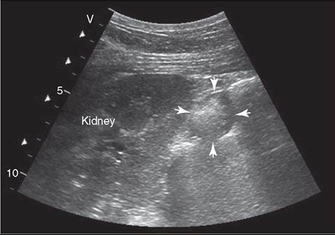
FIG. 32.24 Ultrasound view obtained from the right fifteenth intercostal space in a horse. The left side of the image is dorsal. The duodenum (arrows) can be seen ventral to the right kidney. This still image, captured when a peristaltic wave distended the luminal diameter to approximately 2.5 cm, was obtained with 3.5-MHz curvilinear probe set to a depth of 11 cm.
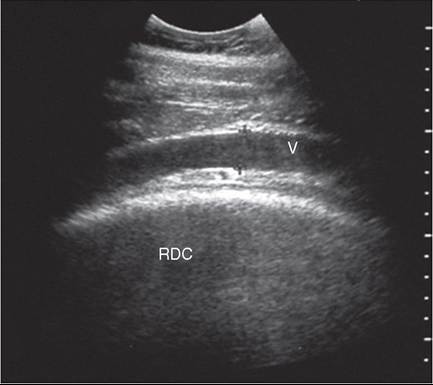
FIG. 32.25 Ultrasound view obtained from the right fifteenth intercostal space in a horse. A distended vein (V) is visible lateral to the wall of the right dorsal portion of the colon (RDC). The horse had a colonic volvulus. This image was obtained with a 3.5-MHz curvilinear probe set to a depth of 17 cm. (Courtesy Science In 3D, Inc. and copyright 2002, University of Georgia Research Foundation, Inc.)
to those of the left portion of the colon, although a wall thickness up to 5.1 mm has been reported in healthy horses.32 The contents of the different portions of the right portion of the colon and their medial walls are normally obscured by luminal gas. The transverse colon is not usually identifiable via transabdominal ultrasonography.12
Right Dorsal Colitis. When diarrhea in a horse is accompanied by panhypoproteinemia and a history of nonsteroidal antiinflammatory drug (NSAID) therapy, transabdominal ultrasonography of the right dorsal portion of the colon can be a useful diagnostic aide.32 In patients with NSAID-induced right dorsal colitis, the wall of the right dorsal portion of the colon often appears thickened with mixed echogenicity and an irregular mucosal-luminal interface.
Displacement Right Dorsal Portion of Colon. Identification of a large gas-distended viscus in the right caudal abdomen by transrectal palpation with accompanying taut colonic bands is consistent with a diagnosis of right dorsal displacement of the colon. The only major blood vessels normally visible ultrasonographically in the right caudal abdomen are those associated with mesentery of the lateral cecal band.15 Thus the ultrasonographic finding of distended vessels with reduced blood flow in colonic mesentery adjacent to the body wall implies that the colon is rotated along its long axis and would be consistent with a diagnosis of right dorsally displaced colon or a colonic volvulus (Fig. 32.25).24
Cecum. The cecum has a sacculated wall that extends from the right paralumbar fossa to ventral midline, and its motility is similar to that of the different portions of the colon.8,15 A unique characteristic that can be useful for distinguishing the cecum from the right ventral portion of the colon is that the lateral cecal band is oriented dorsoventrally and contains an associated artery and vein, whereas the lateroventral band of the right ventral portion of the colon is oriented caudocrani- ally and does not have associated vasculature.15 The cecal wall is less than 4 mm thick, and gas in the lumen precludes imaging the contents and far wall. Most diseases affecting the cecum can be sufficiently identified by transrectal palpation; however, transcutaneous ultrasonography can be helpful in confirmation of the diagnosis.
Typhlitis. With acute inflammatory disease of the cecum, the cecal wall may be thickened, the mucosal surface may appear irregular or wavy, and movement varies from hypomotile to hypermotile. Gas normally present in the body or base of
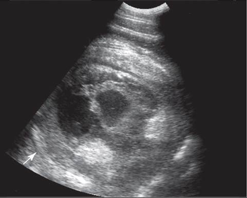
FIG. 32.26 Ultrasound view obtained from the right midflank in a horse with a cecocolic intussusception that appears as a large mass (arrow). The appearance of a “wall within a wall” is most apparent in the near field. This image was obtained with a 3.5-MHz curvilinear probe set to a depth of 24 cm.
the cecum precludes visualization of its contents and distal wall; however, with typhlitis, the cecum is often distended with fluid that is readily seen swirling inside of the lumen, and the distal wall can be visualized, especially toward the cecal apex.
Cecal Impaction. Transrectal palpation is a better method of diagnosing cecal impactions, but transabdominal ultrasonography of cecal impactions may reveal loss of characteristic sacculations.
Cecocolic and Cecocecal Intussusception. On transrectal evaluation, intussusceptions involving the cecum are often palpable as indistinct large, firm masses in the right or midcaudal portion of the abdomen. As with small intestinal intussusceptions, the characteristic ’’bull’s-eye” can be seen in the wall of the cecal apex or body within the lumen of the cecum or right ventral portion of the colon. In chronic intussusceptions, the walls are very thick and may appear abscessed or mass-like (Fig. 32.26).
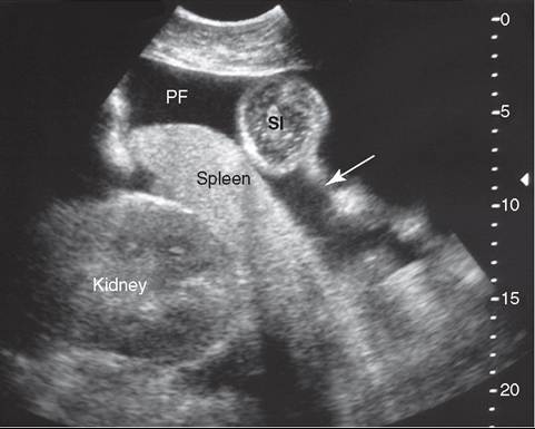
FIG. 32.27 Ultrasound view obtained from the left dorsal sixteenth intercostal space in a horse. The left side of the image is dorsal. Excessive hypoechoic peritoneal fluid (PF) has displaced the spleen from the left body wall and enabled distinct visualization of a single loop of small intestine (SI) and its supporting mesentery (arrow), appearing as a “balloon on a string.” The horse had metastatic mesothelioma. This image was obtained with a 3.5-MHz curvilinear probe.
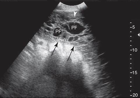
FIG. 32.28 Ultrasound view obtained from the lower right flank in a horse. The left side of the image is dorsal. Numerous hyperechoic fibrinous and fibrous adhesions trapping pockets of hypoechoic peritoneal fluid (PF) are present between the body wall (white arrowhead) and the serosal surface of the right ventral portion of the colon (black arrows). The horse had septic peritonitis. This image was obtained with a 3.5-MHz probe set to a depth of 21 cm.
Right Kidney. Gas in the cecum, right dorsal portion of the colon, or lungs sometimes obscures visualization of the right kidney, which is normally found in the rostral fifteenth to seventeenth intercostal spaces20 (see Fig. 32.24). In Thoroughbred horses, the right kidney measures 6.5 to 9.1 cm dorsoventrally (the slightly oblique transverse plane), 12.1 to 16.2 cm in length craniocaudally (the dorsal plane), and 6.5 to 8.4 cm lateromedially.19 The ureters are usually difficult to identify, but the proximal right ureter sometimes appears as a hyperechoic circular structure at the hilus. The architecture of the right kidney is similar to that of the left kidney.
Peritoneal Cavity. In a healthy horse, only minute pockets of peritoneal fluid should be identifiable in the rostroventral area of the abdominal cavity or adjacent to sacculations of the ventral portion of the colon. Excessive peritoneal fluid displaces organs and greatly facilitates visualization of structures not normally seen on ultrasonographic examination, such as the mesentery of the small intestine (Fig. 32.27).
Peritonitis. Peritonitis should be suspected if volume or echogenicity of peritoneal fluid is increased. In some cases, free-floating or adherent fibrin or fibrous tags may be visible (Fig. 32.28). An abdominocentesis is an integral step for further characterizing the cause of peritonitis.
Hemoabdomen. Excessive blood in the peritoneal cavity displaces organs off the body wall. Blood in the peritoneal cavity appears as a mixed echogenic fluid that, in real time, often appears to swirl as it mixes with less echogenic peritoneal fluid (Fig. 32.29).15 Abdominocentesis confirms the presence of blood.
Intraabdominal Masses. Abscessation, hematoma, and neoplasia are the top potential diagnoses for intraabdominal space-occupying lesions, each of which can be located just about anywhere in the peritoneal cavity, in or attached to bowel, other intraabdominal viscera (i.e., spleen, kidney, liver), or the peritoneum. Space-occupying lesions appear as soft tissue densities of variable shapes and acoustic properties that distort normal sonographic architecture (Fig. 32.30). Although the definitive cause of an intraabdominal mass is often not discernible ultrasonographically, both abscesses and hematomas appear as either uniform echogenic or loculated space-occupying masses of mixed echogenicity. Chronic abscesses may be
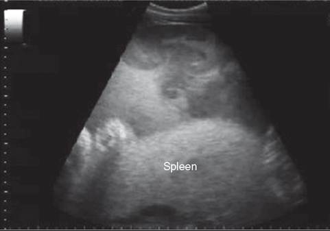
FIG. 32.29 Ultrasound view obtained from the lower left flank of a horse with hemoabdomen. The left side of the image is ventral. Blood displaces the spleen from the left body wall and appears as swirls of mixed echogenicity between the spleen and left body wall. This image was obtained with a 3.5-MHz curvilinear transducer set to a depth of 24 cm.
characterized by the presence of a thick echogenic capsule. Abscesses and hematomas are more commonly confined to a single location, whereas metastatic neoplasia should be considered if space-occupying lesions of varying sizes that distort normal ultrasonographic anatomy are visible in multiple locations. Although the ultrasonographic appearance of a space-occupying lesion may not help determine the cause, ultrasonography can be a useful guide for biopsy or aspiration of the affected tissue.
■ Other Imaging of the Alimentary Tract
RADIOGRAPHY. In horses the alimentary tract is a dynamic and complex environment to evaluate with any modality. Because of the size of the animal, as well as the distinct difference between air and soft tissue, radiographs are a useful diagnostic tool for evaluating the teeth, pharynx, esophagus, stomach, and intestinal tract. Portable radiographic units with maximal kilovolt peak (kVp) settings of 100 and maximal milliampere- second (mAs) settings of 30 make it possible for ambulatory
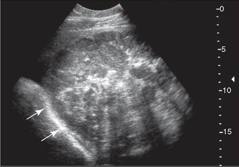
FIG. 32.30 Ultrasound view obtained from the gastrosplenic window in a horse. The left side of the image is dorsal. A large mass of mixed echogenicity occupies the space adjacent to the stomach wall (arrows) in the location normally occupied by the spleen. The horse had abdominal lymphosarcoma. This image was obtained with a 3.5-MHz curvilinear probe set to a depth of 19 cm.
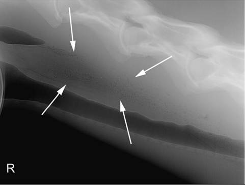
FIG. 32.31 Standing lateral radiograph of a 13-year-old Morgan gelding with an esophageal tear. Note the tubular region of small gas opacities caused by air trapped around the outer border of the esophagus (arrows). An esophageal perforation secondary to an ingested foreign body was confirmed with endoscopy.
practitioners to obtain diagnostic images of the head and cranial portion of the esophagus. However, to obtain images of the thoracic portion of the esophagus and abdomen, a referral clinic with a more powerful radiographic generator is usually required.
For average foals, exposures ranging from 80 to 88 kVp and 20 to 26 mAs have been used in radiography of the abdomen.34 In adult horses, exposures range from 60 to 140 kVp and 20 to 70 mAs.35,36 To perform a complete evaluation of the abdomen, it is recommended that the abdomen be divided into four quadrants (cranioventral, midabdominal, caudodorsal, and caudoventral).35 Large cassettes (35 cm ? 43 cm) and fast screens are also needed to ensure that a diagnostic image is obtained. Because of the large amount of scatter radiation produced from the high exposure and thickness of tissue penetrated, an 8: 1 to 10: 1 grid should be used.34-38 Alternatively, an air gap technique can be used to prevent image degradation.
The advent of computed radiography and digital radiography has greatly increased the diagnostic capabilities of conventional radiographic examinations. Both systems, available to equine practitioners, are considered indirect imaging modalities in which the x-ray photon interacts with an intensifying screen to convert the x-ray photon to light. This light then interacts with an imaging plate: film, as with a conventional radiographic system; within a photostimulable phosphor, as with computed radiography; or within a flat-panel detector, as with digital radiography. Regardless of the method, these images are considered “indirect” because the x-ray photon is first changed to light and then detected by the imaging medium.39 The main benefits of computed and digital radiography are the increased latitude of the film. It is possible to change the contrast and grayscale levels after the exposure if the number of photons is adequate. The digital radiography systems also offer a rapid evaluation of the image because the cassette is directly connected to the computer. This allows the portable systems to show radiographic images within 10 seconds after the exposure is made. In contrast, the computed radiography system requires the cassette to be placed into a reader in order to display the image. Finally, because both computed and digital radiography are generally compliant with the Digital Imaging and Communications in Medicine (DICOM) standard, this allows any specialists with standard medical imaging software to view the images via a compact disk or via the Internet.
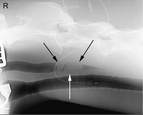
FIG. 32.32 Standing lateral radiograph of a 12-year-old Quarter Horse mare. Note the ovoid mass surrounded by gas just dorsal to the trachea (arrows). This lesion was a mixture of hay and grass.
Survey radiography is generally helpful in evaluating the cervical portion of the esophagus for evidence of rupture, as well as in evaluating the abdomen. Esophageal ruptures secondary to an obstruction or vigorous placement of a nasogastric tube produce a small volume of gas that tracks just dorsal to the trachea (Fig. 32.31). This can be confused with a tracheal laceration; however, with tracheal lacerations, generally the gas accumulates around the trachea, and the volume of gas within the subcutaneous tissues and the cranial mediastinum is extreme. In addition, esophageal obstructions, also called choke, can sometimes be identified on survey radiographs, depending on the material that is causing the obstruction and the amount of air or contrast medium that is able to surround the structure (Fig. 32.32). Although the nature of the obstruction cannot be determined, the extent of the abnormality can sometimes be identified.
Abdominal radiography is useful for evaluating the small and large intestines for sand accumulation, enterolithiasis,
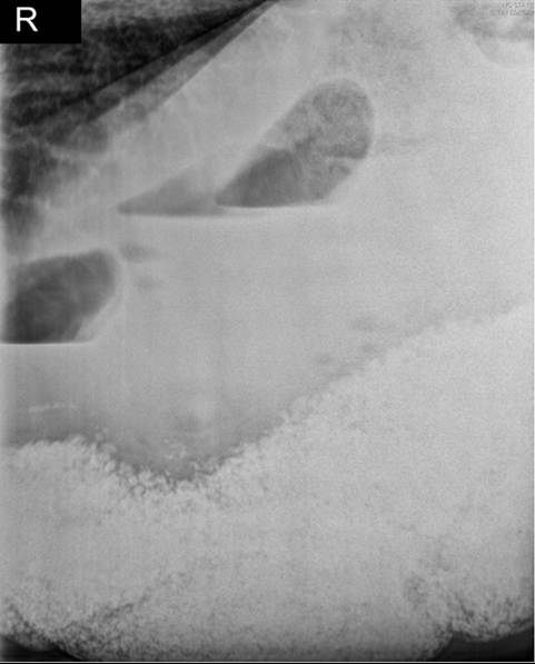
FIG. 32.33 Standing lateral radiograph of a 4-year-old Arab mare with a history of colic. Note the large amount of opaque material within the ventral portion of the colon, probably secondary to sand accumulation.
impactions, or small intestinal disorders in foals. When sand is ingested, it generally accumulates within the large colon along the ventral abdomen10 (Fig. 32.33). Radiography is useful for monitoring the resolution of sand impactions after medical management; however, sequential examinations are needed to verify that the volume of sand has decreased.36 If the volume of sand is large enough, it is difficult to determine whether an enterolith is present because of radiographic summation of the two lesions. Enteroliths are solid concretions of mineral that usually form around a nidus, such as a metallic foreign body (Fig. 32.34). The mineral composition is varied, as illustrated by the different opacities within the enterolith. Radiographs have a 96.4% positive predictive value for detecting enteroliths in high-prevalence areas. In one study, enteroliths were generally found to be within the midabdominal radiograph, and 67% of small colonic enteroliths caused large colonic distention, which was also identified on radiographs.35 Impactions are more difficult to diagnose because radiography usually demonstrates only increased feed accumulation within the abdomen. Although no enterolith or obstruction is identified, granular material can be seen, usually within the ventral portion of the colon near the sternal flexure. This is because pelvic flexure impactions cause the feed material to accumulate orad, causing distention of the left ventral portion of the colon (Fig. 32.35). Intestinal disorders such as functional ileus secondary to enteritis (Fig. 32.36) or obstruction secondary to intussusception or meconium impaction (Fig. 32.37) in foals can also be identified on abdominal radiographs. These images show large dilation of the small intestine, and differentiation between functional and mechanical ileus in foals is generally based on the size of the intestine and the volume of gas that is present.37 Evaluation of the abdomen with ultrasonography may aid in qualifying the small or large intestinal motility, as
FIG. 32.34 Radiograph of enterolith obtained after surgical removal from the small colon. Note the variation in opacities caused by the various types of minerals that are contained within the enterolith.
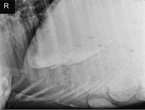
FIG. 32.35 Standing lateral radiograph of a 3-year-old Paint Horse gelding with a pelvic flexure impaction. The radiograph shows the sternal flexure with a large amount of granular material and a small amount of sand accumulation in the ventral portion of the colon.
well as identifying the source of an obstruction if it cannot be determined on radiographs.
Radiography also allows for the use of contrast medium to further outline the alimentary tract and to evaluate pharyngeal function and esophageal motility.40-42 Approximately 60 mL of barium sulfate paste or liquid, administered orally via a 60-mL dosing syringe, for radiographs of the laryngeal region and esophagus provides useful information about swallowing and large obstructions. If the barium liquid is identified dorsal to the soft palate, within the larynx or trachea, pharyngeal function is probably abnormal.40 The barium paste coats the pharynx and esophagus, and this appearance is useful for identifying any ulcerations or irregularities in the mucosal surface. After those procedures have been performed, or if
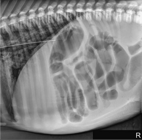
FIG. 32.36 Standing lateral radiograph of a 1-day-old, premature Quarter Horse filly. Note the large amount of gas-distended intestine. Because of the large amount of small intestinal distention, functional ileus was the primary differential diagnosis.

FIG. 32.37 Standing lateral radiograph of a 2-day-old Thoroughbred colt with a meconium impaction. Note the large amount of gas distention of the colon.
there is no evidence of an oropharyngeal dysphagia, esopha- gography is performed. For this procedure, water is added to approximately 200 to 500 mL of barium sulfate liquid to achieve a concentration of 1 :1 or 2 : 1, which brings the total volume to 500 to 1000 mL. This liquid is administered through a nasogastric tube or cuffed endotracheal tube placed within the cranial portion of the esophagus to the level of C2 to C3. If a cuff is available, the cuff can be inflated with approximately 10 mL of room air. A radiograph is then made to verify that the tube is not within the trachea, and when it has been confirmed to be within the esophagus, the dose of barium is administered with a stomach pump. Toward the end of the dose, while the liquid is still being pumped, radiographs of the cranial, mid, and caudal portion of the esophagus are
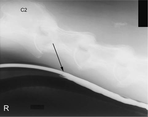
FIG. 32.38 Standing lateral radiograph, showing a normal esophagogram with barium liquid. The arrow marks the region where the nasogastric tube ends. This is approximately at the level of C3.
obtained (Fig. 32.38). The use of the pump provides distention of the esophagus to help identify strictures or irregularities in the esophageal wall.
Positive (barium sulfate) and negative (room air) contrast media, administered orally, have also been used to evaluate the stomach and intestinal tract radiographically,34,43,44 and the rectum and colon have been evaluated through retrograde administration of contrast medium.44 These methods allow for the evaluation of the stomach, intestinal tract, and rectum for regions of obstruction, as well as ulcerations, tumors, motility disorders, and malformations. Although these methods have been described, ultrasonography has virtually eliminated the need to expose personnel and patients to the repetitive, high doses of radiation needed to obtain sequential radiographs of the abdomen.
Computed tomography (CT) and magnetic resonance imaging (MRI) are of little use for evaluation of the alimentary tract (except for the head). This is mainly because of the size of the patient in relation to the size of the gantry and bore in CT and MRI units, respectively. Dental disorders such as abscesses and fractures are clearly visible on CT images, especially on three-dimensional reconstructions (Fig. 32.39), and CT is also useful for detecting pharyngeal and esophageal masses that may not be fully identified with conventional radiographs. CT and MRI can be used in foals that can be placed within the gantry or bore of the magnet; however, because of the motion of the gastrointestinal tract and the long acquisition times used with respiratory gating sequences, MRI has not been used widely to evaluate the thorax or abdomen. A single case report has been published about the use of contrast esophagography and CT to aid in the surgical planning of a persistent right fourth and left sixth aortic arch that caused a vascular ring anomaly in a foal.45 However, the applications for these technologies have yet to be realized.
SCINTIGRAPHY. Nuclear medicine is used widely for imaging the gastrointestinal tract in humans and animals. Although physical obstruction can be assessed with endoscopy, ultrasonography cannot delineate the pylorus in the normal horse because of the peripheral nature of the colon and colonic gas. Nuclear medicine provides a functional evaluation of the pylorus to determine whether gastric emptying is delayed in comparison with that in normal horses. This modality works better than gastric emptying with barium sulfate because of
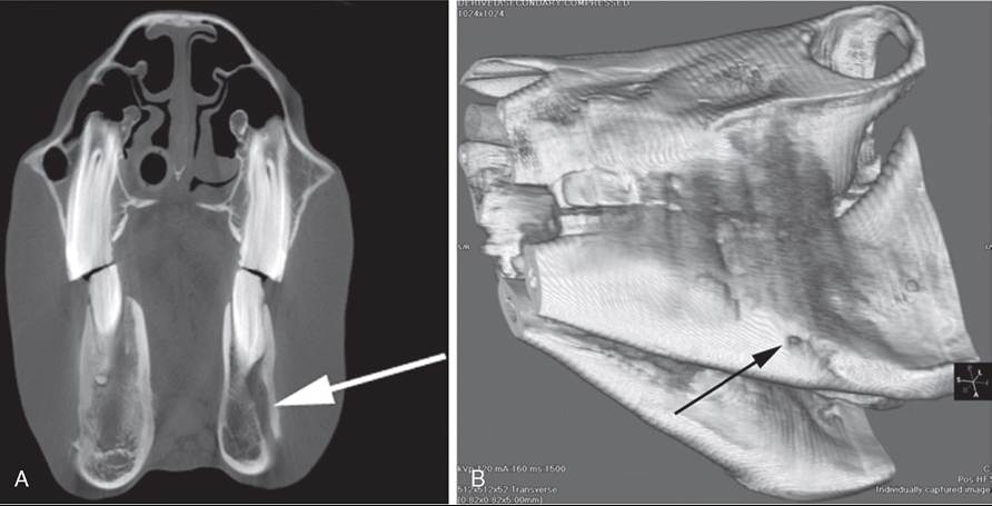
FIG. 32.39 Transverse (A) and three-dimensional (B) reconstructed computed tomographic images of the head of a 4-year-old pony mare with chronic draining tracts from the mandible. The arrows illustrate the tract through the mandible that communicates with the apical portion of the left mandibular first molar (tooth #309).
the minimally invasive nature of the modality and ease of image acquisition. However, because of the need for a gamma camera to generate the images and the licensing requirements to handle radioactive material, this modality is less universally available. The primary use of scintigraphy in evaluating the gastrointestinal tract is to measure gastric emptying time.46-49 Two protocols have been outlined in the references provided: The first entails the use of the readily available technetium-99m pertechnetate (99mTc) bound to disofenin or sulfur colloid.47,50,51 This combination of radioisotope and radiopharmaceutical is fed in a pelleted ration alone or mixed with radiolabeled eggs. The normal range for the t1/2 gastric emptying has been reported to be 1.49 ± 0.17 hours26 and 1.56 ± 1.08 hours.26 The other method is the breath test involving carbon-13 bound to octanoic acid (13C-OABT). This method was compared with the 99mTc sulfur colloid method, and the solid-phase gastric emptying t1/2 was found to be similar.45 The main difference is that the 13C-OABT is measured in the exhaled breath of horses and with spectroscopy, rather than with a gamma camera. The rationale for 13C-OABT is that because a gamma camera is not needed, this test may be more portable and useful for field investigations.50
Another nuclear medicine procedure in the realm of alimentary tract evaluation involves the use of 99mTc with hexamethylpropyleneamine oxime (HMPAO). This procedure allows for radiolabeling WBCs in order to determine areas of inflammation.52,53 The use of 99mTc is a matter of convenience because it is also used in equine bone imaging. Detection equipment, such as a low-energy, all-purpose collimator, is readily available in many practices. HMPAO is used because it binds to granulocytes and therefore should travel to areas of increased inflammation.53 HMPAO also associates with the reticuloendothelial system, enabling localization within the lungs, liver, spleen, kidneys, and urinary bladder. The main drawback is that with the HMPAO bound to WBCs, there is a general lack of anatomic information, which can cause lesion localization to be difficult.53 Although this technique is expensive and labor intensive, the results from the limited studies available appear encouraging.
■ Biopsy The decision about whether to perform biopsy is often based on the ease of obtaining a sample and the relative value of the evaluation that can be made. Very small samples, such as those obtained with an endoscopy biopsy instrument, are relatively easy to obtain, but they provide limited information. Full-thickness bowel specimens, obtained by means of ventral midline or flank laparotomy, are more difficult to obtain, but they provide much more information. Laparoscopic techniques to safely obtain full-thickness intestinal biopsy samples have been described54 and may be more practical than laparotomy.
Obtaining a biopsy sample by endoscopy allows the practitioner to choose the biopsy site on the basis of the appearance of the mucosal surface, which most frequently reflects an inflammatory disorder. Conversely, when a biopsy sample is obtained through laparotomy, the serosal surface of the bowel may not reflect a disorder within the bowel wall. In such instances, obtaining several biopsy specimens may be helpful. Rectal mucosal biopsies are easily performed. Many instruments can be used to obtain the biopsy specimen, and a uterine biopsy forceps works well. A fold of mucosa can be pinched readily between two fingers, and a sample of this tissue is obtained this way. The size of such a sample is adequate for histologic or bacteriologic examination.
■ Fecal Examination Cytologic, biochemical, bacteriologic, immunologic, and electron microscopic evaluations can be performed on fecal samples. In addition, the consistency and color, the presence of foreign material such as sand or gravel, and the presence of parasites should be examined in the examination of the alimentary system. In addition to fecal consistency, fecal particle size can be used to evaluate the efficiency of mastication or the colonic transit time. Increased particle size, with loose or watery stool, is suggestive of decreased colonic transit time.
Cytologic examinations are used primarily to evaluate the parasite burden of the animal. Ova of large and small strongyles, tapeworms, round worms, and Strongyloides westeri are most common. Coccidia are observed occasionally, but their presence is clinically unimportant. Examination of fecal WBCs has been advocated in the evaluation of horses and foals with enterocolitis. Because these cells are very labile, their presence in large numbers indicates that an inflammatory process is present and that the inflammation is in the distal portion of the colon or is associated with decreased transit time.
The presence of fecal occult blood should be determined so as to diagnose gastric ulcers, duodenal ulcers, and other potentially hemorrhagic disorders of the alimentary tract. However, the usefulness of this test has been shown to be quite limited because negative results can be obtained when blood is present in the proximal portion of the gastrointestinal tract.55 The sensitivity of most commercially available tests is poor; they may yield negative results in the presence of severe gastric bleeding.
Fecal culture is an essential component in the evaluation of many patients. In bacteriologic culture techniques for fecal samples, selective media that are designed to isolate Salmonella are routinely used. These media include selenite broth, tetrathionate broth, brilliant green agar, XLD agar, and SalmonellaShigella agar. Less selective media, MacConkey agar, and eosin methylene blue agars are desirable for culture of other potential Gram-negative bacterial pathogens such as Escherichia coli, but the mere presence of E. coli in the feces does not determine its pathogenicity. Enterotoxigenic E. coli strains have been isolated from foals with diarrhea, but special tests, such as polymerase chain reaction (PCR) assays, must be performed to determine whether an isolate produces enterotoxin. Rapid enzyme-linked immunosorbent assays (ELISAs) are available at diagnostic laboratories for detection of enterotoxins of Clostridium difficile (C. difficile Tox A/B Test, TechLab, Blacksburg, Va.) and Clostridium perfringens (C. perfringens enterotoxin test, TechLab, Blacksburg, Va.) in fecal specimens.
The presence of rotavirus in a fecal sample can be determined by ELISA or an agglutination test. Both assays test for the presence of viral antigen in the feces. PCR assays are also available to detect rotaviral nucleic acids. ELISA is reported to be more sensitive than the agglutination test but is less specific. Therefore the agglutination test is likely to yield more false-negative results, and the ELISA test is likely to yield more false-positive results. The ELISA test is more timeconsuming and inconvenient to perform than the agglutination test. When rotavirus is a concern, particularly as a farm problem, a reasonable approach is to screen fecal samples with the agglutination test and repeat testing of samples that yield negative findings with ELISA or PCR.
Some diagnostic laboratories have developed sensitive and specific multiplex PCR tests for panels of gastrointestinal pathogens (equine coronavirus and rotavirus, Lawsonia intracel- lularis, Cryptosporidium, Rhodococcus equi, Neorickettsia risticii, Salmonella spp., coronavirus, rotovirus, and C. difficile toxins A and B), that provide relatively rapid diagnostic information that can be used alone or in combination with culture or antigen detection methods.
■ Absorption and Digestion Tests Tests of the ability of the equine intestinal tract to digest and absorb nutrients have a more limited clinical application in horses than in human or small animal medicine, but they can be useful in the evaluation of chronic weight loss, suspected small intestinal inflammation or neoplasia, gastric and small intestinal partial obstruction, and postoperative small intestinal malabsorptive disorders. For absorption tests to be diagnostic, the intestinal disorder must either be diffuse or affect the delivery to and transit through the small intestine.
Maldigestion tests are performed to evaluate exocrine pancreatic function and disaccharidase activity at the mucosal brush border of the small intestine. Pancreatic exocrine deficiencies have not been described in horses, probably because equine pancreatic secretions consist primarily of water and bicarbonate and have less enzymatic activity than in monogastric omnivorous species. Maldigestion related to disaccharidase activity at the mucosal brush border is relevant in viral and bacterial enteritides of foals, particularly rotavirus and coronavirus enteritides. As a result of these viral infections, there is loss of the superficial villous epithelial cells of the small intestine, in which the disaccharidases lactase, cellobiase, maltase, sucrase, and trehalase are located.56 Lactase levels are highest in young suckling foals, and loss of this enzyme activity, secondary to loss of the mucosal villous cells, leads to lactose maldigestion. Lactose tolerance can be tested by administration of a 20% solution of D-lactose at a dose of 0.5 to 1 g/kg. This dose should result in an approximate doubling of the serum glucose level within 60 minutes of administration.57
Clinically applicable absorption tests include the D-glucose and D-xylose absorption tests. The glucose absorption test has the advantage of being relatively easy to perform and inexpensive. However, cellular uptake and metabolism of glucose, as well as intestinal absorption, influence the results and thus are undesirable variables. The D-xylose absorption test is therefore advantageous because it more directly measures intestinal absorptive capacity. The results of both tests, however, are affected by gastric emptying rate and small intestinal transit time. In the United States, D-xylose is available only through chemical suppliers and only for research purposes; its availability for clinical diagnostic use is restricted.
The D-glucose and D-xylose tests are performed similarly. After an 18- to 24-hour fast, a 10% solution of D-glucose or D-xylose, 0.5 to 1 g/kg, is administered through a nasogastric tube. For the measurement of glucose, blood is collected in sodium fluoride tubes, and for the measurement of D-xylose, blood is collected in heparinized tubes. Samples are taken immediately and 30, 60, 90, 120, 150, 180, 210, and 240 minutes after administration. Peak levels, which normally range from 20 to 25 mg/dL, are reached 60 to 120 minutes after administration and should decrease thereafter. The normal curve resembles an inverted V. Absorption curves vary as a function of each horse's age and type of feed given.58 Delay or flattening of the absorption curve may reflect delayed gastric emptying, increased intestinal transit time, or impaired intestinal absorption.59 Accurate interpretation of the results of these tests depends on the results of other diagnostic evaluations. In addition, different types of diets have been shown to affect the height, although not the shape, of the absorption curves significantly. In general, with diets that have higher digestible energy content, the curve has a lower peak.
■ Breath Tests In humans, dogs, and cats, breath tests are used to assess a variety of intestinal disorders. The hydrogen breath test is used in assessments of intestinal bacterial overgrowth and in determination of carbohydrate digestion and absorption in the intestine. In patients with an abnormal intestinal bacterial population or carbohydrate malabsorption, bacterial fermentation of carbohydrate is excessive, one byproduct being hydrogen. Because hydrogen is freely diffusible from the bowel into the blood and from the blood into the alveoli, measurement of exhaled hydrogen gas can be used to assess the status of intestinal bacterial fermentation of carbohydrates.
In horses, the hydrogen breath test is most applicable to conditions involving carbohydrate malabsorption and thus increased delivery of soluble carbohydrate to the large intestine for bacterial fermentation to occur. This technique has been used in horses: In one report, different carbohydrate substrates were evaluated in seven ponies,60 and in another, the hydrogen breath test was used in conjunction with D-xylose absorption in nine horses with a variety of clinical disorders.61 In the study with ponies, fasting resulted in negligible levels of breath hydrogen excretion. Sustained increases in breath hydrogen concentration to more than 10 parts per million (ppm) were observed in all ponies after they ingested oats or wheat flour, in three ponies after the administration of glucose and xylose, and in two ponies after the administration of lactulose and lactose. The pattern of breath hydrogen excretion varied among animals after the ingestion of identical test meals. In the clinical study, the diseased horses showed higher fasting breath hydrogen levels (range 7.5 to 61.5 ppm) than did normal horses (range, 0 to 5 ppm). After xylose administration, none of the healthy animals showed an increase in breath hydrogen production, and five of diseased animals showed increases in breath hydrogen. In this group of patients, abnormalities in hydrogen breath measurement were more apparent than abnormalities in D-xylose absorption.