Interdigital Necrobacillosis (Foot Rot) in Cattle
Meredyth L. Jones
■ Definition and Etiology Interdigital necrobacillosis, also called foot rot, foul-in-the-foot, and interdigital phlegmon, is a bacterial infection of the interdigital tissues, leading to moderate to severe lameness in one or more feet of cattle.
Animals at high risk include those housed in moist, muddy lots; pastures with standing water; or areas where sharp gravel, concrete, or stubble exists.1 F. necrophorum, a gram-negative anaerobe, is most often associated with this syndrome. Other bacteria, including Prevotella melaninogenica, Porphyromonas levii, and Dichelobacter nodosus, may induce similar lesions alone or in concert with F. necrophorum.2'-4■ Clinical Signs and Differential Diagnoses Early cases of foot rot show mild to moderate lameness in one or more feet, most commonly the hindlimbs. On examination of the foot, there is symmetric swelling of the coronary band region that may progress upward to the level of the fetlock. The interdigital space will also be swollen, spreading the toes. One or more fissures will form in the interdigital space (Fig. 38.31), where tissue necrosis is apparent. The characteristic odor of anaerobic infection with necrosis is apparent. Exudate is present, and there may be pseudomembrane formation.
Severity of lameness may progress such that animals may refuse to bear weight on an affected limb. Young calves may hold up a limb and shake it or, when multiple limbs are affected, may walk in such a way as to appear ataxic from neurologic disease. Invasion of the tendons and synovial structures may occur if the disease is not treated appropriately early, worsening the lameness and the prognosis for recovery. Mild fever may occur, and affected cattle will increase laying time, which leads to reduction in feed intake. Animal performance is diminished, with reductions in weight gain or milk production.
Differential diagnoses include septic arthritis, sole abscess, tenosynovitis, pedal osteitis, interdigital fibroma, laminitis, and trauma.
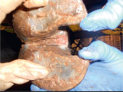
FIG. 38.31 Interdigital fissure formation and necrosis are the hallmark of interdigital necrobacillosis. The lesion will also have a foul odor and variable amounts of swelling of the interdigital space and coronary band region.
■ Pathophysiology The bacteria implicated in foot rot are normal inhabitants of the ruminant gastrointestinal tract and are therefore ubiquitous in the environment in which ruminants live. The syndrome occurs when there is a breach in the skin of the foot, usually through maceration from prolonged exposure to moisture or trauma. Oxygen tension is reduced by early microbial invaders, creating an ideal environment for the anaerobic pathogens of foot rot.5,6 From there, several mechanisms contribute to virulence and evasion of the immune response. The two subspecies of F necrophorum currently recognized are biotypes A and B. Biotype A appears to be more virulent and is more frequently isolated.7,8 Major virulence factors associated with this organism are a high-molecular-weight leukotoxin and lipopolysaccharide (LPS).9,10 The leukotoxin is cytotoxic to ruminant neutrophils and, when combined with LPS, protects the organism from phagocytosis.7 P levii does not stimulate a significant chemotactic response in bovine macrophages in vitro, and suppression of phagocytosis occurs even when the organism is present in low numbers.3 These properties of P levii may facilitate a local tissue environment that allows other anaerobic organisms, including F. necrophorum, to colonize and evade host defense mechanisms.
Bacterial production of proteases and the products of white blood cell action and destruction further destroy local tissues, allowing for deeper invasion and necrosis.
Swelling and pain result from tissue necrosis. Tenosynovitis, septic synovitis, and septic arthritis may all occur as the infection advances.Systemic inflammatory responses do occur, and serum haptoglobin levels are increased in cattle infected with foot rot.11 Other work has shown more extensive changes to plasma protein profiles, including those involved in pathways of inflammation, complement, and coagulation.12
■ Diagnosis The diagnosis of interdigital necrobacillosis is usually clinical, based on typical swelling pattern, interdigital fissures, and odor. Lesion culture may be performed but is limited in utility due to mixed populations of environmental and fecal contaminants. Histopathology may reveal intralesional spirochetes, but this is not consistent. Serodiagnosis has been reported with indirect ELISA for the detection of leukotoxin A antibodies using the recombinant protein PL2, yielding relatively high sensitivity and specificity.13 Lack of expected response to parenteral therapy should result in reevaluation of the diagnosis and more extensive evaluation of the foot.
■ Treatment and Prognosis Most uncomplicated cases of foot rot will respond to parenteral antimicrobial therapy within a few days, with return to full soundness. Antimicrobial selection should be based on animal type, withdrawal time, and dosing logistics. In the United States, commercially available preparations of oxytetracycline, florfenicol, ceftiofur, tylosin, sulfadimethoxine, and tulathromycin carry label indications for foot rot. A comparison of ceftiofur crystalline free acid (6.6 mg/kg subcutaneously [SC] in the base of the ear once) and ceftiofur sodium daily for 3 consecutive days revealed no statistical difference in recovery rates; however, the ceftiofur sodium resulted in more labor and time required for multiple administrations.14 Another study comparing ceftiofur and oxytetracycline found no difference in the recovery rate between the two antimicrobials, concluding that the difference in withdrawal time should be considered in the treatment choice.15
Local treatment is generally unnecessary in most cases, and the use of bandages should be considered in light of the environment to which the animal is returning.
A wet bandage can only worsen the situation. Some cases may benefit from wound cleaning and debridement of necrotic tissue.Antiinflammatory and analgesic therapy may result in improved appetite and ambulation, improving welfare and lessening production losses. A transdermal preparation of flunixin is approved in the United States for pain control due to foot rot.
Regardless of the medical treatments used, animals should be returned to a clean, dry environment for recovery. They should be closely monitored for return to soundness and reduction in swelling. In cases treated early, improvement is expected in 2 to 4 days after parenteral antimicrobial treatment.16 Animals that do not respond as expected should have their diagnosis reevaluated by thorough examination of the foot to determine if another lesion is present or if the infection has extended to deeper tissues.
■ Control and Prevention The bacterial agents of foot rot are residents of the alimentary tract and the environment of the ruminant. Their ability to cause pathology requires damage to the skin barrier in the form of moisture leading to softening and maceration of the skin or abrasions from rocks, concrete, or stemmy or frozen forage.
General principles for control involve evaluation of the environment for potential risk factors, including low-lying areas with standing water and congregation points that collect urine and manure. Hygiene should be improved wherever possible through the use of drainage systems, manure removal, and regrading areas or by avoiding certain pastures during wet times of year.
Some owners and veterinarians may be motivated to control foot rot using in-feed antimicrobials or other additives touted to improve control. No feed additives are approved in the United States for treatment, control, or prevention of foot rot, and extralabel use of feed additives is prohibited by the Animal Medicinal Drug Use Clarification Act.
The utility of footbaths is limited by the availability of efficacious compounds, environmental and occupational safety, contact time, and the ability to maintain the hygiene of the footbath.
It can be difficult to get cattle to stand in the footbath long enough to achieve adequate bacterial killing, whereas standing too long results in contamination with fecal material, eventually leading to inactivation.7 Antibacterial agents and astringents, including 3% formalin, 2% copper sulfate, oxytetracycline, and lincomycin-spectinomycin formulations, traditionally have been used. Dry footbaths have also been reported and include 10% copper sulfate and slaked lime.6 Practitioners must check state and local laws regarding the use of these substances that can cause environmental contamination and occupational health hazards.Commercially available vaccines against F. necrophorum exist for use in cattle. Independent studies regarding the efficacy of these vaccines are limited. One study evaluating the incidence of liver abscess and foot rot in vaccinated versus nonvaccinated feedlot cattle showed reduced incidence of both syndromes with vaccination, but that difference was confounded by diet, with improvement only occurring in cattle fed free-choice forage.17 The role of this dietary influence on foot rot is unknown.
Trace mineral nutrition is an important consideration when evaluating foot health on any operation. Iodine, copper, selenium, zinc, and biotin support overall foot and hoof health. Variable results have been achieved in controlling foot rot using supplementation of trace minerals and biotin.18,19
More on the topic Interdigital Necrobacillosis (Foot Rot) in Cattle:
- Miscellaneous Bovine Rickettsial, Bacterial, and Viral Disease Vaccines
- Bacterial Diseases
- Smith Bradford P., Van Metre David C., Pusterla Nicola (eds.). Large Animal Internal Medicine. Part 2. 6th edition. — Elsevier,2020. — 2279 p., 2020