Medical Disorders of the Large Intestine
Samuel L. Jones
Acute Diarrhea
Diarrhea in horses can be defined as the passage of fecal material that has increased water content. It can vary from soft, formed stools with a mild to moderate increase in water content to projectile fecal passages that contain little solid matter.
The passage of excessive water in the feces reflects disruption of the normal balance of fluid and electrolyte secretion and absorption in the intestinal tract. In adult horses, diarrhea results almost exclusively from disorders of the large intestine, although it may be a feature of some disorders of the descending small colon. Diarrhea can result in significant losses of water, electrolytes, and plasma protein and is often accompanied by local and systemic inflammatory responses.Diarrhea disorders in adult horses can be divided into those characterized by inflammation of the cecum and large intestine (typhlitis, colitis) and those in which there typically is no inflammatory response. Inflammatory disorders include those characterized by an acute inflammatory response (salmonellosis, Potomac horse fever [PHF], clostridiosis, and equine coronavirus, described later), disorders associated with endoparasitism (small and large strongyle larval migration or encystation), disorders arising from toxicity (cantharidin toxicity, described later, and NSAID toxicity), and disorders included under the umbrella term inflammatory bowel disease (IBD). Disorders that can manifest with diarrhea but are not characterized by colonic inflammation include those in which intestinal hydrostatic pressure is increased (congestive heart failure, cirrhotic liver disease), those in which intraluminal osmolarity is increased, and poorly defined disorders in which fluid secretion may be stimulated by the enteric nervous system.
Colitis is active inflammation within the colon that is usually associated with myriad local and systemic pathophysiologic events.
Diarrhea is an important problem in horses with colitis, but sepsis resulting in impaired cardiopulmonary function, coagulopathies, and other sequelae of activation of inflammatory mediator cascades can be life-threatening. Other complications that occur in patients with colitis and sepsis, regardless of cause, include hematogenous organ colonization by bacteria, immune suppression and susceptibility to superinfection with bacteria or fungi, cecum or colon infarction, jugular vein thrombosis, and laminitis.A variety of inflammatory cells and mediators affect the equine colon. In acute colitis the neutrophil is the effector cell, and the cascade of activation of inflammatory mediators associated with acute colitis is designed to bring neutrophils to where chemical signals indicative of bacterial infection have been detected. Signs of sepsis frequently accompany, or even precede, diarrhea in horses. Severe tissue inflammation can cause ulceration of the mucosal epithelium, which results in chronic malabsorption and protein-losing enteropathy. Malabsorption of volatile fatty acids (VFAs) and metabolic alterations that result from excessive production and release of inflammatory mediators can lead to an energy deficit and catabolism of body tissues.
Specific diseases affecting the equine large colon (e.g., salmonellosis, PHF, clostridial and equine coronavirus colitis, and cantharidin toxicity) may have different activators of local and systemic inflammatory responses, and they can produce unique toxins that further contribute to tissue injury. Regardless of the cause, the clinical problems of affected horses are often similar, and the clinician must consider treatments that modify inflammatory changes and replace losses of fluid, electrolytes, and plasma protein. In many cases, the cause of the diarrhea is not determined. Descriptions of important causes of diarrhea in horses are as follows.
■ Salmonellosis Salmonella bacteria possess an array of virulence factors that confer attributes of mucosal adhesion and invasion, produce enterotoxins that stimulate intestinal fluid secretion, activate local inflammation (including recruitment of inflammatory cells and the release of their mediators), cause local cytotoxic effects, and initiate systemic responses attributable to LPS and other microbial molecules.
EPIDEMIOLOGY. Numerous Salmonella serotypes have been associated with equine colitis, and overall more than 2500 serotypes of Salmonella have been described. Salmonella enterica serovar Typhimurium is the most frequently isolated serotype in horses; dozens of other serotypes are isolated sporadically. Many clusters of cases of equine salmonellosis in which specific Salmonella serovars predominate have been described. Nosocomial infections associated with S. enterica serovars Krefeld,1-3 Typhimurium,2 Anatum,4 Infantis,5 and Newport6 have been reported. Pertinent features of these bacteria are the ability to withstand a wide range of environmental conditions, the ability to rapidly invade and spread within the host (and thus be shed into the environment through the feces), and the range of severity of illness that results from infection.
Other key elements that influence whether clusters of cases will occur if an S. enterica serovar is present in the environment are availability and population density of susceptible hosts and the size of the infective dose of the pathogen. For these reasons, veterinary hospitals, breeding farms, and other facilities that may have a high density of horses are most vulnerable to the development of Salmonella outbreaks. In several studies at veterinary teaching hospitals, horses that were at greatest risk for developing salmonellosis or shedding Salmonella in their feces were those treated with antimicrobial drugs or admitted for treatment of colic.7-9 Such horses are the majority of patients in any equine referral hospital and account for a significant biosecurity concern in equine hospitals and potentially in the home stables of shedding patients after discharge if susceptible animals have contact with discharged horses that are shedding.10 Breeding farms are susceptible to Salmonella outbreaks (or other enteric infections) because of the concentration of large numbers of immunologically immature newborn animals.
In either case the particular Salmonella organism involved in disease outbreaks may not have to be especially virulent because the inherent susceptibility of the host provides the microbe the opportunity to colonize and invade the host. Also of importance is the ability of the organism (inherent or acquired) to persist in the environment, lying in wait for both susceptible hosts and environmental conditions that favor its propagation and dissemination.Serovars or strains of Salmonella that have newly acquired virulence plasmids can rapidly become established and spread among farms, sales areas, veterinary clinics, and veterinary teaching hospitals. Often the origin of these “new” bacteria is undetermined, but in some cases the organism can be traced to contaminated feed,11 a specific shedder or index case introduced into the environment,6 or domestic, feral, or wild animals.
Salmonella organisms are ubiquitous in the environment, and the prevalence of fecal shedding varies by the group of horses sampled and the method of detection. Prevalence of fecal shedding, based on fecal culture, in asymptomatic horses admitted to teaching hospitals varied from 1% to 5%.12,13 In a nationwide survey of prevalence of Salmonella in fecal samples from horses on farms and ranches, Salmonella bacteria were cultured from fewer than 1% of horses.14 The incidence of Salmonella shedding in feces has been reported to be 17% among horses admitted to a teaching hospital for lameness examination and 35% to 60% or higher in hospitalized horses with gastrointestinal disease.9,15 In these reports PCR was determined to be specific for Salmonella DNA, but the test cannot differentiate between shedding of live bacteria and that of DNA from dead organisms.
Horses are not considered to be carriers per se of Salmonella because no host-adapted Salmonella spp. have been identified in horses. Salmonella abortus equi has disappeared in the United States.
Horses that shed Salmonella spp. in the feces usually do so transiently for several days to weeks; infrequently, horses shed Salmonella bacteria for several months.In acutely affected horses, large numbers of highly infective Salmonella organisms can be shed in the diarrheic feces. Susceptible animals such as young foals, hospitalized horses receiving antimicrobial drugs, and horses under stress can become ill after becoming infected by the bacteria in numbers far lower (1% to 0.1%) than the number necessary to infect immunocompetent normal horses. Thus particular care should be taken in the management of horses and foals with diarrhea in environments in which there are other animals at risk (e.g., hospitals, breeding farms, show stables, racetracks). Asymptomatic shedders generally pass relatively small numbers of Salmonella organisms in the feces and do not appear to pose an important threat to healthy horses not being treated with antibiotics,10 but asymptomatic shedders have been responsible for outbreaks of salmonellosis in hospitals and on breeding farms.
CLINICAL FINDINGS. Salmonellosis typically is characterized by an acute colitis that results in profuse diarrhea and occasionally abdominal pain. Many horses with salmonellosis have signs of sepsis and potentially cardiovascular shock, vascular leak syndrome, and coagulopathies. Horses are usually febrile, tachycardic, moderately to severely obtunded, and dehydrated. With other clinical syndromes of Salmonella infection, diarrhea is not a feature. These syndromes include fever and leukopenia, colic, and proximal enteritis with gastric reflux.
DIAGNOSIS. Salmonellosis is most commonly confirmed through bacteriologic culture or quantitative PCR detection of Salmonella bacteria. Other diagnostic tests such as lateral flow immunoassay and DNA hybridization are becoming commercially available and may become useful for rapid detection of Salmonella bacteria.16 Multiple fecal cultures or quantitative PCR, or both, for Salmonella spp.
should be performed on all horses with diarrhea, particularly if they have signs of systemic inflammation (fever, neutropenia or leukopenia, tachycardia, or tachypnea). It is recommended that at least three to five fecal samples collected 12 to 24 hours apart be submitted to increase the sensitivity of culture.17 Samples with little solid matter often yield negative culture results, even when horses are infected with Salmonella. Formed fecal samples from infected horses are more likely to produce a positive culture result. A 5- to 10-g amount of feces should be submitted for culture in selective media such as tetrathionate broth or selenite broth and brilliant green agar or XLD agar. Culture of a rectal mucosa biopsy sample may be a useful adjunctive test for diagnosing salmonellosis. PCR is a sensitive screening tool for detection of fecal Salmonella DNA and is important for environmental monitoring and biocontrol measures in hospitals. Quantitative PCR is very sensitive and specific in comparison with conventional culture and has a high positive predictive value for individual patients that have clinical signs consistent with salmonellosis.18TREATMENT AND PREVENTION. In most cases of salmonellosis, aggressive treatment facilitates resolution of the severe diarrhea and associated metabolic disorders within 7 to 10 days of the onset of illness. Intravenous administration of polyionic fluids is necessary to replace fluid and electrolyte losses and to augment preload in horses with poor venous return (see the Fluid Therapy for Horses with Gastrointestinal Diseases section in this chapter and also Chapter 44 for more comprehensive information about fluid therapy for diarrhea). Plasma may be needed to replace lost plasma proteins and increase plasma colloidal pressure. Alternative colloidal fluids, such as hetastarch, may be useful and more cost-effective than plasma in horses with vascular leak syndrome and hypoproteinemia. Flunixin meglumine is often used to treat inflammation in horses with signs of sepsis. Flunixin and other NSAIDs should be used with caution and at the lowest dose possible to prevent worsening of colonic mucosal damage or inhibition of mucosal repair.
Other antiinflammatory strategies may be used to treat sepsis associated with salmonellosis. Total or partial parenteral nutritional support is often indicated to provide adequate calories and amino acids during the most debilitating period of the illness. Antimicrobials are not universally administered to horses with suspected or known salmonellosis, but antimicrobial administration may decrease the spread of Salmonella bacteria to other organs and may have some direct effect on Salmonella bacteria in the colon. Traditionally, use of antimicrobial drugs such as chloramphenicol, trimethoprimsulfonamide, gentamicin, and cephalosporins has not appeared to accelerate resolution of signs of colitis. Fluoroquinolones, such as enrofloxacin, although potentially arthropathic in young horses, may be more effective because of high lipid solubility and bactericidal activity against Salmonella bacteria. The prevalence of infection with and shedding of multidrug-resistant Salmonella strains appears to be increasing in high-risk facilities such as equine veterinary hospitals, which potentially affects treatment success and zoonotic transmission.6,19
Horses that have severe diarrhea and sepsis for 10 days or longer are unlikely to survive, even with intensive therapy, because many have extensive ulceration of colonic mucosa and chronic, severe inflammation within the wall of the colon. Ulceration may be exacerbated by administration of NSAIDs.
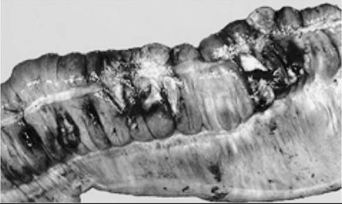
FIG. 32.69 Colonic infarction in a horse with salmonellosis. Tenesmus and rectal prolapse were early clinical signs of colonic infarction. (Courtesy Dr. M.J. Murray.)
Complications such as catheter-associated thrombophlebitis, colonic infarction (Fig. 32.69), colonization of other organs by Salmonella spp. or other enteric bacteria, and laminitis can occur.
Measures designed to prevent the spread of Salmonella in an environment with potentially susceptible hosts need not be excessively laborious or expensive. The goal is to minimize the size of the infective dose of an enteric pathogen to which a susceptible host may be exposed. Thorough cleaning of areas where fecal contamination is likely and prevention of mechanical distribution of contaminated material are the most important measures. Extensive use of disinfectants may not be necessary if cleaning measures are adequate. Cleaning must include removal of organic debris, which can be accomplished with several products designed for that task. Areas that require particular attention are stalls, including water buckets or automatic watering systems, drains, and cracks in the floors and wall; stall implements; surgical areas, including drains; and nasogastric tubes and pumps.
If a diarrheic horse is not hospitalized, it should be isolated to the greatest degree possible, and biosecurity measures should be instituted to prevent transmission and to detect and monitor any potential outbreak.6 Bedding material should be removed frequently to minimize accumulation of potential enteropathogens. Personnel entering the stall should be restricted to the professional staff, and they should wear gloves, disposable coveralls, and disposable plastic boots. Footbaths with disinfectant are effective only if they are kept clean and fresh because they quickly accumulate organic material that interferes with the action of the disinfectant. Vacated stalls should be thoroughly cleaned, allowed to dry, and disinfected, and culture or PCR testing of selected sites in the stall (floor, drain, waterer) must yield negative results for Salmonella. Biosecurity protocol compliance is crucial for preventing an outbreak.
■ Potomac Horse Fever PHF is an infectious enterocolonic disorder caused by N. risticii (formerly Ehrlichia r⅛ftαz).20-22 The organism is an obligate intracellular parasite, infecting peripheral monocytes and macrophages, colonic and small intestinal epithelial cells, and colon mast cells.23 The pathophysiology of the disease is incompletely understood, although many horses infected with N. risticii have clinical signs and complications similar to those in horses with salmonellosis. Two to 4 days after experimental inoculation with N. risticii, horses had a mild, transient fever.24 By 10 to 14 days after experimental infection, horses had become febrile, had a poor appetite, and exhibited mild to severe gastrointestinal signs ranging from mild colic and soft stool to profuse diarrhea.
In clinical cases of PHF, there are signs of sepsis, including fever, leukopenia, congested mucous membranes, and hypercoagulability. The early fever observed in experimental cases is usually not detected by owners, and when clinical signs are seen, it is presumed that the affected horse was infected 10 to 14 days previously. Hypoproteinemia is a frequent finding in horses with clinical cases of PHF, which reflects loss of serum protein through inflamed intestinal mucosa. Of interest is, however, that the magnitude of intestinal inflammation is typically much less than with salmonellosis, and yet the magnitude of hypoproteinemia (total protein concentration < 3 g/dL) can be as severe.
Laminitis is a frequent sequela and may occur in 30% in horses with PHF. N. risticii has also been associated with abortion in mares, although this is an unusual occurrence.25 In some horses that show significant seroconversion to N. risticii, laminitis is the only clinical sign.
MODE OF TRANSMISSION. Although originally described as a disease of horses living near the Potomac River in Maryland and Virginia, horses with serologic evidence of exposure to N. risticii have been reported in most U.S. states. Because early research on PHF demonstrated that infection was readily transmitted through blood,24 it was presumed that the natural mode of transmission involved an insect or arthropod vector. However, several studies have failed to demonstrate any such vectors.26,27
The association between an affected horse and proximity to a river (within 5 miles) remains strong. Investigators have found a possible link between this association and how the disease may be transmitted. N. risticii and Neorickettsia helminthoeca were found to share a high degree of DNA homology.28 N. helminthoeca is transmitted to mammals by a trematode that parasitizes fish and aquatic snails.28 This discovery prompted investigators to search for evidence that N. risticii might reside in trematodes and their hosts found in riverine inhabitants. N. risticii DNA was detected in operculate and pulmonate snails (Juga spp. and Planorbella spp., respectively) collected from stream water in northern California, where PHF is enzootic.29,30
Moreover, N. risticii was detected in trematode stages found in the secretions of freshwater snails and in aquatic insects.31,32 The sequences of these genes were virtually identical to those of the genes of an equine N. risticii strain isolated from horses located on a property near the snail collection site. Further work demonstrated N. risticii DNA in two trematodes, Acan- thatrium spp. and Lecithodendrium spp., found in bats and swallows, which suggests that these animals may serve as reservoirs and that trematodes are a vector for the organism.33 Oral transmission of N. risticii with infected cell cultures has been produced experimentally,34 which indicates that ingestion of N. risticii, rather than blood transmission, is the route of natural infection. Oral transmission and clinical signs of PHF were demonstrated in horses fed N. risticii-infected aquatic insects (caddisflies), which suggests that ingestion of infected aquatic insects may be the natural route of infection.35,36 Ingestion of water containing infected trematode stages released from aquatic snails may also be a route of infection.
DIAGNOSIS. Accurate confirmation of PHF can be difficult because clinical signs of disease are nonspecific and available diagnostic tests are not entirely reliable. The conventional recommendation is that paired acute and convalescent blood samples be submitted for indirect fluorescent antibody or ELISA testing for antibodies to N. risticii. However, serologic evaluation to confirm the disease is not as straightforward as in many other infectious diseases. A fourfold increase in titer between acute and convalescent sera is considered to confirm infection with N. risticii, but failure to seroconvert does not rule out infection. Because the onset of clinical signs can be delayed as long as 14 days after infection, horses may seroconvert by the time an acute sample is obtained.34 The magnitude of titer is not always correlated with active infection, inasmuch as many horses in endemic areas have high titers but no disease. Vaccination also can affect the titer. There is significant interlaboratory variability in results of indirect fluorescent antibody tests, and false-positive results occur frequently.37 Therefore it has been suggested that indirect fluorescent antibody testing for antibodies to N. risticii not be performed in areas that are not endemic for N. risticii.37
The current standard for diagnosis of PHF used by most clinicians is quantitative PCR testing of whole blood samples to detect N. risticii DNA in leukocytes.38 PCR testing appears to be sensitive and specific in horses with compatible clinical signs.
TREATMENT AND PREVENTION. Treatment with oxytetracycline (7 to 11 mg/kg IV twice daily for 4 days) effectively eliminates N. risticii from horses in experimental infections. Fever should resolve within 48 hours of beginning treatment with oxytetracycline, and diarrhea typically resolves within 24 to 72 hours of beginning treatment. In one horse the author treated, clinical signs persisted until the dose of oxytetracycline was increased to 15 mg/kg twice daily. Orally administered doxycycline (10 mg/kg q12-24h) may be effective, although horses with severe gastrointestinal signs may not absorb doxycycline efficiently. Doxycycline must not be given intravenously because it will cause a horse to collapse. Some horses have had clinical relapses 2 to 3 weeks after initial resolution of clinical signs that were responsive to tetracycline. Horses do not remain chronic carriers of N. risticii.
In most cases of PHF, intravenous administration of polyionic fluids is necessary to replace fluid and electrolyte losses and to augment preload in horses with poor venous return. Plasma may be required to replace lost plasma proteins. Other colloidal fluids may also be used in patients with hypoproteinemia or sepsis. Because horses improve rather quickly once treatment with oxytetracycline has begun, parenteral nutritional support is usually unnecessary.
Vaccination has appeared to diminish the incidence and severity of disease, but PHF may still develop in vaccinated animals. Experimental results with vaccine were mixed, and the duration of immunoprophylaxis was very limited.39 Disease severity has appeared to be less in vaccinated animals, although in the summer of 1994 many vaccinated horses developed severe cases of PHF. All of these horses were located in the area where PHF was originally described, and a new strain of N. risticii was identified.40 PCR analysis has revealed multiple strains of N. risticii, which may account for the unreliability of the vaccine.41 Horses in endemic areas should be vaccinated in the early spring and early to mid-summer on an annual basis.
■ Clostridial Diarrhea Enteric clostridiosis was reported as a clinical problem in the 1970s, and clostridial bacteria were implicated as the causative agents of colitis X.42 Difficulty in substantiating clostridial bacteria as the cause of enterocolitis in horses led to a deemphasis of this pathogen as a potential cause of diarrhea in adult horses. In the 1990s, with improved laboratory techniques for identification of clostridial toxins, clostridial bacteria were confirmed to be common causative agents of colitis in foals and adult horses.
The two most important clostridial species affecting the equine intestinal tract are C. difficile and C. perfringens. Most reported cases in adult horses have involved C. difficile, although in one report the prevalences of C. difficile toxin A and C. perfringens enterotoxin in fecal samples from horses with diarrhea were similar, and in some horses toxins from both clostridial species were identified.43
PATHOGENESIS. C. difficile is a Gram-positive sporulated obligate anaerobe responsible for most cases of antibiotic- associated diarrhea in many species, including horses, and for a substantial proportion of nosocomial infections in humans and horses.44,45 C. difficile produces two important toxins, named A and B. Most isolates of C. difficile produce both toxins, but some isolates produce only one of these. Toxin A can elicit both fluid secretion and a pronounced inflammatory response in the bowel. Toxin A has intestinal secretory and cytotoxic effects,46,47 increases intestinal permeability,48 and can activate epithelial cells, neutrophils, mast cells, monocytes, and macrophages to release a multitude of proinflammatory cytokines and vasoactive mediators.49-51 Two interesting features of toxin A are its induction of the neurotransmitter substance P in both the intestine and dorsal root ganglia and its apparent dependence on substance P for full expression of its pathologic effects in the intestine of rodents.52 Thus the effects of toxin A appear to be mediated both through direct effects on intestinal cells and through the enteric nervous system.
Toxin B exhibits enterotoxigenic (secretory) activity and has also been shown to have potent cytotoxic effects on human colonic epithelium.53 Toxin B has little relevance to the pathogenicity of C. difficile in animal models, however, and the roles of toxins A and B in the pathogenesis of equine clostridial enterocolitis are not known.
Strains of C. perfringens are classified on the basis of the toxins that are produced; at least a dozen have been identified to date.54 C. perfringens type A and type C have been recovered from equine specimens,55 and several C. perfringens toxins have been identified in equine specimens.56,57 C. perfringens type A is the most frequently isolated type, and its enterotoxin is released on sporulation of C. perfringens within the intestine. Other exotoxins of C. perfringens have phospholipase activity (alpha-toxin), necrotizing cytotoxic effects (beta-, epsilon-, and iota-toxins), and hemolytic effects (theta-toxin).
CLINICAL FEATURES. Clostridial enterocolitis affects foals and adult horses. In some reports, toxigenic C. perfringens organisms were isolated from more than 50% of foals with diarrhea.57,58 The clinical presentation in foals with C. perfringens is predominantly hemorrhagic diarrhea with sepsis. C. perfringens can cause septicemia and is often cultured from the blood of foals with hemorrhagic diarrhea. In some foals, classic necrotizing enterocolitis is manifested by gas- or fluid-distended intestines and thickened intestinal mucosa. These changes can be appreciated radiographically and ultrasonographically, and intramural gas produced by clostridial bacteria may be detected with ultrasonography as hyperechoic areas within the bowel wall. C. difficile infection is also a cause of acute enterocolitis in foals.59
Adult horses with clostridial enterocolitis frequently have diarrhea, but abdominal discomfort or fever may be the primary presenting problem. There are no clinical features that consistently distinguish clostridiosis from salmonellosis, and a spectrum of clinical signs exists, from moderate illness to severe toxemic colitis. Most horses with clostridial enterocolitis develop diarrhea, but in some cases enteritis manifested by ileus and gas distention of the small intestine may be the primary problem. Although both C. perfringens and C. difficile and their toxins have been detected in a significant number of adult horses with colitis, C. difficile appears to be more common.60-62 In fact, C. difficile appears to be the most common cause of antibiotic-associated diarrhea and is a very significant cause of nosocomial diarrhea in horses.45,63 The most common antibiotics associated with C. difficile colitis in horses are erythromycin, trimethoprim/sulfonamides, β-lactam antimicrobials, clindamycin, rifampicin, and gentamicin. Strain differences among isolates of C. difficile cultured from the diarrheic feces of horses in intensive care units are associated with toxigenicity, severity of disease, and metronidazole resistance.45,64 C. difficile has been implicated in DPJ,65 but a cause-and-effect association has not been proven.
Similar to salmonellosis, clostridial enterocolitis can develop into a widespread problem affecting several animals in a hospital or equine facility.66 Risk factors for developing nosocomial clostridial enterocolitis are similar to those for nosocomial salmonellosis: antimicrobial administration, concurrent gastrointestinal disease, and age susceptibility (foals). In addition, Clostridium spp. are well suited to persist in the environment because of the production of spores that are resistant to environmental extremes and many disinfectants.
DIAGNOSIS. Diagnosis of clostridial enterocolitis requires identification of toxigenic clostridia from intestinal contents or tissue. Several direct and indirect methods are used to detect toxigenic clostridia; these methods include culture, identification of toxins, and identification of toxin genes. Culture of C. difficile or C. perfringens requires anaerobic conditions, and the ability to culture these organisms from ingesta or fecal specimens rapidly diminishes with increased time from collection to arrival at a laboratory. It is recommended that samples be transported, chilled (not frozen) on ice, immediately or by overnight delivery for best recovery of clostridial organisms. Tissue specimens submitted for culture, toxin identification, or toxin gene identification should be handled similarly. Because nonpatho- genic clostridia are common,62,67 isolates should be tested for toxin production either by PCR or with bioassay before a definitive diagnosis is made in horses with clinical signs consistent with clostridial enterocolitis. Commercially available tests for clostridial toxins include an ELISA for C. difficile toxin A/B, a latex-agglutination test for C. perfringens enterotoxin, and an ELISA for C. perfringens enterotoxin. C. difficile A/B toxin tests have the advantages of being rapid, sensitive, and specific. Tests for C. perfringens enterotoxin are not as reliable for definitive diagnosis in most species. Also available is quantitative PCR for C. difficile toxin A/B genes and several C. perfringens toxin genes that can be applied directly to intestinal content or fecal samples without prior culture and isolation of the bacteria.68
TREATMENT. Supportive care may be required, as with other cases of acute diarrhea in horses. Treatment with metronidazole (15 to 20 mg/kg PO q6-8h) appears to be effective in eliminating enteric clostridial infection in most cases. In one veterinary teaching hospital, isolates of C. difficile were resistant to metronidazole,69 and vancomycin was used with reported success. Large-scale resistance to metronidazole has not been reported elsewhere, and it should be the first choice in the treatment of suspected clostridial enterocolitis. Saccharomyces boulardii is a nonpathogenic yeast used in the treatment of C. difficile diarrhea and colitis in humans. The yeast releases a protease that specifically degrades C. difficile toxins A and B, and this has been shown to be protective in experimental C. difficile colitis in rats and to prevent damage to human colonic epithelium by C. difficile toxins A and B in vitro.70 Although S. boulardii (25 g PO q12h) has been shown to colonize the intestinal tract in horses,71 its efficacy in preventing or treating enterocolitis in horses is not clear. S. boulardii decreased the severity and duration of enterocolitis in one small study72 but not in a larger study of antibiotic-associated diarrhea in horses.71 Di-tri-octahedral smectite has been shown to bind clostridial toxins73 and may be useful for treating clostridiosis in horses. Di-tri-octahedral smectite is available as a powder or paste and should be administered according to the manufacturer’s instructions for 3 to 5 days.
■ Equine Coronavirus Enterocolitis Equine coronavirus is an enteric pathogen that causes fever, lethargy, colic, and diarrhea primarily in adult horses.74 Infections have been reported in horses housed in boarding, racing, and show stables and on breeding farms. Rates of morbidity in outbreaks range from 10% to 83%.75-78 Infections last a week or less and are usually self-limiting unless accompanied by potentially fatal complications such as sepsis or hypoammonemia. Equine coronavirus is shed in feces for several days; shedding may persist as long as 24 days.77
PATHOGENESIS. Equine coronavirus is a primary pathogen in adult horses and appears to be involved in co-infections with other enteric pathogens in foals, at least in one study in the United States.79
Equine coronavirus infection in adult horses causes an acute necrotizing, neutrophilic inflammation that has been well documented in the small intestine.80 Disease in the large intestine was not significant in a limited study,80 but it presumably also occurs in the large intestine, at least in horses with diarrhea. Epithelial cell necrosis and intestinal inflammation result in damage to the mucosal barrier, and subsequent sepsis probably results from absorption of microbial molecules. Equine coronavirus can be recovered from the respiratory tract in infected adult horses, but it is not clear whether clinically relevant respiratory disease accompanies enteric infections.81,82 Fatal hyperammonemia has been reported in some cases of equine coronavirus-infected horses,77,78,80 probably as a result of changes in the mucosal barrier and intestinal microbiome.
CLINICAL FEATURES. The most common clinical signs in adult horses with equine coronavirus infection are fever, lethargy, and anorexia.77 Encephalopathy characterized by circling, ataxia, head pressing, nystagmus, seizures, or a combination of these is a less common clinic feature and appears to be associated with hyperammonemia in at least some cases.77 Clinicopatho- logic findings include neutropenia and band neutrophilia (especially early in the course of disease), hyperfibrinogenemia, elevated serum amyloid A concentration, electrolyte and acidbase changes typical of acute enterocolitis, elevated concentration of unconjugated bilirubin, transiently elevated liver enzyme activities, and prerenal azotemia.74 Hyperammonemia may be 7778
present in encephalopathic patients.77,78
DIAGNOSIS. Diagnosis is currently based on quantitative PCR testing of feces while the animal is alive or intestinal tissues or contents after its death. Fecal quantitative PCR is quite sensitive.77,78 Shedding of equine coronavirus begins after 3 or 4 days, at least in experimentally infected horses,83 so quantitative PCR test results may be falsely negative very early in the course of disease. Fecal shedding can persist from 3 to 24 days, and so infected horses may test positive after clinical signs resolve.
TREATMENT. Most cases are self-limiting and necessitate no specific treatment. Intravenous isotonic fluid administration with appropriate electrolyte supplementation may be required in some cases if diarrhea is persistent and severe. NSAID therapy may be indicated if fever and lethargy persist longer than a day or two. Analgesic drugs are indicated if colic is a problem. Complications such as sepsis and hyperammonemia may occur and necessitate intensive treatment and management as described elsewhere.
■ Cantharidin (Blister Beetle) Toxicity
PATHOGENESIS. Cantharidin is the toxic principal found in beetles of the genus Epicauta, commonly known as blister beetles.84-86 Ingestion of the beetles in contaminated alfalfa hay results in absorption of cantharidin through the gastrointestinal tract. Blister beetles feed on the flowers of alfalfa and are incorporated into baled alfalfa hay if the hay is cut and processed simultaneously.84-86 Large swarms of beetles may be found in relatively small portions of hay. The lethal dose of cantharidin is less than 1 mg/kg, but the concentration of cantharidin varies from species to species of blister beetles.84,85 Therefore ingestion of as many as 100 to as few as 6 to 8 beetles may be lethal. Often more than one horse is affected. The fatality rate may be 50% or greater.84,87
Cantharidin is a potent irritant, causing cell damage and 848687
necrosis on contact.84,86,8' Ihe mucosa of the gastrointestinal tract is most commonly affected in horses because they ingest the toxin. Ulceration throughout the alimentary tract has been observed in natural and experimental cantharidin toxicity. Diarrhea probably results from the severe ulceration and inflammation of the large intestine, causing increased secretion of water, electrolytes, and protein. Large volumes of fluid and protein are lost in the gastrointestinal tract, which leads to hemoconcentration and profound hypoalbuminemia in some 848587
cases.84,, Cystitis, nephrosis, and myocarditis occur in natural and experimentally produced cases of cantharidin toxicity.84,86,87 Cystitis and nephrosis result from the high concentration of cantharidin in the urine. The cause of the myocarditis and myocardial necrosis is unknown, but they may also be direct effects of the toxin on the myocardium. Plasma CK activity is often elevated, and this elevation has been postulated to arise from the damaged myocardium.84,85 Affected horses have a characteristically stiff gait, but histopathologic evidence of skeletal muscle injury that explains the elevated plasma CK activity has not been observed.85 Hypocalcemia and hypomagnesemia are characteristic features of cantharidin toxicity in 848587
horses that have not been explained.84,, Hypocalcemia may result from hypoalbuminemia, but the ionized calcium concentration is often decreased, along with the total calcium concentration, which indicates that hypoalbuminemia is not responsible for the hypocalcemia.85
CLINICAL FEATURES. Cantharidin toxicity can cause a range of clinical signs, from mild depression and abdominal discomfort to fulminant signs of toxemia and rapid death, depending on the ingested dose of toxin.84,85,87 Most commonly, clinical signs include depression, sweating, irritability, abdominal pain, elevated heart and respiratory rates, fever, polyuria, polydipsia, and profuse diarrhea.84,85,87 Blood is rarely seen in the feces. Affected horses frequently posture to urinate; indeed, stranguria and pollakiuria are characteristic of cantharidin toxicity.84 Signs of hypocalcemia include synchronous diaphragmatic flutter and tremors. Gait may be stiff and stilted. Neurologic signs include head pressing, swaying, and disorientation.87 Signs of systemic inflammation from sepsis may occur in severe cases. Some affected horses develop severe depression and toxemia and may die within hours after ingestion of cantharidin without developing diarrhea.84,87 Hematologic abnormalities are similar to those of other causes of acute diarrhea, reflecting dehydration and sepsis. Hypocalcemia (both ionized and total calcium concentrations) and hypomagnesemia are characteristic biochemical features of cantharidin toxicity and may be profound. Azotemia with a urine specific gravity in the hyposthenuric 848587
range is common. ,, Microscopic hematuria and mild proteinuria may be evident.
DIAGNOSIS. Tentative diagnosis is based on clinical signs and the finding of blister beetles in the hay. Determining the species of the insects may be necessary to estimate the amount of cantharidin ingested. All species of Epicauta contain cantharidin, but some have small amounts. Definitive diagnosis requires the measurement of the cantharidin concentration in 8488 gastric or intestinal contents and urine.84,88
TREATMENT. Fluid therapy and maintenance of electrolyte balance are important, as in all cases of acute diarrhea. Particular attention should be paid to the degree of hypocalcemia and renal function as a fluid therapy plan is developed. Specific therapy is limited. Administration of absorbent powders via nasogastric tube is important early to prevent further systemic absorption of cantharidin and local toxicity. Mineral oil administration may also help reduce absorption of the toxin. Pain control may be needed for colic or urinary pain. NSAID therapy should be used judiciously to avoid further injury to the intestinal mucosa and renal toxicity. Alternative analgesics, such as butorphanol or a lidocaine controlled-rate infusion, may be better choices.
■ Antimicrobial-Associated Diarrhea The onset of acute diarrhea in horses has been associated with the use of several antimicrobial drugs. Lincomycin administered orally and tetracycline administered parenterally have been demonstrated to induce severe diarrhea in horses.89,90 The oral administration of trimethoprim-sulfonamide, kanamycin, erythromycin, metronidazole, and penicillin and parenteral administration of ceftiofur and gentamicin have been implicated in onset of diarrhea, including fatal colitis, in horses. In one report there was no association between trimethoprimsulfonamide and diarrhea.91 However, in other reports, prior administration of antimicrobial drugs was positively associated with onset of colitis and negatively associated with prognosis for survival in horses with colitis.92
Antimicrobial-associated diarrhea is presumed to be secondary to disruption of normal colonic microflora and the proliferation of an enteropathogen, such as Salmonella spp., C. perfringens, and C. difficile. Of these, C. difficile is the most common cause of antimicrobial-associated diarrhea in horses.45 Of interest, it was reported that mares whose foals were treated with erythromycin for R. equi infection developed severe, acute colitis, from which C. difficile and its toxins were isolated.93 Erythromycin was detected in the mares' feces, and exposure to the erythromycin was presumed to have occurred when the mares licked erythromycin from the foals' faces.
■ Other Causes of Acute Diarrhea NSAID toxicity has been associated with diarrhea secondary to damage to the colonic mucosa. Grain (carbohydrate) overload may cause overproduction of lactate in the colon, which leads to acute diarrhea. A combination of hyperosmolarity of the luminal contents and damage to the colonic mucosa account for the pathophysiologic process grain overload. The clinical signs are similar to those of other acute diarrheal diseases in horses and depend on the severity of the overload. Hyperlactemia, metabolic acidosis, and sepsis are key features of grain overload, and laminitis is a common sequela. Acute diarrhea in adult horses has also been associated with conditions such as lymphosarcoma enterocolitis and other IBDs, intestinal lymphosarcoma, peritonitis, heavy metal intoxication, anaphylaxis, and stress.
■ Clinical Assessments of Patients With Acute Diarrhea The diagnostic evaluations performed on a horse with acute diarrhea are intended to provide the clinician with information to accurately assess the horse's condition and thus direct therapy toward specific requirements. The first part of the evaluation is a thorough physical examination, with particular attention to the horse's hydration status (skin turgor, gum moisture, capillary refill time), evidence of sepsis (fever or hypothermia, hyperemic mucous membranes, prolonged capillary refill time), cardiovascular system (heart rate and rhythm, character of peripheral pulse, capillary refill time), and signs of laminitis (lameness, digital pulse, palpable temperature of hoof walls). Many horses with colitis are moderately to severely dehydrated, with either purplish or brick-red mucous membranes. Purple mucous membrane color reflects venous congestion and poor venous return, whereas brick-red mucous membrane color reflects venous congestion and poor venous return plus arteriole and venule shunting and poor tissue oxygen exchange.
Blood pressure should be monitored; direct measurements via an arterial catheter are the most accurate, but indirect pressure measurements obtained with the use of a Doppler transducer placed over the coccygeal artery are satisfactory. Hypotension and hypertension occur in horses with colitis, and the blood pressure status of a patient often is unpredictable. Blood pressure can be monitored as frequently as labor allows and should be monitored hourly if vasoactive pharmaceutical agents are used.
Laboratory tests that should be performed include CBC and measurements of plasma protein and total solids. Total hemoglobin and PCV are used to assess hydration status. Total protein concentration is used to assess hydration status and degree of protein loss through inflamed intestinal mucosa and, in more chronic cases, through protein catabolism. Clinical hydration, PCV, and total protein concentration are compared to determine the extent of protein loss, and daily evaluations can be used to determine the rate of protein loss. Colloid oncotic pressure measurements are also useful, particularly if colloidal fluids other than plasma are given.
Total WBC count, WBC differential, and WBC structure are examined to assess severity of sepsis; plasma fibrinogen is examined to assess the severity of inflammation. Typically, the total WBC and neutrophil counts decrease initially. This is attributable primarily to bacterial endotoxins and the host's mediators of systemic inflammation, and it occurs in most cases of acute colitis, not just in those caused by Salmonella spp. The structure of the WBCs reflects the severity of the inflammatory response. “Toxic” changes such as basophilia, granulation, vacuolation of the cytoplasm, and scalloped borders of the cell membrane or adherence of neutrophils to RBCs do not reflect injury to the neutrophils by toxins but reflect the cells' responses to stimulation by proinflammatory agents (TNF-alpha, IL-1) and the production of inflammatory mediators by the neutrophils that are toxic to bacteria. The degree of these changes in circulating neutrophils can be used to assess the severity of disease and also to assess the progress the horse is making. Often the initial sign that the horse is improving is a decrease in the “toxic” appearance of the neutrophils and a regenerative neutrophil response. A horse that continues to have severe neutropenia with a degenerating left shift or neutrophils, with cytoplasmic vacuolation, granulation, and basophilia, for more than 10 days has severe colitis that is unlikely to resolve.
Serum chemistry tests that should be performed include measurements of electrolytes (sodium, chloride, potassium, and calcium), BUN and creatinine, and blood lactate concentration, as well as assessment of acid-base status (blood pH and bicarbonate, or total CO2). Many horses with diarrhea are hyponatremic, hypochloremic, and hypokalemic. With decreased feed intake, hypocalcemia occurs. A high-anion gap metabolic acidosis with hyperlactemia may be noted, particularly in horses with sepsis. The severity of these electrolyte disturbances should be monitored, in many cases daily, to allow for appropriate therapy. Parameters of renal function (BUN and creatinine) are frequently increased in horses with diarrhea for several reasons. Prerenal azotemia resulting from dehydration and decreased filtration across the glomerulus accounts for some of the increase in these parameters. Hyponatremia and hypochloremia can cause a decrease in glomerular filtration and an increase in BUN and creatinine levels that are secondary to tubuloglomerular feedback. Many horses that are adequately hydrated and yet moderately hyponatremic (serum sodium 120 to 128 mEq/L) remain azotemic until sodium levels increase above 130 mEq/L. In addition, many horses with toxemic colitis have damage to renal parenchyma, presumably the result of the effects of inflammatory mediators and alterations in renal blood flow.
The acid-base status can be evaluated by estimating serum bicarbonate on the basis of the total CO2 or directly from a venous or arterial blood gas analysis. A venous blood gas sample helps in assessing perfusion and oxygen extraction. An increased venous oxygen partial pressure (>60 mm Hg) is indicative of poor capillary perfusion and oxygen delivery to the tissues. Affected horses usually have brick-red mucous membranes.
■ Principles of Therapy for Acute Diarrhea Because the pathophysiologic process of equine colitis is complex, treatment is often multifaceted. Many of these treatments provide well-documented benefit, whereas with others the efficacy is based only on empirical judgment. Outcome is determined not only by the severity of the primary disease causing colitis but also by complications that may arise from the disease.
In cases of acute colitis, fluid administration remains the treatment of primary importance. Most patients require intravenous administration in the early stages. The fluids used must replace fluid, sodium, chloride, and potassium losses. In many cases, large volumes are required for several days. (More specific guidelines for fluid therapy in colitis patients are covered in the Fluid Therapy for Horses with Gastrointestinal Diseases section and in Chapter 44.)
Most horses with colitis become hypoproteinemic as a result of protein leakage through the inflamed colon and catabolism of albumin secondary to negative energy balance. Hypoproteinemia frequently leads to edema formation in several areas of the body, including the intestinal tract, and can compromise the clinician’s ability to keep the patient properly hydrated through fluid administration. Intravenous plasma therapy (6 to 10 L) is often beneficial as a colloidal fluid and as a protein replacement fluid. Other colloidal fluids may be more costeffective for increasing colloidal oncotic pressures in hypoproteinemic horses but do not have the additional properties of plasma that may be beneficial. Plasma contains other proteins besides albumin and therefore can be of benefit beyond improvement of plasma oncotic pressure. Fibronectin is essential to the normal function of the monocyte-macrophage system in the processing of a variety of antigens. Other plasma proteins such as elastase and proteinase inhibitors, complement inhibitors, AT-III, and other inhibitors of hypercoagulability may be beneficial to patients with colitis.
The nutritional requirements of patients with colitis need to be considered, particularly in a case that may be protracted. Horses with colitis are typically anorexic, and the disruption of normal physiologic processes in the inflamed cecum and colon limits the effectiveness of these organs to digest and absorb nutrients. In addition, several mediators of inflammation and septicemia alter protein and calorie metabolism, which results in a catabolic state. Therefore even if an affected horse eats, it is likely to be in a severe caloric deficit for some time. Normally an average horse requires approximately 15 MCal/ day. An septic horse may require 25 MCal/day. In a catabolic patient, muscle and fat tissue are mobilized and used in lieu of ingested nutrients. The plasma protein pool, including albumin and immunoglobulins, also is catabolized. In many cases of colitis, the decrease in plasma protein may be as much a result of catabolism as of leakage through the inflamed colon. A variety of products can be used for enteral feeding (see Chapter 50).
Colitis is an inflammatory disease, and limiting inflammation is desirable when treating patients, particularly those with severe disease. Moreover, horses with acute colitis, regardless of the cause, have signs compatible with systemic inflammation associated with sepsis. Flunixin (0.25 to 0.5 mg/kg IV q6-8h) is the most commonly used antiinflammatory therapy in horses with colitis. However, the use of flunixin should be judicious and must be carefully monitored to avoid unwanted side effects on the gut mucosa and kidneys. Other antiinflammatory therapies, such as COX-2 selective firocoxib or lidocaine administered by controlled-rate infusion, may also be beneficial with less detriment to the gastrointestinal mucosa.
Sepsis is a common clinical feature in horses with acute colitis. Most of the clinical signs of sepsis are probably attributable to the effects of endotoxin. Therefore specific therapy for endotoxemia may be warranted.
The use of antimicrobial drugs in the treatment of colitis is controversial. In cases of colitis caused by N. risticii, the efficacy of tetracycline (6.6 to 11 mg/kg IV once or twice daily) is documented.94,95 In other cases of colitis, including Salmonella colitis, in which specific antimicrobial sensitivities to the Salmonella spp. have been established, the efficacy of antimicrobial administration is not as well documented. Many clinicians believe that the use of an antimicrobial for which the Salmonella spp. have demonstrated sensitivity, such as chloramphenicol, enrofloxacin, gentamicin, amikacin, or a third-generation cephalosporin, does not significantly alter the course of the disease or hasten the elimination of the organism from the body. However, there is no published evidence either supporting or disputing the efficacy of antimicrobial treatment for salmonellosis, and the use of antibiotic therapy remains a judgment of the clinician. In patients with sepsis, the use of broad-spectrum antibiotics is justified to prevent bacteremia or organ colonization by Salmonella spp. or other enteric organisms. Antibiotics such as chloramphenicol and enrofloxacin that are lipid permeable or are concentrated in cells are a benefit because Salmonella reside, proliferate, and persist intracellularly in macrophages and other cells.
Medications that minimize or abolish colonic fluid secretion are of tremendous benefit in the treatment of equine colitis. Medications such as kaolin, bismuth subsalicylate, and activated charcoal are frequently used in cases of colitis in adult horses, but their efficacy as antisecretory agents in this context is unknown. These medications are more effective in foals with diarrhea, than in affected adult horses, probably as a result of an effect on the small intestine rather than the colon. Absorbent powders such as activated charcoal or di-tri-octahedral smectite (Bio-Sponge) may be useful for absorbing bacterial toxins, particularly in horses with clostridiosis.
■ Chronic Diarrhea Chronic diarrhea is one of the most frustrating disorders encountered by equine practitioners, with regard to both determining the cause and therapeutically managing the diarrhea.96 Chronic diarrhea may be defined as persistent diarrhea of at least a month’s duration. Although there are many causes of chronic diarrhea, these cases can generally be divided into two groups: diarrhea resulting from a chronic inflammatory condition and diarrhea resulting from a disruption in normal physiologic processes. Inflammatory conditions cause histologic changes in the colon mucosa, including pleocytosis (neutrophils, eosinophils, and lymphocytes), mucosal congestion, and mucosal erosion and ulceration. Submucosal edema, capillary congestion, and lymphatic congestion may be present. With physiologic disorders, there are no morphologic changes in the colon, and diarrhea is presumed to result from abnormal VFA synthesis or absorption. A small percentage of horses with chronic diarrhea have a primary disorder of a system other than the intestinal tract, such as congestive heart failure or hepatic disease. A thorough physical examination and evaluation of a minimum database (CBC, serum chemistry profile, urinalysis) should help establish whether a disorder is nonenteric.
CAUSES. Inflammatory disorders that can cause chronic diarrhea include disorders caused by infectious agents such as chronic salmonellosis; chronic parasitism with S. vulgaris, Strongylus edentatus, and larval cyathostomiasis; abdominal abscessation; and, in weanling foals, equine proliferative enteritis caused by L. intracellularis, gastrointestinal R. equi infection, and rotavirus infection.
Noninfectious inflammatory causes include cellular infiltrative disorders such as granulomatous enteritis and lymphosarcoma, as well as sand enteropathy. Sand causes diarrhea through continued irritation of the mucosal lining of the colon. In weanling foals, gastric ulceration and gastric emptying disorders have been associated with chronic diarrhea that resolved when H2 antagonist therapy was started. NSAIDs can cause chronic diarrhea, which is accompanied by varying degrees of pathologic change in the large intestine.
Noninflammatory chronic diarrhea of colonic origin is thought to be a result of abnormal fermentation of cellulose by the resident bacteria in the large intestine. In vitro fermentation of feces from normal horses and horses with chronic diarrhea revealed that feces from the diarrheic horses produced more gas, acetate, and propionate than did feces from normal horses.97 Whether this reflects fermentative activity within the colon is not known, but an abnormal increase in acetate could lead to fluid retention within the colonic lumen because acetate inhibits colonic absorption of sodium and water.
DIAGNOSIS. The diagnostic approach to cases of chronic diarrhea should be based on an attempt to differentiate inflammatory from physiologic causes. The evaluation can be extensive and expensive, and the owner should be prepared for the cause of the diarrhea to remain undetermined. Horses with chronic diarrhea may be adequately hydrated if water consumption has matched water losses; however, many such horses are brought to the veterinarian in a condition of mild to moderate dehydration. Moderate weight loss also has often occurred. On physical examination, signs of toxemia (injected mucous membranes, congested or hyperemic mucous membranes) should be noted.
A CBC should be evaluated for signs of chronic inflammation. Such changes include a decrease in the RBC count and PCV as a result of decreased erythrogenesis secondary to sequestration of iron by bone marrow macrophages (anemia of chronic inflammation). The WBC count may be normal or moderately increased. The fibrinogen levels can be normal or increased. Changes in WBC count and fibrinogen levels are influenced by the degree of inflammation and whether the inflammatory response is localized. Therefore a normal CBC does not rule out an inflammatory cause of the chronic diarrhea.
Peritoneal fluid analysis may reveal an increase in protein or WBCs, which is indicative of an inflammatory process within the peritoneal cavity. Often, however, colon inflammation is not reflected by alterations in the peritoneal fluid.
Serum chemistry values vary in horses with chronic diarrhea. Many affected horses have evidence of hyponatremia, hypokalemia, hypochloremia, azotemia, and metabolic acidemia. Other horses, particularly those with less severe chronic diarrhea, may have no serum chemistry abnormalities.
With a chronic inflammatory disorder of the colon, the total serum protein concentration is usually decreased because of protein leakage from the capillaries and disruption of the colonic mucosal integrity. This is usually reflected by hypo- albuminemia. In some cases, hyperglobulinemia occurs, and total serum protein concentration may be normal.
Increases in hepatic-associated enzymes, including sorbitol dehydrogenase, GGT, and aspartate aminotransferase, and serum bile acids indicate that hepatic disease is present. Hepatic changes and dysfunction such as inflammation, fibrosis or fatty infiltration, or biliary inflammation can be associated with diarrhea.
Feces should be examined for parasite ova; cultured for Salmonella spp., C. difficile, and C. perfringens; and tested for clostridial toxins. Weanlings should be tested for L. intracellularis and R. equi. In cases of acute diarrhea, it has been recommended that five consecutive fecal samples be cultured for Salmonella spp., but in cases of chronic diarrhea, many more are often necessary. As many as 15 fecal cultures may be needed to get a positive Salmonella spp. culture. In addition, a rectal mucosal biopsy sample should be cultured. In weanlings, the feces should be examined for rotavirus by transmission electron microscopy or ELISA. Although it is an unusual cause of diarrhea in weanlings, rotavirus should be considered when several foals on the same farm have chronic diarrhea.
An oral glucose absorption test can be done to determine whether there is small intestinal malabsorption, which would indicate a widespread small and large intestinal disorder if both diarrhea and glucose malabsorption are present.
A rectal mucosal biopsy may provide evidence of a widespread inflammatory disorder, such as one of the IBDs.98 Biopsy specimens should be evaluated by a pathologist experienced in examining equine tissue specimens, and some caution should be used to avoid overinterpretation of the presence of few lymphocytes, plasma cells, and eosinophils.
Frequently the results of the previously mentioned diagnostic procedures do not determine the cause of the chronic diarrhea. In such cases, exploratory laparotomy may be warranted. This is particularly true if episodes of abdominal discomfort accompany the chronic diarrhea. In addition to exploration of the abdomen for the presence of masses or abscesses, the colon and cecum should be thoroughly examined. Biopsy samples from several sites of the colon, cecum, and mesenteric lymph nodes should be submitted for histopathologic study and culture for Salmonella spp.
TREATMENT. Treatment of horses with chronic diarrhea is often empirical because either a cause has not been determined or the cause is not amenable to treatment. With inflammatory causes such as lymphosarcoma and granulomatous enteritis, the disease is usually untreatable. Some cases of eosinophilic colitis have been treated successfully with corticosteroids.
Chronic parasitism may be resolved with appropriate anthelmintic therapy, although damage to the mucosa may have become too extensive to allow normal absorption to occur. Administration of larvicidal doses of fenbendazole (15 mg/kg PO daily for 5 days) is usually effective in horses with nonresistant small strongyle infections. Treatment with ivermectin (0.2 mg/kg PO) or moxidectin (0.4 mg/kg PO) may be indicated when benzimidazole resistance is encountered. Concurrent administration of prednisolone (1 mg/kg PO once daily for 5 to 7 days) may minimize inflammation secondary to dying migrating larvae within the vasculature and mucosa of the colon.
Chronic salmonellosis does not lend itself to specific treatment because antimicrobial therapy generally does not resolve Salmonella infection in horses.
Administration of products containing bismuth subsalicylate is effective in some cases of chronic diarrhea. The action of bismuth subsalicylate is mediated through inhibition of prostaglandin synthesis and possibly by other undefined mechanisms. In full-size horses, a large volume, 1 to 4 L/day, must be administered to be effective.
Iodochlorhydroxyquin administered orally is effective in managing some cases of chronic diarrhea caused by maldigestion of cellulose by colonic microorganisms.99 The actual mechanism of action of iodochlorhydroxyquin in resolving the diarrhea is not known. The drug was originally administered to horses with chronic diarrhea because an increase in fecal trichomonads was observed. However, this observation probably reflected the fact that trichomonads were washed out of the cecum and colon rather their being the cause of the diarrhea. Iodochlorhydroxyquin has minimal effect on colonic protozoal populations. It is not uniformly effective, and in many cases its effectiveness is only transient. Stools may initially become formed, but the diarrhea often recurs within several days. An initial dose of 20 mg/kg/day is recommended. If diarrhea recurs, decreasing the dose to 10 mg/kg/day is sometimes effective. If the medication is effective, it must be continued because if it is discontinued, the diarrhea resumes.
Changes in diet occasionally are helpful in horses with noninflammatory chronic diarrhea. A complete pelleted feed may positively affect the constituent VFAs produced in the colon and thus facilitate water absorption. Alternatively, trying different types of roughage may result in selecting one that creates a more favorable metabolic environment in the large intestine.
The removal of sand from the colon by nonsurgical means is difficult, and in one report the administration of psyllium to remove experimentally introduced sand was not effective.100 Other treatments used with anecdotal, but undocumented, success include fecal transfaunations, probiotics, cultured yogurt, and brewer's yeast.