Neoplasia of the Liver
Geoffrey W. Smith
Primary neoplasia of the liver is uncommon in large domestic species. Only 0.011% of slaughterhouse animals seen in one study had liver tumors.1 Metastatic tumors such as lymphoma are more common (Fig.
33.11), but the clinical signs of these are more likely to reflect changes at the primary site. Metastasis of lymphosarcoma in cattle is most common, but signs produced by growth in other organs (e.g., lymph nodes, abomasum, heart, uterus, spinal cord) are more predominant. In the horse, lymphosarcomas and carcinomas have metastasized from the 2digestive tract.2
Horses
Although uncommon, a number of different primary hepatic neoplasias have been reported in horses.3,4 The nomenclature seems inconsistent throughout the years of reporting, and the signs are usually more consistent with neoplasia in general
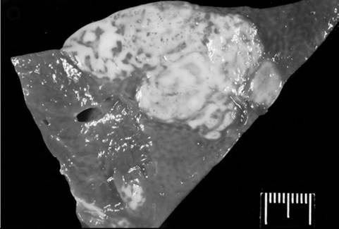
FIG. 33.11 Lymphoma in the liver of a horse. (Photo courtesy Luke Borst, North Carolina State University.)
than with hepatic failure. Weight loss, weakness, anorexia, lethargy, and occasionally colic may be seen in these animals. Serum concentrations of liver-derived enzymes are elevated in many cases. The tumor can often be located by ultrasonography, but the definitive diagnosis of the type of tumor is based on histopathologic examination of biopsy tissue.
Hepatoblastoma has been reported in foals, young horses, and an equine fetus.4,5 Young horses present with weight loss or failure to grow, lethargy, anorexia, and possibly icterus. Serum concentrations of several hepatic enzymes are increased. Ultrasonography reveals hepatomegaly and a heterogeneous appearance of the hepatic parenchyma. Erythrocytosis due to a paraneoplastic syndrome and increased production of erythropoietin is seen in some cases.
Hepatocellular carcinoma is seen in both old and young horses. These horses lose weight and are listless. Serum concentrations of LDH, GGT, and AST are often elevated. Erythrocytosis with a normal concentration of serum protein is seen in most cases as a result of paraneoplastic syndrome with elevated erythropoietin.3,4,6
Cholangiocarcinoma or cholangiocellular carcinoma arises from the intrahepatic bile duct epithelium and appears to be a disease of older horses.4,7 Lethargy, fever, abdominal pain, anorexia, and ventral edema have been reported with this tumor. Hypoglycemia has been associated with hepatic cholangiocar- cinoma as a suspected paraneoplastic syndrome.8 Some of these animals have anemia rather than the erythrocytosis reported with hepatocellular tumors. GGT and ALP concentrations may be elevated, but SDH comes from hepatocytes and is usually within normal range.
Cattle
In addition to the lymphosarcomas, primary hepatocellular carcinomas have been described in cattle.9 The most common presenting clinical signs are anorexia and weight loss of weeks to months in duration.10 Liver-derived enzymes are generally elevated, and polycythemia (erythrocytosis) is a common laboratory finding in these cases.10,11 Ultrasound of the liver may be helpful in diagnosing hepatic tumors in cattle.10
Hemochromatosis
Hemochromatosis is a disorder caused by deposition of hemosiderin in the parenchymal cells, resulting in tissue damage and dysfunction of the liver and other tissue. It is most frequently seen in humans and mynah birds, but hemochromatosis has been described as a new disease of Salers cattle12 and has been reported in three horses.13 In humans, the types include idiopathic hemochromatosis, an autosomal recessive familial condition associated with increased iron stores, cirrhosis, and saturation of the iron transport capacity.14 Both horses and cattle show increased iron deposits in the liver; histopathology of liver biopsy specimens reveals brown pigment that stains for iron in the hepatocytes as well as Kupffer cells.
An increased concentration of iron can be measured in the liver, and there is fibrosis and elevated liver-derived enzymes.In Salers cattle the condition appears to be a homozygous recessive condition more like the human familial type. There is an inappropriate absorption of iron by the GI tract, with subsequent hepatic storage (iron overload) and eventual loss of hepatic function. It has been reproduced by experimental breeding in Salers cattle.15 The primary clinical signs in cattle are decreased weight gain, poor body condition, dull hair coat, loss of incisor teeth, and sometimes diarrhea. Serum concentrations of liver enzymes are elevated, and there is marked hepatic fibrosis, in addition to the hemosiderin deposits in the liver. Total serum iron, total iron-binding capacity (TIBC), and saturation of transferrin (>60%) are increased, similar to the familial disease in humans. Liver iron concentration will be greater than 5000 μg∕g (ppm) on a wet basis (normal herdmates of affected cattle, 84 to 100 ppm).
Horses with hemochromatosis present with evidence of liver disease. Serum concentrations of the liver enzymes ALP, GGT, and AST are elevated, and serum total bile acids are greater than 40 μM∕L. In the cases reported, total serum iron was not elevated, and unlike the idiopathic human condition or the cattle cases, there was no saturation of the iron-binding capacity. Total liver iron has been as high as 6700 ppm (normal, 100 to 300 ppm).13
Hemochromatosis should be suspected in animals with emaciation or elevated liver enzymes and in Salers cattle with greater than 60% saturation of transferrin. It can be confirmed by histopathologic examination of the liver, with excessive iron accumulation in hepatocytes. Clinicians should differentiate hemochromatosis from hemosiderosis, in which iron accumulates in the reticuloendothelial system and not hepatocytes, and which can be caused by hemolysis and other conditions.
In humans, hemochromatosis is treated by reducing the iron stores through phlebotomies and blood removal. The one horse on which this was tried had advanced disease with severe cirrhosis, and it succumbed a few days after the blood removal. Removal of 160 L of blood over 12 months failed to reduce liver iron concentration in one heifer12 but produced some improvement in other calves.15 Deferoxamine is given to some human patients to induce a negative iron balance and reduce the rate at which iron accumulates.