Ophthalmic History and Examination
Renee T. Carter
A full ophthalmic examination should be conducted for patients presenting with a primary ocular complaint and also as part of a minimum workup for patients with neurologic changes or other unknown systemic disease processes.
Prior to conducting a complete ophthalmic examination, basic information including the patient's signalment (species, breed, age, coat color, gender), use, and medical history should be obtained. A complete ophthalmic examination is then conducted in a stepwise methodical pattern to ensure that all aspects of the eye are evaluated and important diagnostic steps are not overlooked.Reasons for presentation for ophthalmic examination include:
• Abnormal appearance of one or both eyes (i.e., asymmetry or color change)
• Presence of ocular discharge
• Presence of ocular pain
• Reduced vision or blindness
• Change in the performance or behavior of the patient
• Prepurchase examination
• Follow-up of a previously diagnosed ophthalmic condition
• Examinations for inherited ocular disorders
Ophthalmic History
A series of questions regarding the clinical signs observed, the duration and clinical course of the condition, changes in activity or behavior, previous eye problems, and whether other animals have been affected should be asked. The clinician should also obtain information on medications the owner has used to treat the eye (topically or systemically) and the animal's response to therapy. Potential causes for an ophthalmic or visual problem, including any possible relationship to neurologic or iatrogenic (e.g., drug-induced) disease, toxin exposure, or systemic illness, should be explored.
Instruments and Materials
A few basic instruments and materials facilitate an efficient and thorough examination. These include a focused light source (a Finoff transilluminator is preferred), direct ophthalmoscope with a cobalt blue filter, magnifying loupes, and a small pair of fixation forceps.
Sterile fluorescein dye strips, Schirmer tear test strips, culture swabs, physiologic saline solution (flushing solution), topical anesthetic (0.5% proparacaine), tonometer, local anesthetic (2% lidocaine), and mydriatic solution (1% tropicamide) are often also necessary. For fundic examination, a direct or PanOptic ophthalmoscope may be used, or indirect ophthalmoscopy can be performed using a handheld lens and light source. To irrigate the nasolacrimal ducts, polyethylene tubing (5-French) should be available.Ophthalmic Examination Procedures
General Inspection
Prior to manual restraint, the animal's activities and movements in its normal environment should be observed. This provides insight into the general well-being of the patient and its visual status. The examiner should study the animal's body condition, attitude, posture, coordination, head carriage, and ability to navigate in its normal surroundings. Animals with vision loss may be noted to spook or exhibit shying behavior on the affected side.1 As the animal is approached, closer inspection reveals whether facial and ocular symmetry and normal eye movements are present. Signs of ocular pain (e.g., blepharospasm, apparent photophobia, epiphora) are noted, as well as size and position of the globes and the presence of ocular or nasal discharge, opacities, or masses.
Restraint
Adequate restraint is essential to conducting a complete ophthalmic examination in large animal patients. Manual restraint of small ruminants and neonates is usually adequate. For cattle, restraint in a chute with a head catch or in a stanchion with the head pulled laterally by nose tongs or a halter is ideal; restraint of a horse in stocks with a halter is recommended. Chemical restraint may be necessary in cattle, and use of a nose twitch and/ or chemical restraint is often needed for horses. The animal's health and duration of the examination should be considered prior to the administration of chemical restraint.
Chemical restraint may consist of a combination of injectable sedative (e.g., xylazine or detomidine for horses), with or without an injectable analgesic (e.g., butorphanol for horses). An auriculopalpebral (and occasionally frontal) nerve block using a local anesthetic agent such as 2% lidocaine facilitates examination, especially in patients with a painful ophthalmic condition. Neuro-ophthalmic assessment must be performed prior to administration of sedatives, analgesics, or local anesthetics (see below).Neuro-Ophthalmic Assessment
An evaluation of the integrity of cranial nerves associated with normal ocular function is conducted (see Chapter 8).
• Menace response (a blink response) may not be present in neonates; use tracking or avoidance behavior to evaluate vision. Assess both the nasal and the temporal visual fields for each eye.
• Evaluate eyelid position and palpebral reflexes. A complete blink response to medial and lateral tactile stimuli should be present in both eyes.
• Direct and consensual pupillary light reflexes.
• Normal ocular motility when the head is moved side to side (oculocephalic reflex).
Detailed Examination
Detailed examination of ocular structures should then be performed in a predetermined pattern (i.e., anterior to posterior).2-4 A set pattern of examination is recommended to avoid interfering with diagnostic tests that may have to be performed (e.g., Schirmer’s test, culture). After evaluation of the animal in its normal environment, it is best to move the patient to an environment that is quiet and well lit for extraocular examination and can be darkened for intraocular examination.
Standing in front of the patient, the clinician should evaluate for alterations in the normal symmetric appearance of the eyes and periocular structures (Color Plate 39.1). Attention should be paid to lash height, pupil size and position, and globe position. If lashes are pointed downward, this may indicate ocular pain, enophthalmos, or ptosis of the eyelid; if lashes are directed upward, this may indicate exophthalmos (orbital disease) or buphthalmos.
Evaluate the size, shape, and symmetry of the pupils by retroillumination using a focal light source in a darkened environment. To perform retroillumination, stand at arm’s length distance in front of the patient and obtain the tapetal reflex using a bright penlight or transilluminator. With equal illumination of the eyes, the examiner can evaluate for changes in pupil symmetry. This must be evaluated prior to dilation. Direct and indirect (consensual) pupillary light reflexes (PLRs) should then be assessed. For most patients, due to the lateral position of the eyes, it is often necessary to have an assistant aid in evaluating the consensual PLR. The assistant uses a dim light to determine if pupil movement occurs when the opposite eye is illuminated by a bright light source.5 The examiner must recall that PLRs are not a test of vision (i.e., abnormal responses may be observed in visual animals, and normal reflexes may occur in nonvisual animals). Pupillary abnormalities that should be noted are inequality in size (anisocoria), abnormal movements (hippus), abnormal location (corectopia), or abnormal shape (dyscoria) (Color Plate 39.2).
At this point, the examiner determines whether ocular cultures or tear measurements are desired, because these procedures must be completed before further manipulations are performed and before topical pharmacologic agents are instilled.6 Depending on clinical signs, severity of ocular disease, and species being examined, viral, bacterial, or fungal cultures may be indicated. Sterile swabs moistened with saline and appropriate enrichment broth or transport media are applied in direct contact with the tissue surface of interest to be cultured.
Palpation of the boundaries of the orbit for irregularities, asymmetry, masses, or fractures should then be performed. The globe is retropulsed to assess for increased resistance (indicating a space-occupying mass) and to inspect the anterior aspect of the nictitating membrane (“third eyelid”) for abnormalities such as trauma or neoplasia.
Retropulsion is performed by gently pressing the globe back into the orbit with a finger placed over the partially closed upper eyelid (Color Plate 39.3). Retropulsion should not be performed if the cornea is compromised by a deep ulcer or laceration to avoid rupturing the globe.It is helpful to use a lighted otoscope head or, alternatively, a direct light source (transilluminator) and wear an Optivisor™ for further examination of the external structures of the adnexa and globe. The eyelids are inspected for integrity, position, and movement. Each eyelid is digitally everted for inspection of the margins, meibomian gland openings, and palpebral conjunctiva. Paresis, malposition (ectropion, entropion [Color Plate 39.4]), defects, masses, inflammation (swelling, ulceration, exudate), alopecia, foreign bodies, and abnormal lashes are noted.
The nictitating membranes are examined for normal position, integrity, degree of pigmentation, and the presence of follicles or masses (see Color Plate 39.3). To inspect for foreign bodies, topical anesthetic solution (0.5% proparacaine or 0.5% tetracaine) is instilled two to three times onto the ocular surface. The nictitating membrane is then grasped and manipulated with small fixation forceps, and both sides are examined for foreign bodies. Care should be taken not to damage the cartilage of the third eyelid with toothed forceps.
Normally the conjunctiva appears moist, glistening, and semitransparent. Signs of conjunctivitis are chemosis (conjunctival edema), hyperemia, and ocular discharge. Color changes of the conjunctiva usually accompany anemia (blanched, pale) or icterus (yellow, amber). Chemosis may indicate severe hypoproteinemia. Conjunctival lesions noted include focal swellings, follicles, adhesions, or masses. The sclera underlying the bulbar conjunctiva is inspected for color, contour, swellings, masses, pigmented areas, or surface irregularities.
Lacrimal system examination entails evaluation of both secretory and excretory components.
Normal secretions result in a moist, glistening ocular surface. Although not typically used in large animals, Schirmer tear test strips may be used to quantify aqueous tear secretion (see the Ancillary Diagnostic Procedures section later). Schirmer’s tear test is indicated in cases with a lackluster corneal appearance, persistent keratitis, or ocular discharge. To examine the excretory components, the upper and lower puncta and nasal openings of the nasolacrimal system are identified. Any overflow of tears onto the face (epiphora) is noted. Causes of increased ocular secretions (e.g., frictional irritants, foreign bodies, corneal ulcers, ocular inflammation) must be ruled out. Causes for stimulation of lacrimal secretions must be differentiated from causes of outflow occlusion, such as congenital atresia and acquired obstruction of the nasolacrimal system.Application of fluorescein dye can detect corneal ulceration; a cobalt blue light (available on the direct ophthalmoscope or as a filter for the transilluminator) should be used to assess for stain uptake. In addition, fluorescein dye aids in the assessment of nasolacrimal system patency. Passage of dye from the nasal opening of the nasolacrimal duct confirms patency (positive Jones test). Retrograde irrigation of the nasolacrimal duct by inserting a length of 5-French flexible tubing into the nasal punctum and flushing with saline may be necessary to differentiate insufficient drainage from excessive secretions.
The rest of the ophthalmic examination is performed in a darkened area. The examiner should perform retroillumination as previously described. Opacities will block thenormal tapetal reflex (Color Plate 39.5). Lesions can be roughly localized based on the ability to see the iris. Opacities in the anterior portion of the eye (cornea, anterior chamber) will block the ability to see the iris. Opacities in the posterior portion of the eye (lens, vitreous) will still allow the examiner to visualize the iris. A clear view of the tapetal reflex eliminates anterior chamber, lens, and vitreal opacities as the cause for vision loss in a blind eye.
The cornea is then examined by direct illumination for irregularities and opacities. Direct a focal light source across the cornea (parallel) to highlight corneal irregularities. Localization of opacities to the cornea can be conducted using this technique or alternatively with a direct ophthalmoscope or handheld slit-lamp biomicroscope. The cornea is an avascular structure that is smooth and transparent, with a moist reflective surface. Corneal lesions can often be diagnosed based on their color. Corneal edema appears as a hazy, blue corneal opacity and should be characterized as focalor diffuse (Color Plate 39.6). With severe corneal edema, raised bullae (vesicles) may be noted on the corneal surface.
Infectious keratitis is characterized by suppuration, necrosis, and stromal loss. The cornea becomes more densely opaque and acquires a creamy, off-white or yellow appearance when infiltrate is present; the margins of the opacity will be indistinct, an obvious defect in the corneal contour may be noted, and the eye will typically be painful (Color Plate 39.7). Corneal abscesses occur as focal areas of suppuration within the stroma underlying a nonulcerated cornea.
Corneal opacities may also result from focal or diffuse scarring (flat, gray, with distinct margins), areas of corneal degeneration or dystrophy (white, chalky to refractile with distinct margins), or stretching of Descemet's membrane (Haab's striae) from elevation of intraocular pressure or previous trauma (parallel lines, resemble “railroad tracks” [Color Plate 39.8]). Inflammatory cells may aggregate on the corneal endothelium (keratic precipitates). The color corresponds to the cellular composition, appearing as multiple white, brown, or red foci, usually on the ventral aspect of the corneal endothelium. This finding indicates the presence of anterior uveitis.
Intraocular examination begins with evaluation of the clarity and depth of the anterior chamber. This can be performed using the small spot of light or slit-beam on a direct ophthalmoscope (light source directed perpendicular to the cornea and brought close until in focus); magnifying loupes may be needed. Alternatively, a handheld slit-lamp may be used. The anterior chamber is the space between the cornea and iris that contains aqueous humor. Breakdown of the blood-ocular barrier is known as anterior uveitis and results in opacification within the anterior chamber. Opacities include inflammatory products (cells and fibrin), protein (aqueous flare), red blood cells (hyphema), or white blood cells (hypopyon). (see Color Plate 39.5).
Besides the presence of exudates, loss of anterior chamber transparency may result from lens luxation, vitreal prolapse, anterior synechia, or intraocular masses (neoplasia or foreign bodies). The depth of the anterior chamber should also be evaluated. A shallow anterior chamber may be the result of leakage of aqueous humor, iris prolapse, iris bombe (forward bulging of the iris caused by iris-lens adhesions), anterior lens luxation, and phthisis bulbi. The anterior chamber will appear deep in eyes with cornea globosa, hypermature (resorbing) cataracts, buphthalmos, and posteriorly luxated lenses.
The iris is inspected for altered contour, pigmentation, mobility, neovascularization, pupil size and shape, and the presence of uveal masses (including the normal granula iridica). Granula iridica (i.e., corpora nigra) are normal structures present along the pupil margin. These structures may atrophy with chronic uveitis or become cystic. Transillumination of uveal masses may allow differentiation of solid structures (e.g., melanoma) from uveal cysts, although cysts originating from the corpora nigra in horses typically do not transilluminate (Color Plate 39.9). In animals with lightly colored or spotted hair coats, multicolored irides should be recognized as normal variants. Although uncommon, congenital iris thinning (hypoplasia) may be noted as dark, flat, or translucent areas when transilluminated. The lens-iris interface is best evaluated when the pupil is dilated. Iris membranes, adhesions, or strands should be characterized as congenital (persistent pupillary membranes, or PPMs) or acquired (synechiae or remnants of iris atrophy) (see Color Plate 39.2). PPMs are strands that typically originate from the iris collarette (center of the iris), whereas the edges of the iris are involved with anterior synechia.
Obscuration of the pupil space may occur with severe miosis (from acute anterior uveitis), anterior lens luxation, condensation of anterior chamber exudates (fibrin, cells), and synechiae formation (from chronic uveitis) (Color Plate 39.10). In animals with normal PLRs, complete examination of the lens and structures posterior to the lens (i.e., vitreous and fundus) may be achieved only after dilation with a mydriatic agent such as 1% tropicamide, which usually occurs 15 to 30 minutes after instillation. Once dilated, retroillumination of the ocular media to evaluate for any opacities blocking view of the tapetal reflex (as previously described) is repeated; full evaluation of the lens and fundus can also be performed.
Using a focused light, the lens is inspected for a smooth, transparent, convex anterior capsule and normal position (no part of the equator should be visible). When evaluating for lens opacities (cataracts), the examiner should direct the focused light through the axial part of the lens to establish the presence of a fundic reflex. Cataractous changes are observed as dark areas seen within the area of reflected light or white opacities when the lens is directly illuminated (see Color Plate 39.2). Cataracts may be classified according to the extent to which a fundic reflex is obstructed; a partial reflex indicates an incomplete cataract, whereas an absent reflex indicates a complete cataract. If the lens profile appears distorted (wrinkled) with numerous refractile particles present, this is typically indicative of a hypermature (resorbing) cataract.
Ophthalmoscopy must be used to examine the vitreous and fundus. The direct ophthalmoscope provides an upright image and high magnification, but it requires a close working distance and provides a small field of view, making it a poor screening tool. Using the direct ophthalmoscope, the vitreous should be in focus with a dioptric setting between +6 and +1, and the fundus is usually in focus between +1 and -2 diopters. Monocular indirect ophthalmoscopy (Panoptic) provides an upright image and moderate magnification and field of view. Indirect ophthalmoscopy provides an inverted virtual image with a less magnified view, making it a better screening tool.
The vitreous is first examined for congenital remnants (retained hyaloid structures) and opacities, including degenerative materials or exudates. Examination of the fundus begins with identifying the optic disk (papilla) and studying its size and shape. The shape, location, and vascular pattern of the optic disk and the appearance of the fundus vary considerably among species. In ruminants the optic disk margin typically appears irregular and fluffy, indicating myelination of axons entering the optic disk. However, it tends to be horizontally elliptical or kidney shaped and located in the tapetal portion of the fundus.7 An optic disk with extensive myelination may be elevated above the surface of the fundus. In ruminants the major retinal arterioles are large and accompanied by venules that anastomose on the surface of the optic disk. The dorsal arteriole and venule usually intertwine as they course away from the disc over the midtapetum (Fig. 39.1). By contrast, the equine fundus is characterized by a large pink or salmoncolored horizontally elliptical or oval disc located in the nontapetum7 (Fig. 39.2). Horses arepaurangiotic, meaning they have vessels that extend only a short distance from the optic nerve head. The small retinal blood vessels extend radially from the margin of the disc, and no anastomotic venules are visible over the optic disk.
In both ruminants and horses, the fibrous tapetum is present in the dorsal two thirds of the eye and is penetrated by choroidal capillaries; thus the fundus in these species is typified by dark, stippled foci called stars of Winslow (end-on capillaries).
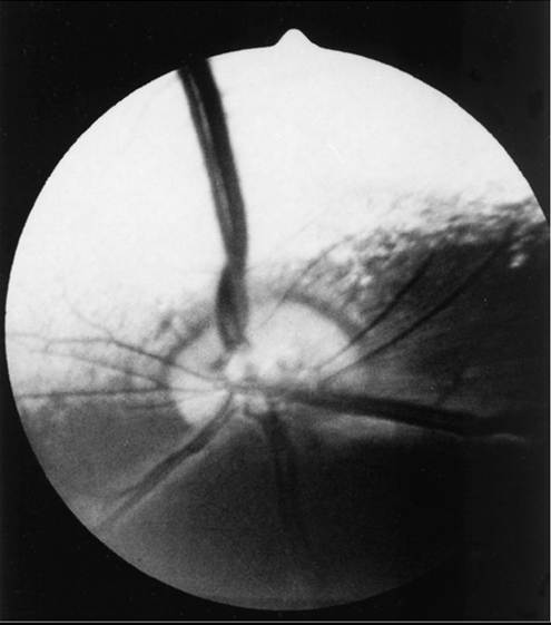
FIG. 39.1 Normal ruminant fundus characterized by a large, kidneyshaped, myelinated optic disk. Note that the large retinal arteriole and venule intertwine as they course dorsally.
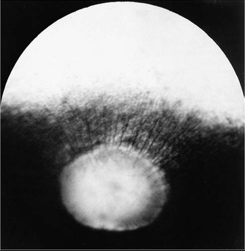
FIG. 39.2 Normal equine fundus characterized by a large, horizontally elliptical optic disk with numerous small retinal vessels entering (arterioles) and exiting (venules) the margin of the optic disk. Note that the optic disk lies in a nontapetal portion of the fundus.
Coloration of the tapeta of large animals also varies considerably and may range from gold to bluish green. In animals with varied coat color, coat color dilution, and/or heterochromia irides (variable iridal color), areas of the fundi may characteristically be devoid of pigmentation and may lack a tapetum. These
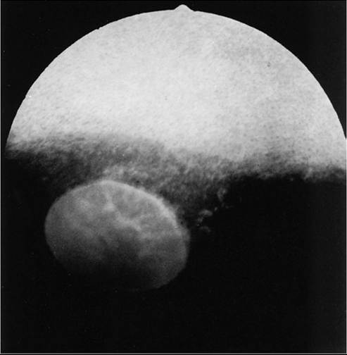
FIG. 39.3 Optic nerve atrophy (equine). Margin of disc is quite distinct because of myelin loss, which is characteristic of optic nerve atrophy. Note absence of retinal blood vessels.
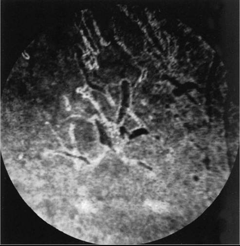
FIG. 39.4 Pigmentary changes after traumatic chorioretinopathy. Irregular linear areas of hypopigmentation and hyperpigmentation are present in the tapetal fundus of a horse after ocular trauma. Pigmentary changes reflect retinal pigment epithelial disturbance from previous hemorrhage and edema. (Courtesy Dr. K.N. Gelatt.)
areas may appear orange or red because of direct visualization of the choroidal vasculature.
Abnormalities of the optic disk include hypoplasia (micropapilla), elevation (papilledema), depression (cupping), degeneration (atrophy [Fig. 39.3]), and vascular changes (e.g., congestion, attenuation, hemorrhage). The tapetal fundus is evaluated for clarity, coloration, pigmentation (Fig. 39.4), and integrity
of the retinal vessels (Fig. 39.5). The nontapetal fundus is evaluated for uniformity of pigmentation. Both tapetal and nontapetal areas are assessed for retinal elevations or separations (Fig. 39.6), hemorrhages, degenerations, disorganization (dysplasia), or scleral defects (colobomas).7
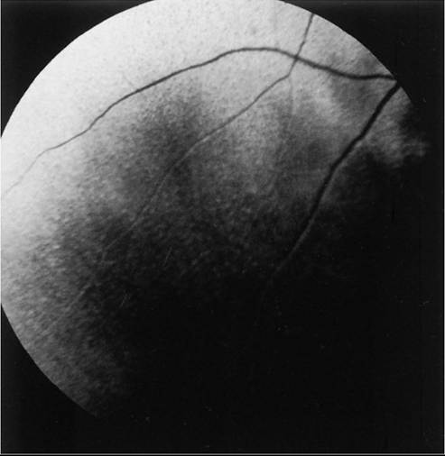
FIG. 39.5 Retinal degeneration (bovine). Peripheral retinal vessels are greatly attenuated near the tapetal-nontapetal junction. Attenuation is accompanied by hyperreflectivity of the tapetal region. These changes are consistent with generalized retinal atrophy.
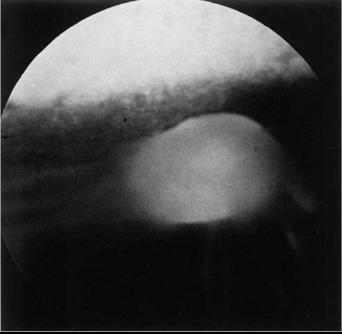
FIG. 39.6 Retinal separation (equine). Gray linear areas radiate from the optic disk into the nontapetal region of fundus. Note that the margin of the disc is quite indistinct in the affected area. Absence of retinal vessels indicates concurrent retinal degeneration.
Ancillary Diagnostic Procedures
Several additional procedures may supplement the complete ophthalmic examination. Although some ancillary procedures require specialized equipment and expertise, many may be performed in general practice.
Fluorescein and rose bengal are ocular surface stains most often used as aids in diagnosing conjunctival and corneal diseases. The tip of a sterile dye strip is moistened with saline or eye-irrigating solution, and a drop of the stain is instilled onto the eye. Fluorescein is a water-soluble dye used to detect exposed corneal stroma resulting from an epithelial defect (erosion), stromal ulceration, or descemetocele and is viewed with cobalt blue light. The pattern of fluorescein staining for a descemetocele is characterized by a donut-shaped area of positive fluorescence, with the perimeter retaining stain and the center or deepest area (Descemet's membrane) not retaining stain. Fluorescein may also be used to evaluate the patency of nasolacrimal ducts because an open duct allows the transmission of stain, which may be observed exiting the duct system at the nasal orifice (Jones test). Rose bengal is retained by devitalized surface cells and is therefore useful in detection of subtle abnormalities like hyperplastic or desquamating cells associated with ocular surface drying, herpetic infection, keratomycosis, or squamous cell carcinoma. Stain uptake appears pink when illuminated with white light. These stains can be performed in any order.
Although tear deficiencies are uncommon in large animals, Schirmer tear test strips may be beneficial to quantify aqueous tear production in selected cases. A sterile filter paper strip (40 ? 5 mm with a notched end) is inserted into the lower conjunctival fornix. In large animals it is sufficient to measure the amount of wetting in 30 seconds (≥20 mm is normal). Tear testing is indicated in patients with a dry, lackluster corneal surface; persistent ocular discharge; or keratitis.
Cytologic evaluation of ocular surface scrapings or intraocular aspirates may differentiate between inflammatory and neoplastic diseases or in some cases may provide a definitive diagnosis. Orbital aspirates may be diagnostic in cases of exophthalmos caused by neoplasia (e.g., lymphosarcoma). Immunofluorescent or polymerase chain reaction (PCR) testing of cytologic specimens may confirm viral (e.g., infectious bovine rhinotracheitis) or Chlamydia infections.3 Bacterial cultures taken from the ocular surface or from ocular aspirates, with subsequent antimicrobial susceptibility testing, may be necessary for definitive diagnosis and appropriate treatment of ocular infections. Bacterial and fungal cultures, as well as corneal cytology, are especially important tests when equine ulcerative keratitis is diagnosed. The diagnostic laboratory performing ocular cultures may offer suggestions on culture procedures, including preferred transport media and handling of samples. It is especially important to consult with the laboratory in advance when anticipating culturing for fungi, Mycoplasma, Chlamydia, or viral agents.
Tonometry, a means of measuring the intraocular pressure (IOP), is useful in diagnosing glaucoma (elevated IOP) and uveitis (low IOP) and in assessing response to therapy for these conditions. Tonometry is indicated in cases with corneal edema, redness, lens luxation, abnormal PLR, and cases with vision loss or a history of glaucoma. Digital tonometry (gently indenting the globe through the upper eyelid) is not considered an accurate measure of IOP; Schiotz tonometry is not applicable to large domestic species. Applanation tonometry using the TonoPen® provides accurate and reproducible IOP readings in large animals and is routinely performed. In addition, the rebound tonometer, TonoVet®, has been used successfully in horses.8 The clinician should be aware that eyelid blocks, sedation, and head position can all affect IOP readings.9
Biomicroscopy, using a portable handheld slit-lamp, is useful for identifying the location and nature of anterior ocular opacities. Focal irritants (e.g., ectopic cilia, small foreign bodies) and small lesions may only be visible with the magnification provided using biomicroscopy. Alternatively, use of a focal light source with magnification in the form of an otoscope or optivisor may be used for close examination.
Fundic examination may be performed relatively quickly and easily using the technique of indirect ophthalmoscopy. Monocular indirect ophthalmoscopy is performed using a handheld light source and a separate 20- or 14-diopter focusing lens for equine and bovine patients or a 28-diopter lens for smaller ruminants.
Other ancillary diagnostic procedures include electroreti- nography (to evaluate retinal function), visual evoked potentials (to evaluate visual pathways), and imaging procedures (radiography, ultrasonography, computed tomography [CT]). Although plain skull radiographs and some contrast studies (e.g., dacryocystorhinography) may be performed in general practice, the remaining procedures require techniques and equipment usually available only at referral centers.