Ovine Hereditary Chondrodysplasia (Spider Lamb Syndrome)
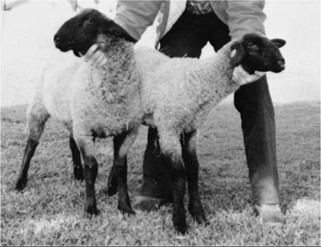
FIG. 38.10 Twin Suffolk lambs (12 weeks old), with normal lamb (78 lb) at left and lamb with spider lamb syndrome (37 lb) at right.
Note extreme height, narrow chest, scoliosis, kyphosis, and facial deformity of the affected lamb.donor females to normal recipient ewes resulted in all offspring with spider syndrome.17
■ Etiology Genetic linkage between SLS and several microsatellite markers have been detected. Mapping the SLS locus was suggested to be the telomeric end of ovine chromosome 6.7 The causative mutation was identified after positional cloning of the ovine fibroblast growth factor receptor 3 (FGFR3).7,18 A transversion at nucleotide position 69 of ovine FGFR3 exon 17 causes a nonconservative amino substitution of nonpolar valine (V) to charge glutamate (E) at residue 700 in the second tyrosine kinase domain of the receptor. The mutation leads to loss of receptor function in homozygotes. FGFR3 acts as a negative bone growth regulator by restricting chondrocyte proliferation and endochondral bone elongation.18,19 Heterozygous (Ss) animals are phenotypically indistinguishable from unaffected, normal (SS) animals. The genotypes of presumed heterozygous carriers of SLS are detectable only after testmating of suspect animals to proven heterozygous carriers with subsequent evaluation of all resulting progeny or by using blood or hair follicles to detect the mutation in FGFR3.
■ Clinical Signs Clinical presentation of the syndrome is also highly variable, with some lambs severely affected at birth and others developing the condition in the first 6 weeks after lambing.1 External symptoms characteristic of the disease include extreme height, fineness of bones, and lack of muscling and angular deformities of the limbs (Fig.
38.10) along with kyphoscoliosis. Affected lambs tend to be thin and arthritic, with poor muscling and insufficient weight gain. The affected individuals rarely survive and are an economic liability in market lamb production.The abnormalities are most frequent in the forelimb (81% to 87%), hindlimb (63% to 69%), spine (52% to 62%), and skull (19% to 35%).20 No differences between Suffolk and Hampshire lambs were detected in the type, extent, or incidence of lesions.20 The forelimbs and hindlimbs were characterized by mild to severe carpus (Fig. 38.11) and tarsus valgus, respectively. The most common spine abnormality was kyphoscoliosis. SLS was observed in aborted fetuses, stillborns, viable neonates, and young lambs less than 6 weeks of age.20
Detection of this abnormality during pregnancy by either radiography or transabdominal ultrasonography even at the
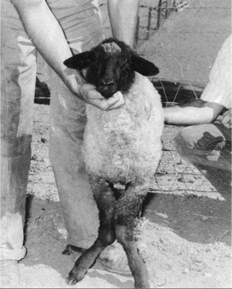
FIG. 38.11 Rotation and deviation of front legs (carpus valgus) characteristic of spider lamb syndrome in a 16-week-old Suffolk lamb.
second or third tiers was not possible.17 Diagnosis was possible only by direct examination of the fetus.17
■ Laboratory Analyses No differences in hematologic and biochemical analyses between chondrodysplastic lambs and normal flockmates were detected. Serum levels of vitamins A and E and liver levels for selenium and vitamin E were within normal parameters.4 Trace elements in serum for iron, magnesium, zinc, and copper were no different compared with normal lambs.4 Serum alkaline phosphatase activity has been reported either high or low in affected lambs compared with healthy lambs.1 The only difference was a trend in a high level of skeletal muscle enzymes in serum.4
Some data suggest that the regulation of insulin-like growth factor 1 (IGF-1) and IGF-binding proteins may be involved in this disorder.
SLS lambs between 50 and 80 days of age showed a significant decrease in serum level of IGF-1 levels and an increase in IGF-binding proteins compared with a control group of lambs of the same age.21No differences in the number of morphologic features of the chromosome were detected, and there was no evidence of deletions, duplications, translocations, or inversions when lymphocytes from clinically normal lambs and lambs affected with ovine hereditary chondrodysplasia were cultured, G-banded, and karyotyped.22
■ Necropsy The primary lesions affect the musculokeletal system. The gross lesions observed were similar to the changes noted radiographically. Lesions were observed in the skull, sternebrae, atlas, elbows, other vertebrae, and physes of the long bones, as well as other areas. The lesion noted in all locations was an excess of cartilage.4 Marked muscle atrophy was also present. The principal lesions in bones are an increase in the width of the zone of proliferation and hypertrophy and an unevenness of the growth cartilage. A cartilage column
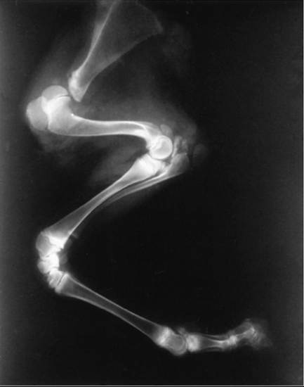
FIG. 38.12 Lateral radiograph of the front leg of a lamb with spider lamb syndrome. Note thick growth plates and multiple ossification centers near the olecranon and distal humerus.
disorder, with failure to form or maintain orderly columns of chondrocytes, is characteristic.1 The chondrocytes and cartilage matrix appear histochemically normal and have proper ultrastructure.23
■ Radiography Chondrodysplastic lambs have characteristic radiographic patterns in the elbow, sternum, shoulder, and spine, with multiple irregular islands of ossification.1 Radiography may be the technique of choice for identifying affected lambs prior to detecting clinical signs or when in doubt.24 A flexed lateral radiograph of the elbow of a suspect lamb appears to be diagnostically reliable when there is a suspicion of the syndrome (Fig.
38.12).1■ Control The mode of inheritance for SLS is autosomal recessive, making the identification and culling of carrier (heterozygous) animals difficult due to their normal phenotype. Eliminating these heterozygous animals from affected sheep flock is possible by identifying individuals that are carriers of the mutation. This can be done by collecting peripheral blood or hair follicles and performing polymerase chain reaction restriction fragment length polymorphism (PCR-RFLP) analysis. The best approach is to perform this test in males, but depending on the objectives of the program, the test also can be run in females. Genetic testing using blood and then PCR-RFLP permits a quick and accurate detection of heterozygotes. The presence of a lamb spider is an indication that both parents are heterozygous for the disorder, so no test is required. Since the introduction of this test in 2000, a significant reduction in the incidence of heterozygous animals in New Zealand and the United States has been reported.25,26
Another possibility is to run breeding tests, which is time consuming and costly and requires adequate numbers of breeding animals to be accurate. In addition, correct identification of the animals is paramount. A ram can be progeny tested by breeding females that produced spider lambs or his own daughters. The number required to be highly accurate is 16 normal offspring from mating with a known carrier's ewes or 32 normal lambs from breeding with his own daughters. However, with these numbers the probability that the ram is a carrier is 1% or less of the cases.27
■ Differential Diagnosis A complete clinical history and physical examination as well as radiologic and pathologic examinations are essential to the diagnosis of this disease. Some of the lesions seen in spider syndrome require differentiation from abnormalities produced by viruses, chemicals, toxic plants, and nutritional factors. Infection with bluetongue and Akabane virus may result in arthrogryposis and hydranencephaly. Border disease can cause abnormalities of the nervous system, skin, fleece, and skeleton. Cache Valley virus and bluetongue can also produce these kinds of anomalies.28 Benzimidazoles, antiparasitic drugs, have been implicated as a cause of skeletal deformities when used in pregnant ewes during specific periods.29 Toxic plants such as locoweeds are known to cause limb deformities in utero.30 Deficiency of vitamin D or phosphorus can cause skeletal deformities or rickets.31 Deficiency of either zinc or manganese during gestation could lead to chondrodysplasia.32