Parasitic Skin Diseases
Stephen D. White
Pediculosis
Lice are obligatory ectoparasites that are generally host specific. Adults and nymphs are seldom able to live for more than a few days away from their host.
Large animals suffer from infestation with several species of lice that belong to the orders Mallophaga, the biting lice, and Anoplura, the sucking lice (Table 40.2).1Clinical infestations are most apparent during the winter months and reflect efficient louse reproduction during the late fall and the fact that summertime temperatures on body
■ TABLE 40.2
Lice Associated With Large Domestic Animals
| Host | MaUophaga (Biting) | Anoplura (Sucking) |
| Horse | Bovicola (Damalinia) equi Werneckiella equi equi | Haematopinus asini |
| Cattle | Bovicola (Damalinia) bovis | Haematopinus eurysternus Haematopinus quadripertusus Haematopinus tuberculatus Linognathus vituli Solenopotes capillatus |
| Goats | Bovicola (Damalinia) caprae Bovicola (Damalinia) limbatus Bovicola (Damalinia) crassipes | Linognathus stenopsis Linognathus africanus Linognathus vituli |
| Sheep | Bovicola (Damalinia) ovis Bovicola (Damalinia) caprae | Linognathus pedalis Linognathus ovillus Linognathus africanus |
areas exposed to sunlight are too high for lice. Apparent carrier animals within a herd maintain populations during the “off” season and serve as a source for reinfestation of the herd during the fall.1 The hallmark of infestation is pruritus, and many clinical changes result from self-trauma.
The neck and tail are typically affected first,2 but infestation and clinical signs may become generalized. The coat becomes dry and scaly. Patchy alopecia and crusted ulcerations result from excoriation. A heavily infested animal may become anemic. Significant hide damage occurs as a result of excoriation, and hairballs may accumulate in the GI tract from self-grooming. Reduced productivity and weight loss result from decreased feed intake associated with restlessness. Diagnosis is usually by examination; a hand lens is useful (e.g., otoscope without cone). Interestingly, when a horse is exercised and becomes warm and sweaty, the lice will climb out toward the tip of the hair and are easier to find.3Several treatment approaches exist. Ivermectin at 200 μg/ kg every 14 days SC for 2 applications is effective for sucking lice but not biting lice. Application of an appropriate topical insecticide to infested animals and to all contact animals at 2-week intervals for 2 or 3 treatments is usually curative for both types of lice. Repetition of treatment is necessary to break the louse life cycle because eggs are not killed by insecticides and will hatch despite therapy. Effective topical agents include pyrethroids, permethrins, selenium sulfide, imidacloprid, phoxim, and fipronil.3,4 Development of resistance is also possible.5 Appropriate attention should be paid to withdrawal times in food animals. Cleaning of fomites like blankets, brushes, and rope halters is probably indicated, although lice do not live long off the host.
Trombiculidiasis
Trombiculidiasis is caused by the larval stages of mites commonly called “harvest mites” or “chiggers.” The most common species affecting large animals are Eutrombicula alfreddugesi (North America) and Neotrombicula autumnalis (Europe). The adults and nymphs are free living. The larvae feed on mammalian hosts, secreting substances in their saliva that hydrolyze the epidermis and allow extraction of tissue fluids.
The individual larval stage is short, but the infestation may be ongoing with the acquisition of new larvae from the environment. In the northern hemisphere, E. alfreddugesi is active from late spring through early fall, whereas N. autumnalis is more common in late summer to mid-fall. The mites may be found in wooded areas, grass, and hay.3,6The clinical signs of trombiculidiasis are crusts and papules, especially on the face, neck, and extremities. Pruritus is variable.6 Rarely, this parasite has been implicated in headshaking in horses.7 Diagnosis may be made early in the course of infestation by careful inspection (e.g., hand lens) and finding the minute red-orange larvae in the center of a papule. The six-legged larvae have round bodies and may be identified specifically on skin scrapings or acetate tape preparations.3 Although the larvae remain on the animal host for only several days, clinical signs may persist longer, so trombiculidiasis should be suspected even in the absence of mites in an animal with appropriate signs in the summer and fall.
The disorder is in theory self-limiting because of the short time of attachment of the parasite. However, if the pruritus is severe, corticosteroid administration is indicated.3 Various products have been used on the horse to eliminate the larvae; 5% lime sulfur, fipronil, and permethrin are generally the safer parasiticides to use.
Mange
Mange is a general term for infestation by any mite species.
Psoroptic Mange
Psoroptic mange has been recognized in horses, cattle, sheep, and goats (and rabbits). The host specificity of Psoroptes equi, Psoroptes ovis, Psoroptes natalensis, and Psoroptes cuniculi is controversial; two reports state that the mites are all genetically homogeneous with little or no host specificity,3,8 whereas another report documents an inability to transfer P ovis to goats.9 The mites do not affect people.
Psoroptic mange is common in cattle but has been eradicated from horses and sheep in the United States; it is still a concern for sheep production in the United Kingdom and elsewhere in the world. The mites have a 2-week life cycle on their host but can live away from the host for up to 3 weeks. Psoroptes mites live on the surface of the epidermis and do not burrow. Symptomatic infestation tends to be more prevalent during the cooler months.The hallmark of infestation in all species is pruritus. In cattle, crusted papular lesions are typically first apparent on the withers but then generalize, resulting in weight loss, secondary infections, and decreased production.10 Horses develop papules, crusts, and alopecia most often at the base of the mane, tail, ears, and intermandibular area, with subsequent spreading to the trunk.3 An otitis externa may be present, seemingly with P. cuniculi.3 In sheep, typical lesions include papules and crusts in wooled areas, and secondary infection with S. aureus may occur.11 The intense pruritus can be debilitating. Certain sheep (Merino) and cattle (Belgian Blue) breeds are more susceptible to infestation than others.9,12 The immune response in sheep has been studied extensively, with early innate and longer-term adaptive cutaneous immunoinflammatory responses as well as mite antigen-directed IgE being reported.10 Goats usually have lesions restricted to the ears, although infestation and symptoms may spread to the neck and body.13,14
Diagnosis is based on demonstrating mites in skin scrapings and ear swabs from affected animals. Psoroptic mites are recognized by their round bodies and long, segmented pedicles.13 Recently, serologic assays have been useful in diagnosis, especially in identifying subclinically infested animals.15
A number of topical insecticides have been recommended for treatment and should be applied to all affected and contact animals according to the manufacturer’s guidelines.
These include deltamethrin, coumaphos, diazinon, malathion, toxaphene, and lime sulfur.6,13,16 The contaminated environment should also be treated. Ivermectin, doramectin, and moxidectin have been used for Psoroptes spp. in large animals at 0.2 mg/kg,6,17-19 usually for two to three times, given subcutaneously but treatment does not always eliminate live mites from all animals. There may be lesser efficacy when using generic formulations of ivermectin.20 It has been recommended that P. ovis-infested cattle be isolated for a minimum of 14 days after treatment to prevent transmission to susceptible contact cattle.18 Psoroptes-infested ears in horses should not be treated topically because of the resulting discomfort; reliance is on systemic treatment. Psoroptic mange is a reportable disease in the United States.Chorioptic Mange
Chorioptic mange, also known as leg mange, is common in cattle and sheep, uncommon in goats, and seen with variable frequency in horses, depending on geographic area. Draft horses may be more susceptible than other breeds. Chorioptes spp. are relatively host specific and include Chorioptes bovis, Chorioptes texanus, Chorioptes ovis, Chorioptes equi, and Chorioptes caprae.6,vi'2i The mites do not penetrate the epidermis6 and do not affect humans.18 They have a life cycle that spans 2 to 3 weeks and can live off the host for only a few days.16
This disease is variably pruritic, more so in ruminants than in horses, and less so than infestations with Psoroptes or Sarcoptes mites. Lesions consisting of papules, erythema, scaling, crusting, ulceration, and alopecia result from self-trauma. In cattle the lower aspects of the hindlimbs, the perineum (particularly the perianal fossa), tail, and scrotum are usually affected. Sheep typically demonstrate involvement of the lower limbs and scrotum. Goats have involvement of the lower limbs. Horses typically have lesions on the pasterns, although the ventral abdomen may become involved in severe cases.
In all species, infestation can become generalized.3,6,18Mites are often numerous and readily demonstrated with skin scrapings. Cattle may be asymptomatic.18 Chorioptic mange will respond to treatments described for psoroptic mange, although ivermectin and related compounds are more effective in ruminants than horses; in horses, lime sulfur or fipronil applied once weekly for at least 1 month is recommended.3,22-27 One study in horses showed efficacy using a single dose of moxidectin 2% oral gel (Equest [Fort Dodge Animal Health, Fort Dodge, Iowa]) at the manufacturer’s recommended therapeutic dose of 0.4 mg/kg body weight.26 Other studies have not found it to be effective in eliminating the parasite in a multihorse situation.28 Chorioptic mange is a reportable disease in the United States.
Sarcoptic Mange
Sarcoptic mange is an uncommon contagious disease of horses, cattle, sheep, and goats. The etiologic agent is Sarcoptes scabiei; several subspecies are relatively host specific but can be transmitted to humans. The mite burrows into the epidermis, where the egg is deposited, and its life cycle is complete in 10 to 17 days. Transmission is usually by direct contact, but the mite has a variable survival time off the host, so environmental and fomite transmission are possible.3,6
Clinical signs are all referable to the severe pruritus caused by the mite. Lesions include papules, scaling, crusting, ulceration, and alopecia. The head (especially the ears) and neck are usually the initial areas of involvement, although lesions become generalized.3,6,16 Horses in areas frequented by infected wild foxes may have lesions affecting the legs and ventral abdomen.3 Dairy cattle may have an associated udder cleft dermatitis.29 Sheep tend to have initial involvement of the nonwool areas. The ears should always be scraped for mites; the mite is identified by its rounded body, terminal anus, short legs, and long unsegmented pedicles.3,6,16
Because mites may be present only in small numbers, negative skin scrapings do not rule out the disease. Diagnosis should be based on clinical suspicion and response to therapy.3,6,16,30
Topical acaricides have been recommended for treatment and are applied to all affected and contact animals at 10- to 14-day intervals for 4 to 6 treatments. These include lindane (not recommended owing to toxicity in humans), coumaphos, diazinon, malathion, toxaphene, and lime sulfur.3,6,16 Ivermectin 0.2 mg/kg SC at 2-week intervals for 2 to 4 injections (ruminants) or orally (horses) has been shown to be effective, as has injectable moxidectin and topical doramectin. The contaminated environment should also be treated. It is recommended that the state regulatory agency for livestock disease control be consulted for methods of treatment and products to use because sarcoptic mange is a reportable disease.
Demodectic Mange
Demodectic mange is a rare disorder, although it has been recognized in all large domestic species. The mites live in hair follicles and sebaceous and sweat glands and are host specific. They are not contagious between members of the same species, but Demodex spp. are presumably transmitted from mother to offspring during the first few days of life by direct contact with the dam. Little else is known about the life cycle of the mite.3,6 Two species of Demodex are recognized in the horse.3,30 Demodex caballi is a normal inhabitant of the pilosebaceous apparatus of the eyelids and muzzle and may be found in skin scrapings of these areas on horses in the absence of skin lesions. Demodex equi inhabits the pilosebaceous apparatus of the remainder of the body and on horses is the species that has been associated with disease. The species found on cattle, sheep, and goats are Demodex bovis and Demodex ghanesis, Demodex ovis, and Demodex caprae, respectively.3,6,16,18,31
Clinical signs are variable depending on the species affected. The disease is quite rare in horses, which may develop alopecia of the head and trunk; we have seen one case of concurrent demodicosis and chorioptic mange on the legs of a draft horse. Underlying pituitary dysfunction of the pars intermedia has been seen in some horses.3 Pruritus and secondary pyoderma are variable. Goats and cattle usually develop nodular lesions that involve the face, shoulder, and neck.32,33 In goats the nodular contents are white and caseous; microscopic examination for mites differentiates the lesions from C. pseudotuberculosis.33 Sheep tend to develop periocular nodular lesions.
Diagnosis is based on recognizing mites with microscopic examination of skin scrapings or exudates obtained from nodular lesions. The mites are elongated and have short, stubby legs.3,6,16
No treatment has proved to be consistently effective in large animals, although relatively few case reports exist in the literature. Amitraz has not been successful in the treatment of caprine demodicosis.33 Horses sprayed with 0.025% amitraz developed somnolence, depression, ataxia, muscular weakness, and progressive large intestinal impaction, suggesting that amitraz is contraindicated in equids.34 Daily ivermectin has been suggested as a successful treatment for equine demodicosis, but dosages have not been codified. Oral ivermectin (0.67 mg/ kg once weekly for 12 weeks) and pour-on eprinomectin (0.5 mg/kg) each successfully treated one goat with generalized demodicosis.35 A combination of ivermectin injections of 200 μg/ kg body weight twice weekly and amitraz dip once weekly has been reported as successful in cattle.36
Culicoides Hypersensitivity
The females of various Culicoides fly species (“no-see-ums,” “punkies”) may feed on either the dorsal or the ventral surface of the horse, affecting the mane, saddle, and rump or causing a ventral midline dermatitis in a diffuse pattern. Alopecia, papules, crusts, and erythema may all be present (Fig. 40.12). The insects induce a hypersensitivity response through salivary antigens; this condition is termed Culicoides hypersensitivity (Queensland itch, sweet itch) and is recognized in various areas of the world. Evidence indicates a hereditary predisposition to develop the hypersensitivity. Histopathology of lesions frequently reflects a hypersensitivity response.37 Although many species of Culicoides exist, the three most commonly suspected of causing the allergic response are Culicoides variipennis, Culicoides obsoletus, and Culicoides nubeculosus.
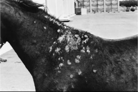
FIG. 40.12 Severe Culicoides hypersensitivity in a horse, showing typical distribution of withers, shoulders, and head (not visible).
Numerous studies have attempted to define the nature of the hypersensitivity response. One study found serum antibodies to Culicoides salivary glands in both healthy horses exposed to Culicoides bites and horses with insect dermal hypersensitivity.38 In contrast, no antibodies were detected in serum from native Icelandic horses that had not been exposed to Culicoides. Anti-salivary gland IgG antibodies were detected in both groups of horses, whereas IgE antibodies were detected only in clinically affected horses.38 Another study compared two groups of affected Icelandic horses: those born in Iceland (no previous exposure to Culicoides) and transported to Europe, where they were then exposed to the flies, and those born in mainland Europe. The former had IgE and IgG antibodies to a greater number of the insects' salivary proteins.39 Further investigation of immunoglobulins in relation to Culicoides found significantly more IgE-bearing cells in the dermis and epidermis of acute and chronic lesions than in skin biopsies from healthy horses.40 Further evidence for a hypersensitivity response was demonstrated in a study in which intradermal injection of a Culicoides antigen extract induced T-lymphocyte and eosinophil accumulation in the skin of affected horses.41 Other studies have supported the immunologic nature of this disease.42-46 Two recent reports have shown some linkage between insect bite hypersensitivity (IBS) and airway disease.47,48
The hallmark of disease is pruritus. Although no gender predisposition has been noted, Icelandic horses may have a higher incidence of allergic reactions to the Culicoides insects than other breeds. The disease is uncommon in horses younger than 1 year of age, with an onset usually between 2 and 4 years. Culicoides hypersensitivity may be seasonal, at least during the first few years of life, in temperate climates. Diagnosis is primarily by clinical signs. Culicoides antigens (commercially available in the United States by Greer, Lenoir, N.C.) seem useful in both diagnosis and treatment,49-51 but this must be documented in large case studies.
Therapy is aimed at insect control, especially the following:
1. Stabling horses at sunrise and sunset, peak Culicoides feeding hours.
2. Ultrafine setting or screens placed in windows (60 squares to the square inch).
3. Fly control, especially keeping horses away from standing water and using permethrin repellents, usually 2% permethrin sprays. Frequently, sprays must be applied more often than the label recommends (i.e., daily, at least at first). We have had success using a nonpesticide herbal spray or roll-on (MedZone [VetShield & Sheen, Sun City, Ariz.]).
4. Overhead or stall fans (drafts interfere with insects' flight).
5. “Dresses” that physically obstruct the insects from reaching the skin.
6. Hyposensitization is controversial; success may vary with the presence of an adjuvant or the actual antigen used.
7. Oral prednisolone to manage the pruritus.
Ventral Midline Dermatitis of Horses
This is a reaction pattern in horses to ectoparasites, and not necessarily specific to any one arthropod. Culicoides hypersensitivity, Onchocerca cervicalis infestation, and black flies (Simu- liidae) may cause generalized ventral midline dermatitis, whereas horn flies (Haematobia irritans) cause a focal dermatitis. Severe chorioptic mange may also involve the ventral midline. In all cases the lesions have variable alopecia, pruritus, and crusts. Treatment is based on eliminating or repelling the causative organism.
Other Flying Insects
Stable flies (Stomoxys calcitrans), black flies (Simuliidae), horn flies (H. irritans), horse flies (Tabanus and Hybomitra spp.), deer flies (Chrysops spp.), houseflies (Musca domestica), face flies (Musca autumnalis), and mosquitoes are commonly associated with irritant and allergic skin disease, as well as being vectors of many parasites (Color Plate 40.8). They must be controlled by limiting breeding areas and by repellents; 2% permethrins are often used. It is important to remember the relationship of these various flies to the environments they favor or require for breeding and when they feed, as follows:
| Horse flies, deer flies | Vegetation and water | Daytime feeders |
| Horn flies | Cattle | Daytime feeders |
| Stable flies | Manure and decaying bedding | Daytime feeders |
| Black flies | Running water | Morning and evening |
| Culicoides | Standing water, manure and decaying vegetation | Twilight to dawn |
| Mosquitoes | Water | Dusk to 2 hours after sunset |
Screwworm Infestation
Screwworm flies cause primary myiasis; the species of importance in the Americas is Cochliomyia hominivorax (Callitroga americana). The fly was formerly found throughout the American tropics and subtropics from the southern United States to northern Chile.52,53 It is no longer found in the United States or Mexico because of state, federal, and international eradication efforts.54
The adult fly is about three times the size of a housefly, with a metallic bluish or blue-green color.52,54 Females are attracted to fresh wounds (castration, dehorning, branding, and shearing sites), abraded body orifices, areas soiled by discharges or excretions, and navels of newborns. They lay batches of 150 to 500 white eggs on the margin of damaged tissue in rows that overlap like shingles. Eggs hatch within 24 hours, and the larvae begin to feed in a head-downward position. Larvae are obligate parasites that require living tissue as feedstuff. They cannot develop in carrion. Larval development continues for 4 to 10 days, and at the time of their maturation they may have created a cavity 10 to 12 cm (4 to 5 inches) in diameter.53 Mature larvae are about 2 cm in length, pink, pointed anteriorly and blunt posteriorly. The larvae then drop to the soil and pupate. The life cycle averages about 21 days and is favored by hot, humid weather.
Screwworms do not have a dormant stage in their life cycle and cannot overwinter in cold climates.52 Thus the susceptible stage of development is the pupae, which will not survive soil temperatures below 15° C (59° F). The feeding larvae burrow deeply, creating a cavernous lesion characterized by liquefactive necrosis, profuse brownish exudate, and an objectionable odor. The syndrome is self-perpetuating in that wounds infested by screwworm larvae become increasingly attractive to gravid females. The end result is often death of the host from secondary bacterial infection, toxemia, and fluid loss.53
Treatment of wounds requires clipping and cleansing, as well as destroying all larvae. Dressings containing larvicides and antiseptics should be applied. Preparations containing lindane or organophosphates have been used in an ointment or gel base. The treatment is repeated twice weekly. When large numbers of animals are affected, a 0.25% solution of coumaphos, chlorfenvinphos, or fenchlorphos may be applied to herds with a power sprayer. Calves may become ill from the spray; thus applications should be restricted to the ventral abdomen.53 Another study showed that the curative efficacy of fipronil 1% against C. hominivorax larvae infestation in castration wounds was 100%.55 Even more impressive, doramectin as a prophylactic treatment was 100% effective in prevention of C. hominivorax infestations.56
Control of screwworm flies in the United States has been achieved largely through the release of sterile males, because of the single mating tendency of the female fly. The eradication program has proceeded through Mexico south to the 21st parallel.52 Screwworm myiasis is a reportable disease in the United States, and suspect larvae should be preserved in 70% alcohol for positive identification.57
Blow Fly Strike (Fleeceworms, Woolmaggots, Secondary Screwworms)
Blow flies are found throughout the western hemisphere. The species of importance include Cochliomyia macellaria (secondary screwworm), Phaenicia sericata (green bottle fly), and Phormia regina (black blow fly). The flies tend to be most common in the warmer regions, with the exception of P regina, which is widespread throughout the cooler parts of North America and Europe.52 Blow flies cause serious loss of sheep and wool in many countries.54 Female flies are attracted to decaying animal matter, such as wounds infested by primary screwworms, infected sores, carcasses, and fleeces that are dampened with feces, urine, or bloody fluids.52 They lay eggs in batches of up to 300, which hatch within hours. The larvae feed on necrotic tissue but may invade healthy tissue. Larvae develop for 3 to 5 days and, when fully mature, are white and 6 to 12 mm in length. The larvae then drop to the ground for pupation. As soil temperatures fall, larvae fail to pupate and may overwinter until the following spring. The entire life cycle is generally complete in 2 to 4 weeks under ideal conditions of warm temperatures and high humidity.53
Unlike primary screwworms that feed in pocket-like aggregations, secondary screwworms tend to be dispersed throughout the infested tissue. In wool infestation the larvae may remain on the skin surface, feeding on the decomposing wool, or may penetrate the skin through small abrasions. The most common site of involvement is the breech. Infested tissue attracts more ovipositing females, and thus the syndrome is perpetuated.52 Affected sheep are restless and do not feed. They move with their heads close to the ground, bite or kick at their wounds, and continually wriggle their tails. Affected wool is moist and brown with an obvious odor. Animals may become systemically ill and die.57
Treatment of individual wounds is as described for screwworms. Control involves management practices that decrease the incidence of wounds or skin irritations. Sheep are often clipped below the tail and between the hindlimbs (“crutched”), where wool is likely to become saturated with urine or feces. Castration, shearing, docking, and lambing are avoided during the summer season.52
Cutaneous Onchocerciasis
■ Definition and Etiology Cutaneous onchocerciasis in horses is a common filarial dermatitis with a worldwide distribution. Its incidence in the United States has decreased dramatically since the introduction of ivermectin for routine deworming. In a report from South America, O. cervicalis microfilariae were detected in midventral skin biopsy samples in 215 (17.9%) of 1200 horses examined, and the adult worms were recovered from 200 (16.6%) ligamentum nuchae from the same animals.58
The disease is caused by the microfilaria of O. cervicalis and is seen primarily in adult horses.59,60 Adults typically are found coiled in the funicular part of the ligamentum nuchae, where they produce calcified nodules. Viviparous females may live for up to 5 years and produce large numbers of microfilariae that migrate through connective tissues to the superficial layers of the dermis. Preferential areas of microfilarial localization include the ventral midline, lower eyelid, and lateral limbus
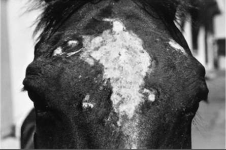
FIG. 40.13 Cutaneous onchocerciasis in a horse.
of the eye.59 The infection is transmitted by Culicoides, which act as an intermediate host. The larvae are ingested by the vector and undergo development into the third-stage (L3) larvae in approximately 2 weeks. The L3 larvae enter the animal host through lesions created by the feeding vector.61 Many horses are infected with the parasite without demonstrating clinical disease.59,61 A form of cutaneous onchocerciasis seen in donkeys in Africa and possibly in other nearby areas causes severe ulceration in the withers and neck region; it is the adult parasite (sometimes extractable by the handful) that causes this problem. This is possibly caused by the species Onchocerca raillieti.
■ Pathophysiology Pathogenesis is believed to involve a hypersensitivity reaction to antigens released by dying microfilariae.59,61 This theory has support because not all infected horses demonstrate disease, neither the presence nor the severity of the dermatitis is correlated with the number of organisms present, and treatment with filaricides often causes a temporary exacerbation of clinical signs.
■ Clinical Signs Clinical signs occur most often in older horses and can include both ocular and cutaneous lesions.59,61 Ocular lesions include uveitis, conjunctivitis, keratitis, and depigmentation of the lateral limbus.62 Cutaneous lesions include diffuse or patchy alopecia, erythema, and scaling. Focal cutaneous depigmentation is common. Lesions tend to occur in regions where microfilariae are typically present in highest concentrations, such as the ventral midline, face (Fig. 40.13), base of the mane, anteromedial proximal forelimbs, and anterior pectoral region. An inflammatory, alopecic, or hyperpigmented area in the center of the forehead is highly suggestive of the disorder.62 The dermatitis is generally reported as being nonseasonal and nonpruritic.61,63
■ Diagnosis and Histopathology Because many normal horses have microfilariae without disease, the finding of microfilariae does not prove that they are the cause of the cutaneous lesions. In addition, because microfilariae tend to “nest” in the dermis, there is a tremendous difference in the number of microfilariae recovered from adjacent skin samples.60,64 Thus although the absence of microfilariae makes cutaneous onchocerciasis unlikely, it does not definitively exclude it as a diagnosis. Differential diagnoses should include ventral midline dermatitis caused by the horn fly H. irritans, hypersensitivity reaction to Culicoides, dermatophytosis, and infestation with mange mites.
Ultimately diagnosis should be based on history, supportive clinical findings, exclusion of other differential diagnoses, and most importantly, response to treatment.
A positive Onchocerca microfilarial saline preparation demonstrates slender, delicate microfilariae that are approximately 8 ? 220 μm. Typical histopathologic changes include an eosinophilic and lymphocytic perivascular dermatitis, a finding that is nonspecific and common to many other equine parasitic dermatoses (see Chapter 11). Aggregations of microfilariae may be found in the superficial dermis or perifollicular region.59,61,
■ Therapy Ivermectin or moxidectin are the treatments of choice and are administered at 0.2 mg/kg PO.65,66 Most horses improve within 2 to 3 weeks. Minor adverse reactions, including fever and swelling of the periorbital, facial, and ventral midline regions, may occur in up to 25% of infected horses treated with ivermectin; moxidectin did not cause posttreatment dermal reactions in one study. Severe reactions may benefit from treatment with corticosteroids, but most reactions resolve within 24 to 72 hours. Because there is no effective adulticide, recurrence may be noted as soon as 2 months after therapy. Most animals remain free of clinical signs for 6 to 12 months.63,65 Re-treatment is recommended at 4-month intervals. Interestingly, ivermectin has been reported as having both greater and lesser GI absorption in donkeys than in horses. This awaits more clarifying studies.
In cattle, onchocerciasis has been caused by the microfilariae of Onchocerca gutturosa and Onchocerca Iienalis and, in Africa, Onchocerca ochengι.68,69 Flies of Simuliidae are the vectors. The disease causes nodules and crust on the skin, including the teats; adult worms surrounded by neutrophils were detected free in the teat canal. Ivermectin and moxidectin are effective treatments.
Stephanofilariasis
Hypoderma (Warbles)
■ Definition and Etiology Infestation with the larvae of Hypoderma spp. is a common and serious economic problem in cattle and is recognized sporadically in horses that are pastured near cattle. Occurrences have also been reported in sheep, goats, and humans. Warble flies are present in the northern hemisphere between 25 and 60 degrees latitude in more than 50 countries of North America, Europe, Africa, and Asia. Hypoderma spp. are not established south of the equator.60,74 The U.S. cattle industry loses millions of dollars annually because of cattle grubs.
Two species of Hypoderma parasitize cattle: Hypoderma bovis and Hypoderma lineatum. The latter prefers warmer climates and is the only Hypoderma species present in the southern United States. Each of the species occurs in the northern United States and Canada.60 H. lineatum and H. bovis have very similar life cycles, but the stages of H. lineatum tend to occur 3 to 8 weeks earlier than those of H. bovis. Timing of the life cycle also varies with local geographic and climatic factors.60,74
The adult flies are beelike in appearance, covered with dense chin bristles (hair), and are 12 to 188 mm in length, and they have nonfunctional mouthparts. They have a short life span, only 1 week. Adult flies are active in the spring to early summer, with H. lineatum appearing 3 to 4 weeks before H. bovis. Female H. lineatum flies deposit eggs on the hairs of the legs or lower body, whereas H. bovis tends to deposit eggs on the rump or upper parts of the hindlimbs. Total egg production by a single female has been estimated to range from 500 to 800.75 The eggs hatch in 3 to 7 days, after which the larvae penetrate the skin and begin their migration through the connective tissues. H. lineatum larvae migrate to the subcutaneous connective tissues of the esophageal wall, whereas H. bovis larvae migrate toward the spinal canal. The first-stage (L1) larvae remain in this location for 2 to 4 months during autumn and early winter. Between January and February the larvae begin a final migration through the connective tissues to the subdermal tissue of the back of the host, where they form a breathing hole through the skin. Cysts (warbles) develop around L1 larvae. Within the cyst the larvae undergo two molts over a 4- to 6-week period. The L3 larvae (grubs) emerge from the breathing pore, fall to the ground, and pupate. Adult flies emerge from the pupae in 1 to 3 months, and emergent adults are ready to mate almost immediately.57,75 The life cycle is complete in 1 year. Most larvae fail to reach normal size and complete their life cycle in the horse.74,75 A recent study suggests a humoral response from the host against the larvae while they are intact. Once the larvae are destroyed, cellular response occurs, isolating and destroying the remains of the larvae.
■ Clinical Signs
CATTLE. During the spring and summer when adult flies are active, “fly worry” may be a serious problem. Cattle exhibit a stampeding behavior called “gadding” when chased by ovipositing females, even though the flies do not bite or sting. This fear reaction results in self-injury and decreased feeding and milk production.75 Diagnosis of warble flies in goats by ELISA has been reported, but it is not known whether this is applicable to cattle.
Clinical lesions are not usually observed in association with migration of L1 larvae unless the larvae die along the migratory path.74 Lesions that have been associated with infestation of L1 larvae of H. bovis include fat necrosis and inflammation of the connective tissues surrounding the spinal canal. Secondary periostitis, osteomyelitis, and rarely paralysis or other nervous disorders may occur. Similarly, infestation with L1 larvae of H. lineatum in the submucosa of the esophagus may cause inflammation and edema in the surrounding tissues. Swallowing and eructation may be hindered, resulting in bloat and subsequent respiratory failure.74
Lesions most often observed with Hypoderma infestation are attributed to L3 larvae. Warbles occur along the back from the shoulders to the tailhead and from the dorsal midline to a point about one third the distance down the sides. The lesions may be firm to fluctuant, raised, and painful to the touch. They measure approximately 3 cm in diameter and contain a breathing pore that usually exudes a yellowish serum. Excisional biopsy of a nodular lesion reveals the larvae within a cystlike structure that contains yellow fluid. The surrounding tissue is necrotic. Secondary infection of the cysts can result in large suppurating abscesses. The number of warbles in an infested animal may range from 1 to 300. Infestations are most serious in younger animals and become progressively less severe with age. Emergence of the grub results in healing, although carcasses retain evidence of infestation and are devalued at slaughter.60,74 Diagnosis is usually by direct clinical examination, but immune diagnosis using pooled serum and/or milk sample is also possible.78
HORSES. Most horses have only one or two grubs, but heavy infestation is present in rare cases. Lesions associated with L3 larvae include small nodular swellings that develop dorsally and frequently in the region of the withers. Most lesions have a breathing pore.57 Differential diagnosis is most often eosinophilic granuloma (nodular collagenolytic granuloma, nodular necrobiosis), but mastocytoma, sterile nodular panniculitis, and amyloidosis should also be considered. Posterior paralysis associated with involvement of the spinal cord has been reported in both horses and cattle.75
The season, location of the lesions, and presence of a breathing pore are usually diagnostic. Larvae can be recovered by enlarging the breathing pore with a scalpel and extracting the grub.
■ Therapy When small numbers of cattle are affected with just a few grubs, manual removal of the grubs is possible by simply enlarging the breathing pore. This is usually the preferred treatment in horses as well if only one or two lesions are present. Care must be taken to remove the larvae in their entirety, because breaking the larvae and rupturing the cyst during removal can result in a severe systemic reaction.60,'4
Systemic insecticides are the only means of eliminating the migrating larvae. Organophosphates or the macrocyclic lactones (doramectin, eprinomectin, ivermectin, moxidectin) have been used for this purpose and are applied on the midline of the back.74,79,80 Administration of insecticides should be timed to provide treatment in early autumn after all eggs have hatched and larvae are in the early stages of their connective tissue migration. L3 larvae are less susceptible to insecticides, and destruction of larvae in later stages of migration increases the risk of serious secondary illness. It is advisable not to administer systemic insecticides later than 8 to 12 weeks before the anticipated first appearance of grubs along the back.60,74 In the northern United States and Canada, treatment of cattle with systemic insecticides is not recommended from October 1 to March 1; this is when H. bovis larvae are in the epidural space and H. Iineatum larvae reside in the esophagus.60
Sheep Keds
The sheep ked, Melopbagus ovinus, is a wingless fly approximately 6 to 7 mm in diameter with a ticklike appearance. Sheep keds have a worldwide distribution. They primarily parasitize sheep and occasionally goats that are kept under poor management conditions.60,81 Recently the sheep ked has been shown to be a vector of Rickettsia raoultii and Rickettsia slovaca (of the spotted fever group of organisms).82
The entire life cycle is spent on the host. Females live 4 to 5 months, individually laying 10 to 15 larvae that are cemented to the wool or hair. The larvae pupate in 12 hours, and the adult emerges 3 weeks later. Adults feed on blood and do not survive longer than a few days off the host. Transmission is by direct contact, and infestation is more common in the winter months 60,61,81
Clinical signs of infestation include pruritus with subsequent self-trauma, wool stains from the flies’ fecal material, and, in severely parasitized animals, anemia. Multiple firm nodules (cockles) develop because of repeated puncture of the skin as the keds feed.60,61 Infestation results in economic loss from a reduction in dressed carcass weights of lambs, reduced clean dry weight of fleece, wool staining, and reduced value of sheep skins because of nodular defects.60,81,83 Diagnosis is based on demonstration of the parasite.
Therapy involves shearing all sheep in the affected flock, followed by two topical applications of malathion, diazinon, or coumaphos at 14- to 21-day intervals to kill emerging adults.60 Because the larvae are attached to the wool or hair some distance above the skin surface, many larvae and pupae are removed by shearing.61,83 Imidacloprid has been shown to be effective in killing the parasite.84 All new animals should be isolated and treated before introduction to prevent reinfestation. A control program includes annual treatment of the entire flock.
Cutaneous Habronemiasis (Equine Summer Sore)
Habronemiasis (“summer sore”) is a granulomatous disease caused by the deposition of Habronema microstoma, Habronema muscae, or Draschia megastoma larvae by flies at the site of wounds or natural body moisture (sheath, eyes).85,86 Adult parasites normally reside in the stomach, where they cause little tissue reaction, with the exception of D. megastoma, which produces gastric nodules of varying sizes near the margo plicatus. Females are viviparous, and larvae are passed in the feces, where they are then ingested by the larvae of flies acting as intermediate hosts. H. muscae and D. megastoma develop in the housefly, M. domestica, and H. microstoma develops in the stable fly, S. calcitrans.60 The L3 larvae are then deposited around the horse’s mouth and swallowed to pass to the stomach, where they mature to adults. Cutaneous lesions occur when the larvae are deposited in damaged skin or areas of natural body moisture. The larvae cannot penetrate normal healthy skin.61 In these aberrant locations, they are unable to mature to adults, and the resulting proliferative lesions are thought to represent a hypersensitivity reaction to the dead or dying larvae. In temperate parts of the world, habronemiasis is a seasonal disease, occurring in warm, wet weather.
Arabians, gray horses, and horses with a dilute hair coat seemingly have a predilection for this disease. The medial canthus of the eye, male genitalia, nictitating membrane (“third eyelid”), and distal extremities are the most common parts of the body affected.85 Lesions consist of ulcers and nodules. Diagnosis is based on clinical signs, history, and the presence of calcified concretions (sulfur granules) and is confirmed by biopsy. The nodular lesions may take on the gross appearance of neoplasia. Alternatively, the larvae may infest a preexisting tumor, especially squamous cell carcinoma of the male genitalia, and a biopsy is crucial for accurate diagnosis. Characteristic histopathologic findings include granulation tissue with a diffuse infiltration of eosinophils and an ulcerative epithelial surface. The granulation tissue usually contains focal areas of coagulation necrosis surrounded by a dense eosinophilic infiltrate. Crosssections of larvae can often be identified within some of the necrotic foci.85 Rarely, larvae are found in scrapings from the lesions.60 Larvae are not always found in the biopsy sections85; in these cases, if neoplasia has been ruled out, presumptive treatment for habronemiasis should be performed. A PCR assay for diagnosing habronemiasis has been developed and may hold promise in the future for cases where larvae are not found on biopsy.87
Treatment in the past has been either corticosteroids or organophosphates, topical or systemic. Ivermectin (0.3 mg/ kg PO) has been shown to be effective and is considered the treatment of choice by many clinicians. Moxidectin (0.4 mg/kg PO) may also be used. These medications may be used once to twice in treating this disease. Systemic corticosteroids (e.g., prednisolone 1 mg/kg PO once daily for 10 to 14 days, then tapered over 2 weeks) or intralesional or topical corticosteroids are often used because of the hypersensitivity-reaction nature of the disease process. In severe cases, surgical removal or debulking of the lesion should be considered.86
Habronemiasis has been seen in horses routinely given ivermectin as part of their deworming program.86 Prompt removal and disposal of manure and soiled bedding are important to eliminate vector breeding habitats. Insect repellents should be applied to affected horses. Face guards (fly masks) will also prevent infection. In general, the prognosis for resolution of individual lesions is good if the therapeutic goals are achieved.
Besnoitiosis
Donkeys have a higher reported incidence than horses for infections with the protozoal parasite Besnoitia bennetti.88-91 This organism is the same species that afflicts cattle and is closely related to Besnoitia tarandi, which afflicts reindeer (Rangifer tarandus).s9 The mode of transmission has not been determined. Skin lesions affect the nares, skin, and sclera. Males and younger animals may be at greater risk.91 Diagnosis may be from histopathology, serologic testing, and PCR. Unfortunately there is no known treatment.
Bovine besnoitiosis, due to the cyst-forming organism Besnoitia besnoiti, is widespread in Africa, Asia, and southern Europe, although its range may be expanding.92 Economic losses result from decrease in cattle and milk production, transient or definitive sterility of bulls, laminitis, and mortality.92-94 Goats are also affected by this parasite. Lesions are reported as warm and painful edema progressing to excessively folded skin, primarily on the legs, udder or scrotum, and head.95 The mode of transmission is unknown, although flying insects have been suspected.96 Diagnosis is as with donkeys; the skin of the rump may be the best place to biopsy.9' 99