Physical Examination
Inspection from a Distance
It is often useful diagnostically to observe a group of goats from a distance prior to disturbing them for “hands-on” examination. This is especially true at the time of the initial visit to identify the existence of common problems in the herd or flock.
The animals should be observed at rest, while eating or drinking, and during spontaneous and forced movement. General impressions of body condition, mental attitude, and social hierarchy may be acquired and abnormal behaviors characteristic of certain diseases may be noted. Estimated prevalence of common disease problems such as kid pneumonia, diarrhea, and pinkeye can be roughly assessed, respectively, by counting coughers, stained hindquarters, and runny eyes. Other specific observations that might suggest commonly seen disease problems in goats are briefly discussed below. This is for illustration and is not meant to be comprehensive.Individual goats that appear listless, separate themselves from the herd, or are not actively feeding when others are should be noted and later caught for careful examination, as should animals in very poor body condition. Reluctance to feed may be due to a wide range of systemic diseases or localized conditions such as dental or pharyngeal problems, or be the result of inadequate bunk space or bullying by dominant does.
Latent signs of respiratory disease or anemia associated with parasitism can be brought out by forced movement of a flock. Anemia is manifested by rapid fatigue, increased heart and respiratory rates, and sometimes collapse. Increased respiratory rate, dyspnea, and coughing indicate respiratory problems.
Signs of skin irritation or pruritus manifested as hair loss, fleece biting, or rubbing against fences or other solid objects usually suggest ectoparasitism, though scrapie, pseudorabies, and migrating Parelaphostrongylus tenuis are other possibilities.
Goats scratching at their ears with their hindlimbs or shaking their heads vigorously probably have ear mites.Goats observed resting or walking on their knees often have chronic CAEV infection or sore feet. Any animals with abnormalities of gait or lameness after forced exercise should be carefully examined for evidence of arthritis, fractures, laminitis, and lesions of foot rot, foot scald, or mastitis, as the latter condition may induce the affected animal to alter its gait to avoid brushing the painful udder with its leg.
A variety of clinical signs may be observed in goats with neurologic disease. Among the more common are ataxia, posterior paresis, circling, depression, head pressing, unilateral facial paralysis, and blindness. Details on carrying out a neurologic examination and differential diagnosis for signs of neurologic disease are provided in Chapter 5.
Straining while attempting to urinate, particularly in bucks and wethers, suggests the possibility of urolithiasis or posthitis.
Cutaneous swellings or discharges may be observed. Draining abscesses associated with lymph nodes are highly suggestive of caseous lymphadenitis in the flock or herd. A high rate of subcutaneous swellings is often associated with injection site reactions in goats in response to certain adjuvanted vaccines or bacterins, or when aseptic injection techniques are not followed.
When kids are left with does, careful observation of suckling behavior can indicate kids that are not successfully nursing. Their does should be examined particularly for signs of mastitis or other udder problems.
Direct Physical Examination of Individual Goats
A topographic approach to physical examination is presented here. Because of the small size of goats, rectal palpation is limited to insertion of a finger into the rectum to assess the pelvic structures and to determine the presence and character of feces. When economics permit, physical examination findings should be supplemented through use of appropriate imaging techniques, diagnostic procedures, and laboratory tests.
When numerous individuals in a herd or flock show signs of disease, field necropsy examination may be the most effective way to establish or confirm a diagnosis.General Inspection
Body condition, mental attitude, and the status of the superficial lymph nodes should be noted. The temperature, pulse, and respiration should be recorded. Fleece in Angora goats makes visual assessment of body condition difficult. Digital palpation of the ribs, spinous and transverse processes of the vertebrae, and the loin muscle may be necessary to evaluate condition. A word of caution about condition scoring in goats: scoring systems derived for sheep are not directly applicable to goats, because goats as a species tend to deposit stored fat intra- abdominally rather than subcutaneously. Scoring systems for dairy goats combine palpation of the sternal and lumbar regions and are described in detail in Chapter 19. In general, any palpable back fat in a dairy goat would clas - sify her as obese. A scoring system applied to the Small East African goat in Zimbabwe showed a good correlation between a backbone condition score and changes in bodyweight, with a one-point change in condition score representing an average change of 12% in bodyweight (Honhold et al. 1989).
Normally goats have an alert, attentive, and inquisitive mental attitude. A depressed attitude is characterized by dullness, separation from the flock, and indifference to handling. Depression is present in a wide variety of septic and toxemic conditions, but is a particularly prominent sign in pregnancy toxemia and listeriosis. An anxious or apprehensive state is often associated with urethral obstruction in males, sudden blindness as may occur in polioencephalomalacia, or persistent irritations such as flies and nasal myiasis. An attitude of extreme excitation is most often associated with neurologic diseases such as tetanus and meningitis, which may be accompanied by muscular rigidity, or encephalitic conditions such as pseudorabies and rabies.
Digital palpation of all superficial lymph nodes should always be a part of the physical exam because of the clinical importance of caseous lymphadenitis in goats. The mandibular, parotid, retropharyngeal, superficial cervical (prescapular), subiliac (prefemoral), popliteal, and superficial inguinal (supramammary) lymph nodes should be inspected. Normal-sized nodes may not be palpable in some of these locations, but affected nodes should be readily evident. Any other swellings on the body surface should be noted. Temperature, pulse, and respiration should be measured when the animal is calm, because the activity of catching the animal for examination may elevate all three parameters. When taking the goat's rectal temperature, an accumulation of brown, waxy material may be noted near the anus. This is the normal secretion of the sebaceous gland located below the base of the tail (Figure 1.2).
The normal body temperature of goats is usually reported in the range of 38.6-40 °C (101.5-104 °F). However, the body temperature of a normal Angora goat with a full fleece on a hot, humid day can reach 40.3 °C (104.5 °F) or higher, and goats of lighter bodyweight are more likely to have higher temperatures when exposed to sun than are bigger goats (McGregor 1985). To accurately assess the febrile state of the patient, it is useful to record body temperatures in apparently normal herd mates.
The heart rate can be measured by stethoscope over the heart or by digital palpation of the femoral artery. Normal pulse rate ranges from 70 to 90 beats per minute (bpm) in resting adults, but can be double that in young, active kids. Fetal heart rates up to 180 bpm have been recorded by ultrasound. It may be useful to assess respiratory rate both at rest and after exercise. Any abnormalities of respiration should also be noted, including flaring of nostrils, extension of head and neck, grunting, abdominal press, and so
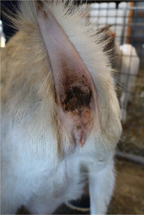
Figure 1.2 Typical waxy secretion found at the base of the tail of goats, which is produced by the sebaceous glands in that area.
This secretion should not be confused with diarrhea, vaginal discharge, or lochia. Source: Courtesy of Dr. M.C. Smith.forth. Normal resting respiratory rate is 10-30 per minute in adults and 20-40 in kids.
Neonates should be inspected particularly for congenital defects. More commonly observed problems include brachygnathia, cleft palate, hydrocephalus, atresia ani, or rectovaginal fistula, and abnormalities of the genitalia associated with the intersex condition as discussed in Chapter 13. A list of congenital and inherited diseases is provided in Table 1.2. Not all of these, of course, will be evident at birth. Up-to-date information on inherited conditions of goats as well as other species is available through the Online Mendelian Inheritance in Animals (OMIA) database at http://omia.org. In 2019, 85 inherited traits or disorders were listed for goats and 37 were considered to be potential models for the study of human diseases.
Goat stature is quite diverse. Many small breeds of goats such as the Pygmy or West African Dwarf goat are in fact achondroplastic dwarfs. They appear disproportionate,
Table 1.2 Some congenital and inherited abnormalities in goats.
Known inherited conditions Known acquired conditions Conditions of unclear status
| Afibrinogenemia in Saanen goats | Arthrogryposis and hydranencephaly | Absence of hair |
| Beta mannosidosis in Nubian goats | caused by Akabane, Schmallenberg, | Atresia ani |
| Bipartite scrotum in Angora goats | and Cache Valley viruses | Atresia coli |
| Brachygnathia superior or inferior | Border disease | Cleft palate |
| Cryptorchidism in Angora goats | Congenital copper deficiency | Congenital goiter of Boer goats |
| Excessive facial hair in Angora goats | Cyclopia due to Veratrum californicum | Double or fused teats |
| Gynecomastia | Freemartins | Entropion |
| Hereditary goiter in Dutch goats | Hydrops | |
| Inherited abortion in South African Angora goats | Patellar luxation | |
| Intersexes associated with polled condition | Precocious milking | |
| Myotonia congenita | Progressive paresis of Angora | |
| N-acetylglucosamine 6-sulphatase deficiency in | goats | |
| Nubian goats (mucopolysaccharidosis IIID) | Rectovaginal fistula | |
| Recessive atrichosis | Skeletal malformations | |
| Robertsonian translocation | Spastic paresis | |
| Short tendons in Australian Angora goats | Sticky kid syndrome of Golden | |
| Sperm granulomas | Guernsey goats | |
| Supernumerary teats Testicular hypoplasia | Umbilical hernia |
with short legs and normal-size torsos.
This may draw visual attention to the degree of abdominal distension present, which, though quite pronounced, is usually normal. Dwarfism because of pituitary hypoplasia is also seen in goats. These small goats are proportionate in appearance; the Sudan goat is an example (Ricordeau 1981).Examination of the Integument
In goats the character of the skin and haircoat is a good indicator of general health. A rough, dry, unglossy coat; excessive dander or flakiness; and failure to shed out in the spring are all suggestive of poor nutritional status, parasitism, or other chronic diseases. The hair or fleece should be parted and the skin examined for lice, ticks, fleas (in the tropics), nodules, swellings, crusts, eczema, necrosis, neoplasia, photosensitization and sunburn, and focal or regional alopecia. The differential diagnoses for these findings are discussed in Chapter 2.
Because many goats are used primarily for cashmere or mohair production, the veterinarian should know something about the nature of goat hair used in textiles. Detailed information on the subject is given in Chapter 2.
Examination of the Head
Many conditions can cause general asymmetry or focal swellings around the head and these abnormalities should be noted. The differential diagnoses for such swellings are discussed in Chapter 3.
Membranes
Inspection of the conjunctivae and mucous membranes of the mouth may reveal paleness due to anemia, icterus resulting from hemolysis or hepatic dysfunction, or hyperemia and congestion associated with acute febrile or toxemic states.
Oral Cavity
Evidence of brachygnathism, cleft palate, mucosal lesions, dental abnormalities, or dysphagia such as drooling, salivation, dropping food from the mouth, or accumulating food in the buccal space should be noted. The differential diagnoses for these signs are given in Chapter 10. Necrotic odors of the breath may occur. They may reflect necrotic stomatitis, alveolar periostitis, pharyngitis, or even pneumonia.
Thorough examination of the oral cavity requires good restraint, a speculum, a towel, and a penlight. All oral structures should be examined and the molar arcades digitally palpated for missing teeth from outside the mouth. If more direct examination of the teeth is required, extreme caution should be taken, because the molars may have sharp, jagged edges and exert a powerful grinding motion. Tranquilization is indicated, because fingers can be badly injured. Wearing gloves during oral examination is a wise precautionary measure, particularly if the goat shows neurologic signs.
Eyes
Facial hair covering the eyes is a heritable trait in Angoiar goats. Affected goats tend to do poorly on range because their ability to selectively browse is impaired. The body condition of such individuals should be noted, as well as their prevalence in the flock. Blindness is initially assessed by testing the menace response, but facial nerve paralysis can render a sighted animal unable to blink. Intact pupillary light responses in a blind goat suggest a cerebrocortical lesion. Lacrimation and hyperemia of the conjunctiva, cloudiness of the cornea, and hypopyon in the anterior chamber should be noted if present. The differential diagnoses for these various findings are given in Chapter 6.
Nares
Both nostrils should be evaluated for symmetry of air flow. If nasal discharge is observed, determine if it is unilateral or bilateral and note its character. Collapse of a nostril may result from facial nerve paralysis. Crusting of the nares occurs when the sick animal does not clean the nostrils, or it may be a sign of specific disease problems. The differential diagnoses for nasal discharge and crusting of the nares are given in Chapter 9.
Ears
Ear mites, if suspected, can be identified by collecting debris from the ear canal on a cotton swab and smearing it on a slide for examination.
Goats are often identified with tattoos applied inside the ear; tattoo numbers may have to be checked against health papers at shows and sales. It may be necessary to clean the inside of the ear with soap and water and use a powerful light source to backlight the tattoo to make it readable. Metal and plastic ear tags commonly tear out of goats' ears and their use should be avoided in pet and show animals. Tags that can rotate around a post are preferred to tags that make a closed loop around the ear, as the latter are more easily torn out. In the United States, scrapie tags are required when selling unregistered goats or moving them interstate, unless the herd identification is tattooed into the ear.
Ear tips may be lost on young goats due to prolonged exposure to freezing temperatures. Goats of the La Mancha breed lack a well-developed external ear. Only a vestigial pinna is present and is referred to as an elf (up to 5 cm, with some cartilage) or gopher (up to 2.5 cm, with little or no cartilage) ear. These animals are usually tattooed on the underside of the tail.
Horns
Goats may be horned or polled. Horn buds may be present at birth or become palpable within several days, or in the case of Nigerian Dwarf goats weeks, of birth. Generally, horned kids have two irregular whorls of hair over the location of the horn buds, whereas hornless kids have a smooth poll with a single central symmetrical whorl of hair. It is important to establish and record which offspring are naturally polled, because homozygous polled goats have a high incidence of infertility. The relationship of the polled trait and the intersex condition is discussed in detail in Chapter 13. Deformed horns, or scurs, are often seen on older goats as a result of incomplete removal of germinal horn tissue at the time of disbudding. Techniques for, and problems with, disbudding and dehorning are discussed in Chapter 18. The glands partially responsible for the characteristic odor of buck goats in the breeding season are located in skin folds just caudomedial to the horn buds.
Examination of the Neck
Traumatic injuries to the pharynx from balling guns and drenching equipment occur in goats. The throat should be palpated for swelling, heat, and pain associated with cellulitis from traumatic injury. A number of normal and abnormal structures and swellings in the neck must be differentiated on physical examination, including goiters, thymus, branchial cleft cysts, wattles, and abscesses. Their differentiation is discussed in Chapter 3. Thorough examination of the neck should include palpation of the jugular furrows for evidence of phlebitis, palpation of the esophagus for evidence of obstruction, and auscultation of the trachea. Prominent distension of the jugular vein, though possibly suggestive of congestive heart failure, is most commonly due to overly tight collars or neck chains in goats. This should be brought to the owner's attention if found.
Examination of the Chest
The extent and severity of respiratory disease are often difficult to assess in goats. To improve the chances of accurate diagnosis and prognosis, careful attention should be given to auscultation. When possible, the animal should be moved to quiet surroundings. The fleece in Angora goats should be parted to place the stethoscope in contact with the skin. The trachea and the lungs should be ausculted to identify the presence of referred sounds. Eliciting a cough by compression of the pharynx may clear the trachea or reveal the presence of bronchial exudate. Care should be taken to listen with a stethoscope placed well forward under the elbow and in front of the shoulder, otherwise cranial ventral pneumonias, which are common, may be overlooked. The intensity of identifiable sounds can be augmented by forced activity of the animal before auscultation or by placing a plastic bag or exam glove over the nares to increase the depth of respiration by rebreathing of carbon dioxide. Radiographic or ultrasound examination is indicated when questions about the severity of lung disease persist.
Normally the heart can be heard about evenly on both the right and left sides of the chest at the fourth or fifth rib space. Mediastinal abscesses may displace the heart, resulting in a shift in intensity of cardiac sounds. Muffling of the heart sounds because of pericarditis is uncommon in goats. Murmurs are rarely heard and are discussed in Chapter 8.
Examination of the Abdomen
The abdominal contour should be inspected to assess conditions such as bloat, advanced pregnancy and false pregnancy in females, and ruptured bladder in wethers and intact males. Characteristic contours and their clinical significance are discussed in Chapter 10. Ballotment may help to detect abdominal fluid accumulations, pregnancy, or rumen impaction. Auscultation of the rumen in the left paralumbar fossa is essential. Normal rumen contractions occur at a rate of one to two per minute. Observation of cud chewing suggests normal rumen activity.
Examination of the Limbs
Locomotor problems are common in goats. Lameness and abnormalities of gait may result from neurologic disease, conformational defects, muscular dysfunction, skeletal trauma, infectious and non-infectious arthritides, and diseases of the foot. Localization of the problem by careful physical examination is the first step in making an accurate diagnosis. Differential diagnoses of locomotor problems are discussed in detail in Chapter 4.
Overgrown hooves must be pared with shears or a hoof knife to adequately assess the health of the foot. Hyperemia and swellings at or above the coronary band should be noted. They may represent either local infections or systemic disease.
All joints should be carefully palpated. Distensions of the joint capsule, heat, pain, swelling, or fibrosis of periarticular structures, limitations on the range of joint motion, and enlargement of bursae should all be noted. The degree of joint enlargement may not necessarily correlate with the severity of lameness.
The vertebral column and the long bones of the legs should be palpated for evidence of fractures in acutely lame or recumbent animals. A major goal of physical examination in recumbent goats is to differentiate musculoskeletal, metabolic, toxemic, and neurologic causes of recumbency. The differential diagnoses for recumbency are discussed in Chapter 4.
Examination of the Reproductive System
Mammary Gland
Careful examination of the udder is always warranted. Visual inspection may reveal weakness of the suspensory apparatus, slack halves, abnormal swellings, and discolorations of the skin. Digital palpation of the gland will identify udder edema, active inflammation, fibrosis, scarring, or abscesses in the parenchyma or teats. In gangrenous mastitis the udder skin may be blue-black and cold, and in time the gland may slough if the animal survives. Patency of the teats should be assessed in lactating animals. Supernumerary teats may be present. Their identification and removal are discussed in Chapter 14.
The milk of lactating does should be observed on a black plate or strip cup to assess color, consistency, and the presence of clots or flakes. Bovine screening tests for subclini- cal mastitis such as the California Mastitis Test must be used cautiously in goats, because normal goat milk tends to have higher cell counts. The somatic cell count issue and interpretation of test results are discussed in Chapter 14.
Vulva
Swelling and hyperemia of the vulva may be signs of heat or impending parturition, but may also be seen in herpes vulvovaginitis in conjunction with vesicles or scabs. Any vulvar discharges should be noted. As females come out of heat, the vulvar discharge, initially serous and mucoid, may become white and tenacious. This is often misinterpreted as a purulent discharge by the inexperienced observer. Often, there is a sanguinous discharge after termination of a pseudopregnancy and neoplasia of the reproductive tract may also result in a bloody vaginal discharge. Speculum examination of the vaginal canal and cervix should be carried out when there is doubt about the source and nature of the discharge. Occasionally, otherwise normal does may have ectopic mammary tissue present at the vulva, which may swell at the beginning of lactation.
Does may show vaginal eversion or frank prolapse in advanced pregnancy or immediately post partum. After uncomplicated births, normal lochia may be discharged for a period of one to three weeks. It is reddish brown in color and odorless. Placentas are usually passed within four hours of parturition and are frequently eaten.
It is important to carefully examine the external genitalia of young does, particularly when there is a complaint of infertility, because of the high incidence of intersexes among polled goats. Malformations of the genitalia range from the clinically subtle, such as a slightly enlarged clitoris, to the overt, such as male phenotypes in genetically female individuals. Accurate record keeping may help to identify homozygous polled individuals.
Scrotum
The scrotum and its contents should be palpated. Normally there is bilateral symmetry of all structures. A bipartite or split scrotum is a common congenital condition that some breeders consider a fault. The differential diagnoses for abnormalities of the scrotum, testes, and spermatic cord structures are given in Chapter 13. Semen samples can be collected for evaluation by electroejaculation or by use of an artificial vagina. These procedures are described in Chapter 13. Gynecomastia occurs in male goats and it is not extraordinary to detect distended teats anterior to the scrotum during physical examination, as discussed in Chapter 13.
Penis and Prepuce
Examining the penis of male goats, especially wethers, can be difficult and is not ordinarily attempted unless there is a history of urinary or breeding problems. Details on special examination and catheterization of the penis are given in Chapter 12.
The preputial opening should be routinely examined, particularly in wethers, for the presence of ulcerative posthitis. The preputial orifice may become occluded in this condition. Crystals or drops of blood may be noted at the orifice in cases of obstructive urolithiasis.
Examination of the Environment
An examination of the environment where goats are raised should include a detailed review of all feeds used, the feeding facilities, water sources and water delivery systems; the yards, pastures, range, or buildings where the animals are kept; and any mechanical or manual equipment used as part of the routine farm procedures. Too often, farmers attempt to make do with equipment and buildings that are inadequate for an expanding operation. Because of prolificacy, goat herds tend to expand faster than owners anticipate. Inadequate feeder space for does, a smell of ammonia in the air due to poor ventilation and/or soaked bedding, and overcrowded kid pens are three examples of common faults found on environmental inspection. Besides the visual inspection of equipment, farmers could be asked to demonstrate how common procedures are carried out to detect if they are using inappropriate techniques.
Equipment used for treatment should be examined for its potential to cause injury. Poorly maintained syringes with either contaminated or overly long, large-gauge needles can result in injection abscesses or systemic infections. Drug and vaccine stocks should be examined to determine if they are appropriate, clean, unexpired, and maintained at proper temperatures. Storage facilities for these items should be properly secured. If the farmer blends his or her own feed or components, the techniques and constituents used should be examined, particularly when toxicoses are suspected. Accidental inclusion of agricultural or other farm chemicals is not uncommon. Milking machines used for goats should also be examined to determine their efficiency and the milking procedures studied if it appears that mastitis is a significant problem. Finally, the facilities available and routinely used to dispose of any dead animals should be particularly noted to see that they are consistent with any legal requirements, do not attract predators, and do not serve as environmental contaminants or as a source of infection for the remainder of the flock or herd.
In recent years, in association with the expansion of international trade in livestock and livestock products and increasing concerns about bioterrorism, there has been a growing emphasis on food safety and biosecurity, with recognition that food safety encompasses all aspects of food production, from the farm to the table. As such, quality management assurance programs have grown in popularity, as have procedures to ensure biosecurity to limit the introduction and spread of disease on and between farms. Environmental inspections should include an assessment of biosecurity and aspects of management and sanitation that can affect food quality.
Special Considerations for Range and Pasture Operations
The quality and quantity of pasture and the availability of supplementary or conserved feed should be assessed. This may require specialized knowledge of pasture species, weeds, or other potentially toxic plants. A list of plants known to be poisonous to goats is provided in Chapter 19. If possible, animals should be observed while feeding. The source of water available both year-round and seasonally, its quality, and its quantity should also be inspected. Feed and water samples should, if necessary, be collected for laboratory examinations.
On range or pasture, goats may congregate in certain shaded or sheltered areas, particularly during periods of high or low temperatures or precipitation, and after shearing. Provision of adequate shelter should be documented. Heat stress can be reduced by ensuring the animals have adequate shade and water when the livestock weather safety index is in the danger or emergency zone. Cold stress is particularly likely in recently shorn animals, and temporary shelter and extra feed are necessary for these animals. If shelter areas are limited or overused, buildup of infectious and parasitic agents can occur, leading to increases in enteric and other diseases, especially in kids. This is aggravated by poor drainage under feeders and water troughs.
Fencing for these types of operations should also be inspected, particularly regarding its effectiveness in keeping goats in and keeping predators out.
Special Considerations for Confinement Operations
In semiconfinement operations, adult animals and sometimes young stock are grazed on pasture during the warmer months, but confined during the winter and early spring. As herds expand, there is a tendency for small pastures to be overused, with poor nutrition and increased parasitism as the result. Stocking rates should be noted and deworming history and efficacy verified.
In confinement systems, the type of buildings used for housing should be evaluated for the following: available area (square feet or square meters) per adult breeding animal; type of flooring and bedding; degree of insulation; presence of supplemental heating if any; adequacy of natural or mechanical ventilation systems; source and availability of water; and the amount of trough or bunk space per goat. Requirements for goats are given in Chapter 9. The distribution of animals within buildings should be noted. Ideally, a separate kidding facility should exist and be hygienically maintained. Artificially reared kids should be in a separate facility from adult stock, and bucks should be housed separately from milking does. Bucklings should be separated from females by three months of age, to avoid the risk of unwanted pregnancies. Methods and times of feeding for all groups within the herd should be noted. If other species of livestock are pre - sent, consideration should be given to them as possible sources of disease for goats.
Unfortunately, many confinement goat operations have both rodent and associated feline populations. Cats are a recognized source of Toxoplasma gondii, a major cause of abortion in goats and a potential zoonotic disease from consumption of meat or milk. Farmers should be encouraged to seek alternate means of rodent control.
Special Consideration for Hobby Farms
The principles and procedures of environmental examination already described also apply to the hobby farm. Special considerations that occur are usually the result of inexperience on the part of the hobbyist. Medications, insecticides, and grain supplies may be inadequately secured, allowing access of inquisitive goats with resultant overdoses, poisonings, and/or grain overload, respectively. In the barn, overcrowding with inadequate feeder space is often observed, along with fecal contamination of feed and water supplies.
In winter, lack of adequate ventilation in closed, overcrowded barns leads to outbreaks of pneumonia. Some owners consistently shut all windows and doors, believing that animals must stay warm in winter. These individuals need to be counseled on the beneficial effects of continuous air exchange on respiratory health. Pasture problems often involve disappearance of animals or predation due to inadequate fencing, bloat due to inadequate adaption to pasture, or parasitism due to placement of young stock on contaminated, overcrowded plots. The veterinarian should examine all elements of the ration being fed on the hobby farm to ensure that the hobbyist understands basic ruminant nutrition.
Field Necropsies and Slaughterhouse Checks
The economic value of most individual goats in commercial herds and flocks is such that most owners can be persuaded to have a necropsy undertaken of an ill or obviously moribund animal. This is an extremely useful adjunct to clinical examination, because necropsy can confirm the suspected diagnosis or suggest new avenues of investigation. All necropsies should be carried out following a systematic procedure, with special regard to the likelihood of any zoonotic infections being transmitted and the safe and legal disposal of carcasses.
Necropsy of all or a random sample of young animals born dead or dying in the first two to three weeks of life is an extremely valuable service that veterinarians can provide to their clients. Categorization of deaths into preparturient, parturient, and postparturient causes can be done with a minimum of laboratory diagnostic techniques and immediately direct the owner to possible ways of reducing these losses.
Whenever possible, goats slaughtered for meat or culled from the herd or flock should be examined post mortem by a veterinarian as an inexpensive and worthwhile means of disease surveillance. Such inspections can provide information on the efficacy of parasite control measures; the presence of subclinical pneumonia; and the occurrence of injection site abscesses, visceral caseous lymphadenitis, and hydatid disease. Opportunities to perform routine slaughter checks on goats at an abattoir are uncommon in the United States because the goat meat industry is decentralized, though this will change as demand for goat meat continues to increase.
More on the topic Physical Examination:
- Medical Record
- PHYSICAL EXAMINATION AND DIAGNOSTICS
- THE PAEDIATRIC CONSULTATION AND CLINICAL CONSIDERATIONS
- Introduction
- Clinical features of Lyme borreliosis in humans in Bulgaria
- Vogelnest L., Portas T. (Eds.). Current Therapy in Medicine of Australian Mammals. CSIRO,2025. — 848 p., 2025
- Abdominal Distention and Constipation
- Smith Mary C., Sherman David M.. Goat Medicine. 3rd edition. — Wiley-Blackwell,2023. — 976 p., 2023
- Pregnancy Loss
- Fluid Therapy for Renal Failure in Horses (Box 44.4)