Pituitary and Hypothalamus
Dianne McFarlane
Physiology of Equine Hypothalamic-Pituitary Axis
The pituitary gland is composed of two embryologically distinct portions: an adenohypophysis, derived from invagination of the pharyngeal epithelium known as Rathke’s pouch, and the neurohypophysis, derived from neural tissue of the hypothalamus.
The adenohypophysis can be further divided into the pars distalis, pars tuberalis, and pars intermedia. The pars distalis contains five different endocrine cell types, each of which is responsible for the release of a unique hormone or set of hormones in response to hypothalamic-releasing factors delivered from the median eminence via the hypothalamic-hypophyseal portal system. The pars tuberalis is a highly vascular band of cells surrounding the pituitary stalk. The pars tuberalis is rich in melatonin receptors and is believed to be involved in regulating the seasonal rhythm of reproductive hormone production.1 The neurohypophysis or pars nervosa is a collection of nerve axons and terminals that originate in the paraventricular and supraoptic nuclei of the hypothalamus. Oxytocin and arginine vasopressin (antidiuretic hormone) produced in the cell bodies of these nuclei are transported into the pars nervosa for storage and eventual release into the systemic circulation.The pars intermedia of the horse incorporates tissue derived from both the adenohypophysis and neurohypophysis.2 It is composed of a single endocrine cell type, the melanotrope. Melanotropes are directly innervated by nerve terminals of the hypothalamic periventricular dopaminergic neurons. These originate in the periventricular nucleus of the hypothalamus adjacent to the third ventricle, project through the infundibulum, and terminate in the pars intermedia (Fig. 41.1).3 These neurons release the neurotransmitter dopamine, which acts to tonically inhibit the release of hormones from adjacent melanotropes.
Dopamine released from the nerve terminals interacts at dopamine (D2) receptors on the melanotropes to inhibit cell proliferation, transcription of proopiomelanocortin (POMC), and release of POMC-derived peptides.4 Additional regulatory signals to the pars intermedia may be delivered by direct systemic arterial supply and from the hypothalamic- hypophyseal portal veins.5 In addition to being under tonic inhibition, melanotropes are also positively regulated by thyrotropin-releasing hormone (TRH), which stimulates hormone release from melanotropes.6 Additional regulatory molecules likely exist but are poorly defined in the horse.The primary product of the melanotrope is the hormone precursor protein POMC, which is also expressed by the corticotropes of the pars distalis. However, due to differential post-translational processing by proteases called prohormone convertases, each cell type secretes a different complement of POMC-derived peptides (Fig. 41.2). Due to the action of prohormone convertase I, POMC in corticotropes is primarily processed into adrenocorticotropic hormone (ACTH). ACTH circulates to the adrenal cortex, where it stimulates secretion of cortisol. Melanotropes contain active prohormone convertase I and II, and therefore POMC in the pars intermedia (PI) is cleaved into the secretory peptides, α-melanocyte stimulating hormone (α-MSH), β-endorphin (β-end), and corticotropin-like intermediate lobe peptide (CLIP). A small amount of ACTH may also be produced. Further processing of the peptides, including cleavage of C-terminal amino acids and N-acetylation, serves to control the activity of the final peptide product. For example, the most abundant form of β-end produced in the normal horse's pars intermedia is Ac-β-end (1-27), which lacks opioid activity. The most abundant β-end in horses with pituitary pars intermedia dysfunction (PPID) is β-end (1-31), which is an opioid agonist.7
The physiologic role of the pars intermedia POMC-derived peptides, α-MSH, β-end, and CLIP, has not been extensively studied in the horse.
In other species, α-MSH has several diverse actions, which are mediated through interaction with one of five distinct G-protein-coupled melanocortin receptors. α-Melanocyte stimulating hormone is named such due to its ability to induce skin pigmentation in amphibians. Its role in pigmentation is through interaction with melanocortin receptor 1 (MC1R), which is predominantly expressed in skin. In horses, mutation of the MC1R gene is associated with the chestnut coat color.8 In white Camarque horses the degree of coat pigmentation is directly correlated to the plasma concentration of α-MSH.9 α-MSH is also an integral mediator in control of energy homeostasis. MC3R and MC4R are both expressed in the central nervous system (CNS), particularly in the hypothalamus, where they function in the leptin-melanocortin pathway, regulating appetite-satiety balance and fat metabo- lism.10 Animals and humans lacking functional MC3R or MC4R are obese, and melanocortin receptor defects are a common monogenetic cause of obesity in humans.11 Plasma α-MSH concentration in obese men has been reported to be higher than in lean men.12 It has been suggested high plasma concentration in obese individuals may be an attempt to maintain homeostasis in individuals with a defect in MC4R. Plasma α-MSH concentration was also found to be positively correlated to obesity in horses.13 Another function of α-MSH is as a potent anti-inflammatory agent.14 α-MSH has been demonstrated to have multiple immune-modulating effects. Its most profound effect is in regulation of cytokine response. α-MSH inhibits activation of NF-κB by lipopolysaccharide (LPS) and interferon-γ (IFN-γ). Data in mice have suggested that α-MSH may suppress LPS-mediated inflammation by facilitating the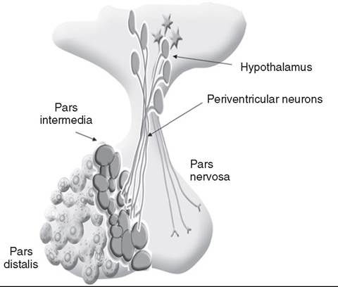
FIG. 41.1 Physiology of the equine pituitary pars intermedia.
The melanotropes of the pars intermedia produce the hormone precursor protein, proopiomelanocortin (POMC), which in the pars intermedia is cleaved into the hormones, α-melanocyte stimulating hormone (α-MSH), β-endorphin (β-end), and corticotropin-like intermediate lobe peptide. Production of POMC in the pars intermedia is under inhibitory control by dopamine released from the nerve terminals of the periventricular neurons. The cell bodies of the periventricular neurons are in the hypothalamus, adjacent to the third ventricle.interaction of IRAK and IRAK-M.15 IRAK is a kinase that functions in activation of NF-κB following TLR-4 (the receptor for bacterial LPS) stimulation.15 IRAK-M is a negative regulator of IRAK. As a result, NF-κB activation and proinflammatory cytokine release of tumor necrosis factor (TNF)-α, interleukin (IL)-1β, and IL-6 are all decreased following α-MSH administration. Fever and other clinical evidence of inflammation are also reduced. In addition, α-MSH has been shown to inhibit human and rodent neutrophil function, including adhesion, chemotaxis, and oxidative burst.16-17 High α-MSH plasma concentration in horses was also associated with decreased oxidative burst and adhesion.18 β-End is a known endogenous opioid. Secretion of β-end provides analgesia and behavioral modification. It also suppresses immune responsiveness and has effects on vascular tone.19-20 Corticotropin-like intermediate lobe peptide (CLIP, ACTH 18-34) has not been extensively studied in any species. In pancreatic islet cells in culture, CLIP was shown to be a pancreatic β-cell secretagogue, stimulating the release of insulin.21 However, when administered to rats by either intraperitoneal or intraventricular injection, ACTH but not CLIP resulted in release of insulin and decrease in blood glucose.22
A distinct seasonal effect on the activity of the pars intermedia of horses and ponies has been demonstrated in horses residing in tropical and temperate environments.23-30 Plasma α-MSH concentration is considerably higher in horses and ponies in the northern hemisphere in August through October, compared with samples collected in the winter, spring, and early summer.23-28 Similarly, horses in the southern hemisphere experience higher plasma α-MSH concentrations during shortening day length, from February to April.29-30 An effect of season on α-MSH concentration has been described for humans, hamsters, and sheep.31-33 The functional importance of the seasonal cycle is unknown, but several physiologic events occur in parallel with the α-MSH cycle.
In sheep, body weight, voluntary food intake, and condition all peak simultaneously with α-MSH, with seasonal maximums occurring in September. Soay sheep with a surgically created hypothalamic-pituitary disconnection have an increase in circulating concentration of α-MSH and chronic increase in body weight.32 These findings suggest that α-MSH or other POMC-derived peptides may play a role in metabolic preparation for winter in sheep. It is possible that horses and ponies have a seasonal increase in POMC-derived peptides to metabolically prepare them for a decrease in accessible food observed in the wild in winter. If so, dysregulation of this pathway might be associated with abnormalities in body weight and fat storage. Weight loss and abnormal fat distribution are two clinical signs associated with equine PPID. Development of a winter coat also begins as length of day decreases in the fall. The development of hypertrichosis in horses with PPID leads one to speculate that the naturally occurring seasonal increase in POMC-derived peptides contributes to development of winter coat growth. This has not been critically assessed in equids.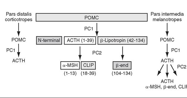
FIG. 41.2 Proopiomelanocortin (POMC) processing.
POMC is cleaved by prohormone convertase I into adrenocorticotropic hormone (ACTH) in the pars distalis and into α-melanocyte stimulating hormone, β-endorphin, and corticotropin-like intermediate lobe peptide by prohormone convertase I and II in the pars intermedia. Only a small amount of ACTH is produced by the normal pars intermedia.
Equine Pituitary Pars Intermedia Dysfunction
Equine PPID is one of the most common diseases of horses and ponies 15 years and older.34-36 PPID was originally termed equine Cushing disease because of features similar to human Cushing disease. However, in contrast to human Cushing disease, PPID affects the pituitary pars intermedia rather than the pars distalis, is typically not a neoplastic condition, and the adrenocortical contribution to the clinical syndrome is of much less importance.
To avoid confusion, equine Cushing disease is now more correctly referred to as PPID.In the past 2 decades the population of aged horses has increased dramatically. This, in conjunction with the vast amount of information now available online to the horse-owning public, has led to a heightened client awareness of age-associated equine health issues and a desire to promote healthy aging in their horses. As a result, diagnostic testing and treatment of horses for PPID has increased. Yet despite increased clinical recognition of this disease, much about PPID remains poorly understood.
Pathophysiology
The pathologic hallmark of PPID is hypertrophy, hyperplasia, and microadenoma or macroadenoma formation in the pituitary pars intermedia with increased secretion of POMC peptides. Horses with PPID develop enlarged pituitaries that may exceed five times normal weight. As the pars intermedia expands, it compresses the adjacent pituitary lobes and hypothalamus, often resulting in a loss of function of these tissues. In contrast, the pars intermedia remains active in horses with PPID, secreting relatively large quantities of POMC-derived peptides into the peripheral circulation. Horses with disease may have as much as a 40-fold increase in plasma concentration of pars intermedia POMC-derived peptides.37 Clinical signs of disease likely result from a combination of increased circulating POMC peptides and loss of neuroendocrine function of adjacent tissues.
Evidence indicates loss of dopamine inhibition is critical in the pathology of PPID. Dopamine and dopamine metabolite concentrations in the pars intermedia of PPID horses are decreased eightfold compared with age-matched controls.7 Systemic supplementation of dopamine or a dopamine agonist to horses with PPID results in a decrease in plasma concentration of POMC peptides.37 Horses treated with the dopamine agonist pergolide show improvement in both clinical signs and biochemical abnormalities associated with disease.38-41 Immunohistochemistry of formalin-fixed tissue showed a fivefold decrease in pituitary dopaminergic nerve terminals and a 50% reduction in the number of dopaminergic periventricular cell bodies in the hypothalamus of PPID animals.42 Considered together, this evidence suggests a loss of functional periventricular dopaminergic neurons—“dopaminergic neurodegeneration”—occurs in horses with PPID (Fig. 41.3). Loss of periventricular dopaminergic inhibition of the pars intermedia in other species results in pathologic changes similar to those of PPID. Surgical disruption of the periventricular hypophyseal dopaminergic tracts in rats results in increased expression of pars intermedia melanotropes.43 In addition, D2 dopamine receptor knockout mice develop pars intermedia lesions similar to PPID.4 These data suggest PPID is primarily a disease of hypothalamic origin, rather than the consequence of a spontaneously forming pituitary adenoma.
One potential cause for dopaminergic neurodegeneration is oxidative stress. Oxidative stress causes modification of cellular components including proteins, DNA, and cell membrane lipids due to excessive exposure to reactive oxygen species leading ultimately to cell death or, in the case of neurons, neurodegeneration. Dopaminergic neurons are particularly vulnerable to oxidative damage because dopamine metabolism
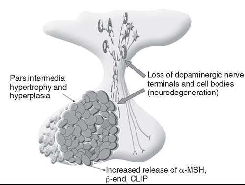
FIG. 41.3 Pathophysiology of equine pituitary pars intermedia dysfunction. Loss of functional dopaminergic periventricular neurons leads to a decrease in dopamine at the pars intermedia. This in turns results in dysinhibition of the melanotropes of the pars intermedia. The outcome is hypertrophy and hyperplasia of the pars intermedia and increased systemic release of the pars intermedia proopiomelanocortin (POMC)-derived peptides, α-melanocyte stimulating hormone (α-MSH), β-endorphin (β-end), and corticotropin-like intermediate lobe peptide.
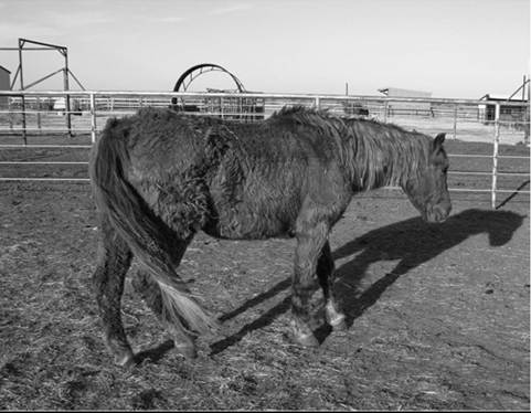
FIG. 41.4 Typical horse with pituitary pars intermedia dysfunction (PPID). This 22-year-old Morgan mare shows obvious hypertrichosis. Other clinical signs of PPID in this horse included laminitis and weight loss despite an excellent appetite.
itself produces free radicals. Horses with PPID have evidence of oxidative damage, including accumulation of pars intermedia 3-nitrotyrosine42 and decreased plasma thiol.44 The oxidative damage in PPID does not appear to be the result of impaired antioxidant capacity because systemic and pituitary antioxidant capacity of affected horses appears unchanged.45
Clinical Signs
Equine PPID affects many aged equids, resulting in a variety of clinical signs including hypertrichosis (hirsutism), muscle atrophy, regional fat accumulation, polydipsia, polyuria, secondary infections, lethargy, infertility, persistent lactation, exercise intolerance, and sweating dysregulation (Fig. 41.4).46-51 Laminitis and metabolic abnormalities including hyperglycemia and hyperinsulinemia also occur in approximately 30% of horses with PPID.48 It is now thought that endocrinopathic laminitis occurs in horses with PPID due to concurrent insulin dysregulation or equine metabolic syndrome (EMS), rather than as a direct result of PPID itself.
The most unique clinical manifestation of PPID is an abnormally long, curly hair coat that fails to shed (previously referred to as equine hirsutism, although more correctly called hypertrichosis'). Often, horse owners may report the horse sheds its winter coat slowly or incompletely in the year(s) before development of a full failure to shed. Hair may be initially retained along the legs or under the mandible (Fig. 41.5). The mechanism responsible for development of hypertrichosis in horses with PPID has not been reported. The onset of hypertrichosis in an aged horse or pony is considered essentially pathognomonic for PPID, although the author has observed similar hair coats in severely debilitated aged animals with confirmed normal pituitary pars intermedia function.
In the author's experience, weight loss due to muscle mass atrophy is the most common and often the earliest clinical sign of PPID (Fig. 41.6). Despite weight loss, PPID horses often have a potbellied appearance and, as a consequence, owners may fail to notice the lost weight. Weight loss and muscle atrophy may result from several factors, including poor dentition, poor nutrition, heavy parasite burden, minimal exercise, and protein catabolism induced by increased cortisol activity. Histologic evidence of type 2 myofiber atrophy has been documented in muscle biopsies from horses with PPID, consistent with corticosteroid-associated muscle atrophy in other species.52 Significant improvement in muscle mass was observed in a large group of horses with PPID after 6 months of treatment with pergolide mesylate, suggesting overexpression of POMC peptides contributes directly or indirectly to the mechanism of muscle atrophy.41
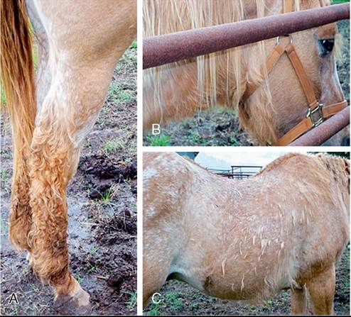
FIG. 41.5 Coat changes vary among horses with pituitary pars intermedia dysfunction (PPID). Retained hair along the lower limbs (A), under mandible (B), and as guard hairs and tufts along trunk (C).
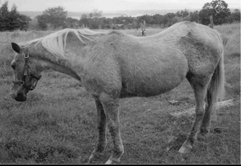
FIG. 41.6 Horse with pituitary pars intermedia dysfunction (PPID). The major clinical sign of disease in this horse with PPID was weight loss. Muscle mass atrophy along the dorsum, with a potbelly appearance, is characteristic of PPID.
Despite weight loss and muscle mass atrophy, horses with PPID often have abnormal accumulations of fat, most notably in the crest of the neck, tailbase, sheath, and superorbital fossa. This fat accumulation typically predates the weight loss and has a similar pattern as that observed in horses with equine metabolic syndrome (EMS). The similarity of the clinical signs and at-risk breeds of these two diseases has resulted in a misdiagnosis of PPID in animals that have EMS. Unlike what was previously postulated, these two conditions, PPID and EMS, are no longer considered mutually exclusive conditions. There has also been speculation that animals with EMS or sustained obesity with hyperinsulinemia may be at greater risk for developing PPID as they age. Although epidemiologic data are currently lacking to support this association, client-provided anecdotal data suggest this may warrant more critical assessment.
Horses with PPID have been reported to be more susceptible to infection, including endoparasitism, bacterial sinusitis, skin infections, abscesses, and respiratory infections.46-47,54-56 The morbidity and mortality of secondary infection in equine PPID have been reported to range from 27% to 48%.49-50,54-55 In the absence of parasite control, a heavy parasite burden is common.56 Routine, quantitative fecal egg counts are recommended to ensure an adequate anthelmintic program. Vigilance on the part of the owner and veterinarian is important in both prevention and early recognition of infections because they may be clinically insidious. Bronchopneumonia was found at necropsy in 7 of 19 horses with PPID.57 Bronchopneumonia should be considered in the PPID horse with fever or tachypnea.
Laminitis secondary to PPID is reported to occur in approximately 30% to 40% of diagnosed cases and can necessitate euthanasia in affected animals.41,49-51 When adult horses
Polydipsia and polyuria (PU/PD) occurs in some horses with PPID and is typically mild in severity.46-47,49-51 The mechanism responsible for PU/PD may include (1) compression of the pars nervosa resulting in decreased arginine vasopressin (antidiuretic hormone) production, (2) osmotic diuresis secondary to hyperglycemia and glucosuria, or (3) cortisol induced. Cortisol is thought to cause PU/PD in other species by interfering with the secretion and/or action of arginine vasopres- sion.53 Evidence suggests ACTH and cortisol may inhibit the renin-angiotensin-aldosterone axis as well. The mechanism of PU/PD in horses with PPID has not been extensively examined. Glucosuria is not a common finding in horses with PPID. In one study, two PPID horses with marked hyperglycemia were found to have similar water consumption to normal horses, suggesting that osmotic diuresis is unlikely a major mechanism of PU/PD in the PPID horse.51 with laminitis of unknown origin were examined, 30% had evidence of PPID based on the presence of clinical signs (abnormal hair coat) and diagnostic test results.58 Although an earlier study suggested 70% of undiagnosed laminitis cases might be due to PPID, this study relied on plasma ACTH concentration measured irrespective of season and in the absence of clinical signs for the diagnosis.59 The reliability of plasma ACTH concentration for diagnosis of presymptomatic PPID, particularly in thrifty horses in the fall, is likely poor. The pathogenesis of laminitis in PPID is not currently well understood, but endocrinopathic laminitis is the subject of ongoing investigation. Multiple lines of study support a primary role for Eyperinsulinemia and insulin dysregulation in the induction of endocrinopathic laminitis.60-65 Current data suggest that PPID is a comorbidity or risk factor rather than a primary inciting factor in the development of endocrinopathic laminitis.48 Therefore, clients should be advised to assess all horses with insulin dysregulation (ID, equine metabolic syndrome) for PPID and all animals with PPID for ID. In horses with concurrent PPID and ID, the utmost care should be taken to manage risk factors for laminitis and minimize dietary nonstructural carbohydrates that could perpetuate ID.
Diagnosis of Pituitary Pars Intermedia Dysfunction
Equine PPID is both common and life threatening; therefore early and accurate diagnosis with appropriate intervention is considered critical. Antemortem diagnosis of PPID currently relies on testing hypothalamic-pituitary-adrenal axis responsiveness or measurement of endogenous plasma concentrations of POMC-derived peptides such as ACTH. These tests have been the subject of recent evaluation, and the search for improved testing strategies is considered an ongoing research priority.66 Accepted best practices for the diagnosis of PPID have evolved significantly over the past 5 years, and further changes are likely to follow.
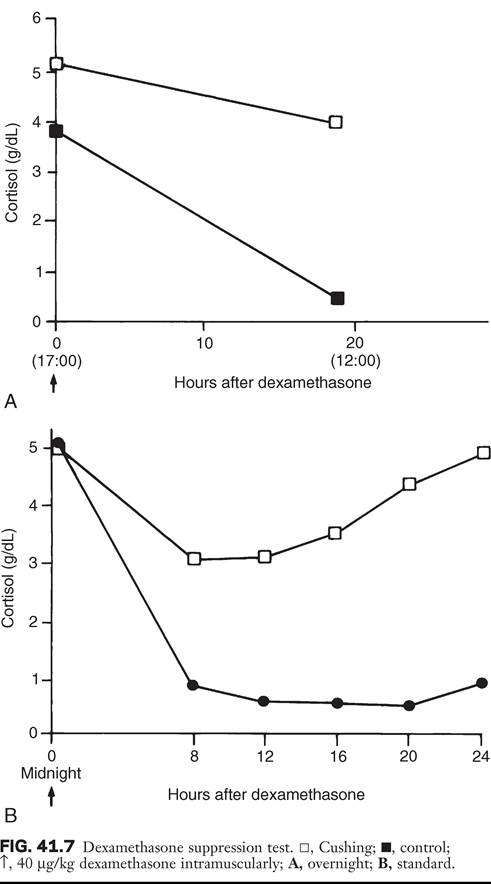
Currently, three antemortem diagnostic tests are most commonly used in practice settings: (1) the overnight dexamethasone suppression test (DST), (2) measurement of endogenous plasma ACTH concentration, and (3) ACTH release following thyrotropin-releasing hormone stimulation. The overnight DST was long considered the “gold standard” method of antemortem PPID diagnosis.67 In the unaffected horse, intramuscular (IM) administration of dexamethasone decreases release of ACTH from the pars distalis, resulting in a serum cortisol concentration of less than 1 pg/dL (27.59 nmol/L) 19 hours after dexamethasone (Fig. 41.7).67 Horses with PPID fail to suppress serum cortisol concentration due to ACTH production from the pars intermedia. Although originally this test was reported to have a sensitivity and specificity of 100%, horses included in that study were selected for having end-stage PPID, resulting in a favorable bias when assessing test performance.67 Although highly specific for PPID, the DST has shown poor performance in identifying horses with earlier PPID. In fact, the DST was only useful in identifying horses with end-stage disease (defined as those with hypertrichosis and grossly enlarged pituitary adenomas) when a random population was assessed using necropsy as the gold standard.68
Measurement of endogenous concentrations of POMC-derived peptides is also useful in the diagnosis of PPID.69 Measurement of plasma ACTH concentration is the most commonly used method for diagnosis of PPID in ambulatory practice because it requires collection of only a single plasma sample and poses no risk to the patient. Difficulty in sample handling was long cited as a limitation of this test; however, more critical evaluation has found equine ACTH to be more stable in plasma than originally assumed, and as long as a plasma sample remains cooled, separation of plasma can be delayed as long as 12 hours without affecting results and without the need for special preservatives (e.g., aprotinin).39,50,70 Measurement of plasma concentrations of ACTH was originally reported to have a sensitivity and specificity of approximately 80% to 90%.50,69 However, evaluation of a larger, randomized equine population has found the test to have a sensitivity and specificity of approximately 70% and 80%, respectively.68 α-MSH concentration measurement, although used in research to similarly assess pars intermedia function, is not significantly better than ACTH concentration in identification of PPID and is not commercially available.27,68-69,71
Seasonal variation in both static and dynamic testing of pars intermedia function has been well documented.23-30,72-73
Testing performed in autumn, between August and October in the northern hemisphere or February and April in the southern hemisphere, yields a significantly greater hormone concentration than testing performed between November and July.23-30,72-73 Initially, these findings led to the recommendation to avoid endocrine testing in autumn. However, further work has demonstrated that the difference in ACTH concentration between normal horses and those with PPID is much greater during autumn.26 As a consequence, provided seasonally specific reference intervals are available, ACTH testing in autumn is more discriminating and therefore preferable to testing during other times of the year.
A positive test result in an animal with clinical signs of PPID is considered strong diagnostic evidence. However, due to the poor performance of the DST and ACTH concentration at identifying early disease, it is not uncommon for a clinician to have a suspicion of PPID in a patient that has a negative diagnostic test result. When this occurs, repeating testing at a later date or further testing using a provocative test is indicated. The dynamic test that has been shown to most accurately identify PPID is the thyrotropin-releasing hormone (TRH) stimulation test with measurement of plasma ACTH concentration.74-76 TRH is known to be a physiologic releasing factor of the equine pars intermedia.6 Although this test was initially performed by documenting an increase in serum cortisol concentration after TRH administration, measurement of plasma ACTH rather than serum cortisol has proven more reliable.6,77 Plasma ACTH response is a more direct assessment of pars intermedia activity and eliminates the influence of adrenal gland response or ACTH bioactivity. It has been demonstrated that in PPID, immunoreactive plasma ACTH is not as bioactive as plasma ACTH from the healthy horse.78-79 Therefore, in horses with PPID the TRH response may be attenuated at the level of cortisol release. The initial recommended protocol for the TRH stimulation test was to measure plasma ACTH concentration at time 0, 10, and 30 minutes after intravenous administration of TRH (1 mg/horse or 0.5 mg/pony).76 However, to reduce cost and improve efficiency, most practitioners choose to collect only the 0- and 10-minute samples, as data are lacking for improved test performance with inclusion of the additional 30-minute sample. Current recommendations suggest limiting use of the TRH stimulation test to nonfall seasons.66 Although several investigators have collected TRH stimulation data during the fall, TRH response is highly variable and may depend on such factors as breed and geographic location.80-81 Although unlikely to be practical in most cases, advanced imaging of the pituitary using computed tomography (CT) or magnetic resonance (MR) has also been examined in a limited number of cases as a method for documenting pituitary or pars intermedia enlargement.82-85 Accuracy of CT at estimating width, height, length, and volume of the equine pituitary gland in disarticulated heads from 25 normal horses was determined to be 81% to 93%, 58% to 71%, 88% to 99%, and 43% to 53%, respectively.83 In a more recent study in which CT measurements were performed in a small number of live horses, accuracy was between 92% and 105%, likely due to the larger dose of contrast agent administered.84 Despite the ability to characterize overall size of the pituitary, CT does not provide information on the internal structure of the gland, including the relative size of the pituitary lobes and presence of microadenomas. Magnetic resonance imaging is the preferred diagnostic imaging modality for pituitary masses in humans, and its usefulness in characterizing the pituitaries of horses is currently being evaluated.85
Necropsy Findings
Postmortem examination of the horse with PPID reveals a grossly enlarged pituitary due to hypertrophy and hyperplasia of the pars intermedia.86-89 The normal horse's pituitary typically weighs between 1 and 3 g; the affected horse's pituitary may be two to five times this size. Enlargement may be due to an adenoma (>1 cm), which often contains areas of hemorrhage and necrosis. Alternatively, microadenomatous (≤1 cm) hyperplasia may be present. Melanotropes in the affected gland are pleiomorphic (polyhedral or spindle shaped) with eosinophilic, granular cytoplasm.86-89 Cells are organized into nodules, rosettes, bundles, or follicular structures separated by fine septal tissue. Pigment deposition (lipofuscin) is common in the pars nervosa, and hemosiderin may be observed when hemorrhage is present.57 Other lesions include compression of the pars distalis, pars nervosa, or in the case of large tumors that outgrow the sella turcica, compression of the optic chiasm or hypothalamus. Associated lesions of other organs are also common, including those related to immunosuppression such as intestinal parasitism, pneumonia, abscesses, or sinusitis.57 Laminitis may be observed in horses with PPID and insulin dysregulation; however, PPID alone is not typically associated with laminar pathology.48 Mild enlargement of the pars intermedia is also observed in the healthy aged horse,89 and moderate enlargement with microadenomas has been observed in clinically normal horses during the autumn.88
Treatment and Prognosis
Treatment of PPID is aimed at improving general health and reducing the risk of disease complications such as laminitis and infections. Management practices should be optimized for care of an aged horse. Diet and feeding practices, dental and hoof care, and deworming schedule should be assessed. Feeding of a pelleted diet designed for senior horses along with strategic deworming and correction of dental abnormalities are useful in maintaining the animal's weight and improving overall general health. Body clipping the horse that fails to shed during warm weather is critical to limit excessive sweating and avoid hyperthermia. Careful observation for evidence of infection followed by early intervention is important to avoid protracted illnesses. Due to the strong anti-inflammatory effects of POMC peptides, clinical signs of infections are often mild and don't reflect the severity of disease. Assessment of insulin regulation and vigilant attention to diet and hoof care are essential to avoid laminitis in those horses with concurrent PPID and insulin dysregulation.
Pharmaceutical interventions for PPID function by decreasing the concentration of circulating POMC peptides and/or cortisol, which theoretically should reduce the risk of disease complications beyond what can be achieved by management alone. Ideally, treatment should also reverse or retard the hyperplastic growth of the pars intermedia, thereby limiting compression of adjacent tissues.
The preferred drug for the treatment of PPID is pergolide, a dopamine agonist, which has been shown to improve clinical signs and diagnostic test results in treated animals.38-40 An initial dose of 0.002 mg/kg orally every 24 hours has been recom- mended.46,66 Response to the drug should be assessed by both clinical improvement and normalization of diagnostic test results. If no response is observed in 4 weeks, the dose is increased by 0.001 to 0.002 mg/kg increments monthly until clinical signs and biochemical abnormalities normalize.46 In some horses, splitting the daily dose into two 12-hour treatments also may improve response. Complications associated with pergolide use in the horse include anorexia, colic, and diarrhea. These are typically dose dependent and often resolve spontaneously following a reduction in dose. Once an effective dose is established, endocrine testing should be repeated every 6 to 12 months to ensure hormonal control is maintained because horses may require periodic increases in dose over time. Although other dopaminergic agonists, such as bromocriptine and cabergoline, have been shown to be effective in suppressing pars intermedia activity, the only approved and well-studied dopaminergic agonist for the treatment of PPID at this time is pergolide (Prascend).90
Cyproheptidine, a drug with antiserotoninergic, antihista- minergic, and anticholinergic activity, was one of the original drugs used to treat horses with PPID. However, several studies using objectively measurable outcomes failed to show consistent efficacy of the drug.38-39 In addition, although historically inexpensive, the cost of cyproheptidine has increased significantly, making pergolide the more rational current treatment choice. Cyproheptidine may be useful as an adjunct therapy in horses resistant to pergolide monotherapy. Cyproheptidine may be added at 0.3 to 0.5 mg/kg orally once daily to horses that show minimal response to a 0.006 mg/kg or higher dose of pergolide.46 Cyproheptidine has been associated with induction of seizures in rodents and therefore may be contraindicated in horses with a history of previous seizure activity.91
The prognosis of horses with PPID is not well documented. Many horses live for years following diagnosis, particularly if receiving optimized management. Reports of PPID horses that have responded to pergolide for more than 5 years suggest long-term effective treatment is achievable.92 As with all diseases, early recognition, appropriate intervention, and avoidance of complications are the keys to a positive outcome.