Ulcerative Posthitis and Vulvitis
Stacey R. Byers
Ulcerative posthitis and vulvitis (enzootic balanoposthitis, pizzle rot, sheath rot) is an ulcerative bacterial infection of the mucous membrane and surrounding skin of the prepuce and vulva.
It is most commonly observed in small ruminants but has been reported in dromedary camels.1Clinical Signs and Differential Diagnoses
In male small ruminants, the infection begins as a moist ulcer, usually at or near the mucocutaneous junction of the prepuce. A thin, loose, brown to red, malodorous scab (Fig. 34.17) covers the ulcer. If the scabs are removed, little or no hemorrhage occurs. Focal swelling is often noticeable at the cranial aspect of the prepuce, and the area is painful on palpation.
If untreated, the infection spreads along the mucosal surface inside the prepuce, creating the more serious internal form of ulcerative posthitis. In such cases the entire prepuce may be
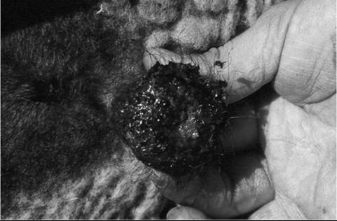
FIG. 34.17 Ulcerative posthitis in a Suffolk ram.
swollen and elongated. Affected animals often show dysuria, and goats may vocalize during voiding. As the local inflammation progresses, ulceration may result in fibrous adhesions between the penis and prepuce. Severe inflammation of the glans penis can cause stricture of the urethral process (pizzle) and restrict urine egress, resulting in signs similar to those of obstructive urolithiasis. Impairment of breeding soundness may result from pain, blood or exudate in the ejaculate, penile adhesions, scarring of the preputial orifice, or suppurative urethritis. Weight loss may occur in chronic cases. Phimosis has been reported in affected camels.1
Ulcerative lesions of similar appearance can develop on the vulva and perineum of affected females. Gross vulvar enlargement may be noticed from a distance.
Dysuria may result from involvement of the urethral orifice. The fibrosis and contracture that develop in chronic, severe cases may distort normal vulvar conformation to the point of impairing copulation or parturition.Ulcerative dermatosis (lip and leg ulcer) in sheep is dermatitis caused by poxvirus related to the contagious ecthyma virus. Infection with this agent may manifest as balanoposthitis and vulvitis, and the crusted ulcers that develop closely resemble those induced by infection with Corynebacterium renale. Ulcerative dermatosis can be differentiated from ulcerative posthitis because lesions are also present on the face and legs with the former condition. The lesions readily bleed and often contain a nonodorous, purulent discharge.
Contagious ecthyma (orf) occasionally affects the genitalia and perineum, although lesions are more frequently found on the lips, face, and udder. These lesions are raised, appear proliferative, and are covered with a thick, durable scab. Secondary bacterial infections and necrosis can occur with concurrent Fusobacterium necrophorum infection.2
Urolithiasis must be strongly considered in the differential diagnosis of any dysuric male small ruminant. Preputial trauma, particularly if resulting from entrapped grass awns, can cause preputial swelling, pain, and exudation within the preputial cavity. External ulcerative lesions would not be expected in such cases.
Caprine herpesvirus 1 (CHV-1) and ovine herpesvirus 2 (OHV-2) infections have been documented to cause preputial,
penile, vulva, and vaginal lesions in goats and sheep, respectively. Ulcers tested positive by PCR for the herpesviruses.3-6
Corynebacterium pilosum, Corynebacterium cystitidis, Mycoplasma mycoides subsp. mycoides large colony type, Actinobacillus seminis, Ureaplasma spp., Mycoplasma ovine-caprine serogroup 11, and Mycoplasma capricolum subsp. capricolum have been implicated in posthitis and vulvitis in sheep.7-9 A possible synergism between Mycoplasma mycoides subsp.
mycoides large lolony type and Trueperella pyogenes has been demonstrated in Dorper sheep in South Africa.8Pathophysiology
C. renale is the bacterium most commonly implicated in ulcerative posthitis. These are aerobic, gram-positive, pleomorphic, club-shaped bacteria that are normal inhabitants of the skin and external genitalia of small ruminants. The bacteria contain the enzyme urease. A high-protein diet results in increased ammonia production in the rumen, which is converted to urea in the liver. The urea is eliminated through the renal system. C. renale proliferates, and the urease hydrolyzes the urea back to ammonia, causing chemical irritation and ulceration of the prepuce and surrounding skin.
Epidemiology
The condition appears to be more common in males than females. Risk factors are associated with increased or excessive dietary protein concentration (usually >16%), such as alfalfa hay or lush legume or legume-grass pastures. However, diets with protein levels as low as 12% have been implicated.7 Rams, pet wethers, club or show lambs or kids, wool and fiber production animals, and feedlot lambs are most predisposed to the disease. Increasing the plane of nutrition in intact males before breeding predisposes to development of this disease, and multiple animals can be affected simultaneously.10,11 Breeds with dense wool or hair, such as Merino sheep and Angora goats, are also at risk owing to urine soaking the coat near the preputial orifice. Blowfly myiasis appears to increase the risk of ulcerative posthitis.11
Disease can occur in individual or multiple animals, since the organism can persist in wool, hair, and scabs for as long as 6 months and can survive freezing in scabs and exudative material. Venereal transmission of the disease has been documented; several months may pass between exposure of ewes to infected rams and development of vulvitis in the ewes. Production losses result from debilitation caused by pain, incapacitation of breeding animals, loss of breeding soundness, and deformation of external genitalia.
Treatment and Prognosis
Reduction of protein and nonprotein nitrogen intake is crucial for successful treatment of affected animals and prevention of additional cases. Dietary changes alone may result in satisfactory cure if the lesions are early in development. Incorporation of grass hay feeding into a program of legume pasture grazing may help limit protein intake.
Affected animals should be isolated to limit venereal or contact transmission. Fly control should be implemented. Wool or hair should be removed from the skin surrounding the prepuce or vulva, and a topical antibiotic may be applied. Caustic antiseptic solutions should not be used. Treatment with systemic antimicrobials is indicated for advanced cases or for outbreaks, in which handling of several animals for topical therapy is not feasible. C. renale is sensitive to penicillin, ampicillin, cephalosporins, and oxytetracycline. Treatment should continue until the lesions have dried and acute inflammation has subsided. Topical therapy of the preputial or vulvar mucosa includes application of mastitis treatment tubes, triple antibiotic ointment, or “petercillin.” Petercillin is prepared by mixing 240 g anhydrous lanolin, 20 mL of procaine penicillin G (300,000 IU∕mL), and 200 mg aqueous prednisolone. A small amount can be warmed up and injected into the prepuce using an AI infusion pipette or small feeding catheter. Oral or injectable NSAIDs can be used to decrease the inflammation and pain associated with the infection.
Surgical treatment of advanced cases is recommended for severe strictures. The ventral preputial tissue can be resected to allow for normal urine flow or be incised along part of its length.
Response to treatment is optimal early in the infection, before deformation of the prepuce or vulva secondary to fibrosis occurs. The chances for full recovery without recrudescence are poor if dietary protein intake is not reduced. Complete recovery of breeding soundness is unlikely in animals with internal ulcerative posthitis. In breeding males, the presence of ulcerative posthitis does not directly influence semen motility or morphology, but since pain is likely to suppress libido, proper treatment and documentation of lesion resolution are recommended in affected males before the breeding season.12
Prevention
Prevention primarily involves limiting both protein and nonprotein nitrogen intake. Breeding males should be body condition scored early enough before the breeding season so that any necessary weight gains can be made slowly rather than as a sudden onset immediately before the breeding season. Whenever possibly, weight gain should be induced by feedstuffs with a protein concentration of less than 12% on a dry matter basis. Separation of affected males will help decrease spread through contact with exudative materials. Shearing, especially at the time of highest protein intake, may be efficacious in reducing disease incidence.