Urinary Incontinence
Elizabeth A. Carr • Dominic R. Dawson-Soto •
Alexandra J. Burton
Risk Factors and Causes
Urinary incontinence in the horse can result from urolithiasis, congenital anomalies or defects of the lower urinary tract, trauma, neoplasia, neurologic diseases accompanied by bladder dysfunction, and decreased urethral tone.
Bladder and urethral calculi frequently result in transient or intermittent incontinence secondary to sabulous cystitis or partial obstruction.1-4 Ectopic ureter and other congenital malformations of the urinary tract generally produce incontinence from birth, although it may not be recognized until several weeks or months of age. Presentation of incontinence in adult horses due to congenital anomalies has also been described.5-8 Trauma-induced incontinence may develop after breeding injury or dystocia in mares, or in both genders after sacral or spinal injury. Incontinence may also develop with severe long-standing lumbosacral or back problems if they make it difficult and/or painful for the horse to posture to urinate. In these instances, over time, incomplete bladder emptying allows the crystals normally present in equine urine to accumulate in the ventral aspect of the bladder. This crystalloid sediment becomes heavy (and in some cases quite firm), further preventing complete bladder emptying. This condition, which is termed sabulous urolithiasis, can accompany bladder paralysis of any cause but also may itself cause myogenic bladder dysfunction, in the absence of any underlying neurologic problem.1,2 The weight of the sediment may induce chronic stretching of the bladder wall, contributing to myogenic dysfunction. Horses with lower urinary tract neoplasia can also present with incontinence, but other complaints (e.g., stranguria or hematuria) are usually reported as well.Neurologic disorders that most commonly result in bladder paralysis and incontinence include equine herpesvirus-1 myeloencephalopathy (EHM), cauda equina neuritis syndrome, polyneuritis equi, sorghum toxicosis, and aberrant larval migrans (Halicephalobus gingivalis) and equine protozoal myeloencephalitis (EPM), dependent on parasite location within the nervous system.9-14 These diseases, along with other problems affecting gray matter of the sacral segments, result in loss of lower motor neuron (LMN) function, whereas lesions of the lumbar or higher portions of the spinal cord result in loss of upper motor neuron (UMN) function.
Loss of detrusor muscle function and overflow incontinence is seen with LMN damage, and a large, easily expressed bladder is found on rectal palpation. Initially, UMN disease is characterized by increased urethral resistance, which leads to increased intravesicular pressure before voiding can occur. Voiding may occur as short bursts of urine passage with incomplete bladder emptying, and rectal examination may reveal a turgid bladder that is variable in size. Although UMN signs are initially different from those of LMN disease, incontinence is not usually recognized until overflow incontinence develops as a result of sabulous urolithiasis and progressive loss of detrusor function. The latter progression can explain why bladder paralysis and incontinence may be found in horses with other neurologic diseases like cervical stenotic myelopathy (CVM), equine degenerative myelopathy (EDM), and even viral encephalomyelitis other than EHM. Presence of other signs associated with LMN dysfunction (e.g., loss of anal or tail tone) or UMN dysfunction (e.g., ataxia) may aid in differentiating the inciting cause of bladder paralysis. Despite many possible causes, the prognosis for recovery from incontinence resulting from bladder paralysis is generally poor because sabulous concretions and UTI rapidly complicate the problem. However, successful management of sabulous cystitis has been described in five horses for up to 3 years with medication in combination with bladder catheterization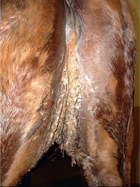
FIG. 34.5 Extensive thickening of the perineum and caudomedial thigh region due to chronic dribbling of urine (incontinence) in a 17-year-old mare with a large cystic calculus (see Fig. 34.10). (Image courtesy Montague N. Saulez.)
and lavage.2 A syndrome of estrogen-responsive incontinence caused by decreased urethral sphincter tone has been reported in a few mares.15,16
Diagnosis
In addition to taking a complete history and performing physical and neurologic examinations, it is helpful to take time to observe the incontinence or any attempts to urinate. Clinical signs often include stranguria and urine scalding of the perineum and hind legs in females, sometimes with tissue thickening if the incontinence has been long-standing (Fig.
34.5). Rectal palpation, transrectal US of the bladder and upper urinary tract, transabdominal US of the kidneys, and cystoscopy are useful to rule out uroliths, neoplasia, cystitis, and congenital anomalies as causes of incontinence. If trauma or sacral bony abnormalities leading to incontinence are suspected, radiographs and pelvic ultrasound should be attempted. Nuclear scintigraphy may further help in identification of a traumatic lesion, especially in large patients where radiography is limited. CT or magnetic resonance imaging (MRI) should be considered in foals and small ponies.17 Although most affected horses remain nonazo- temic (unless significant obstruction or bilateral pyelonephritis has developed), laboratory analyses of blood and urine, including a quantitative urine culture, should be performed in all horses with incontinence, because UTI is a common sequela. Urethral and bladder pressure profiles can be used to assess urinary sphincter and detrusor muscle function. Normal values for both mares and geldings have been reported.18-20 Where an underlying neurologic problem is suspected, cerebrospinal fluid (CSF) collection and analysis may also be of value.Treatment
Treatment for incontinence varies with the underlying cause. Removal of calculi and appropriate antimicrobial therapy are effective treatments for urolithiasis and UTI. Surgical correction is generally required for treatment of congenital anomalies, but owners should be discouraged from using affected animals for breeding. Incontinence due to EHM or EPM carries the most favorable prognosis for recovery, although in one author's (DRD) experience, with EHM, bladder paresis may persist for several weeks to months. To prevent continued distention and further damage to the detrusor function, removal of sabulous crystalloid material by bladder lavage with saline and temporary placement of an indwelling bladder catheter are indicated in cases of recent onset of bladder paresis.
Debate exists in the human literature regarding the preference of intermittent versus continuous urinary catheterization. In animals, both methods carry potential risks—infection and/or trauma from repeated catheterization—and each case should be evaluated individually to choose the most appropriate method.1,2,21 Antimicrobial treatment, ideally based on urine culture results, is also indicated in all horses with bacterial cystitis secondary to bladder paralysis.Bethanechol (0.03 to 0.04 mg/kg SC or IV q6-8h, or 0.22 to 0.45 mg/kg PO q6-8h [poorly absorbed]), a parasympathomimetic agent that has a somewhat selective effect on smooth muscle of the gastrointestinal tract and bladder, has been recommended for improving detrusor tone and strength of contraction in horses with bladder paralysis. However, response to treatment is often disappointing, perhaps because of longstanding paralysis before incontinence is recognized, although bethanecol-responsive bladder atony has been reported in a foal.22 Use of phenoxybenzamine (0.4 mg/kg PO q6h), an α-adrenergic blocker that decreases urethral sphincter tone, has also been recommended in combination with bethanechol in cases with UMN bladder dysfunction. In horses with evidence of urethral sphincter hypotonia, the sympathomimetic agent phenylpropanolamine (0.5 to 2 mg/kg PO q8-12h) has also been used. Dosing regimens for these autonomic drugs have been extrapolated from other species because no pharmacokinetic data are available for the equine species.
In general, treatment with these autonomic drugs has largely been ineffective in controlling incontinence due to bladder paralysis, and overall the long-term prognosis for recovery is usually poor. On a more positive note, treatment of a few mares with urethral sphincter hypotonia with estradiol cypionate or benzoate (5 to 12 μgZkg IM every day for 3 days, then every other day) has been effective at resolving incontinence as long as detrusor function is normal. Historically in the literature, estrogen-responsive incontinence has been reported in a total of three mares.15,16 Estrogen may modulate the effect of norepinephrine on α-receptor activity in the urethral sphincter, thereby improving urethral sphincter tone. There is also a report of successful surgical repair of an incompetent (damaged during removal of a cystic urolith) urethral sphincter in a mare.23