Viral Diseases
Stephen D. White
Papillomas (Warts, Fibropapillomas)
■ TABLE 40.1
Distribution and Appearance of Bovine Warts by Type of Virus
| Virus Strain | Usual Site | Appearance | Comments |
| BPV-1 | Nose, teats, glans penis | Filamentous or frondlike | Can prevent breeding when on penis |
| BPV-2 | Head, neck, brisket, occasionally alimentary tract | Pedunculated or broad-based mass | Most common typical warts |
| BPV-3 | Atypical warts, head, neck, possibly interdigitala | Nonpedunculated protruding growths, delicate fronds with hair between | Persist for years |
| BPV-4 | Alimentary tract, urinary bladder | Pedunculated mass | See Chapters 32 and 34 |
| BPV-5 | Teat | Smooth, white | Persist for years; other types may also occur on teats |
| BPV-6 | Teat | Round and either flat or frondlike | Similar in appearance to BPV-1 when frondlike |
aSuspected, but BPV-3 has not yet been isolated from interdigital warts.
Some authors speculate that interdigital warts may be caused by yet another new BPV strain.BPV, Bovine papillomavirus.
Modified from Hunt E: Infectious skin diseases of cattle. Vet Clin North Am Large Anim Pract 6:163, 1984.
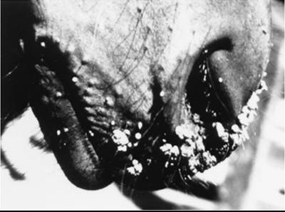
FIG.
40.8 Typical warts (papillomas) on the nose of a yearling horse.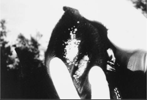
FIG. 40.9 Aural plaques on inner surface of pinna of a horse’s ear.
■ Treatment and Control Small warts can be crushed, pinched off, or surgically removed. Cryosurgery can be used on larger warts. Many regress spontaneously within a few months, even without treatment.
When show animals are involved or when animals have multiple large papillomas, tissue can be removed and made into crude autogenous vaccine (2 mL intradermally 3 times weekly) by homogenizing, grinding, freezing and thawing twice, filtering, and killing virus with 0.5% formalin. Autogenous wart vaccines are variable in their efficacy, as are commercial vaccines.14-16 The latter rarely seem to result in effective regression of existing warts but may prevent development of new lesions if the same BPV strain is involved. Autogenous vaccines can prevent new lesions caused by the same BPV strain in a herd. There is no indication that cattle vaccines have any efficacy in other species. No wart vaccines for horses, sheep, or goats are currently marketed.
Because the viruses can be transmitted directly or via fomites, prevention involves isolation; preventing animals from rubbing on each other; and not sharing halters, brushes, and other equipment. Dipping of dehorning, tagging, and tattooing instruments in a viricidal solution between animals will also slow spread of the virus.
Aural Plaques
Aural plaques is a form of viral papilloma that often affects the inner pinna (Fig. 40.9). Nonpruritic, these plaques may also occur on the genitalia and mammary glands. The color varies from pink to grayish-white. Plaques do not resolve spontaneously, as do the “classic” papillomas seen in young horses. Biopsy or “shaving off” aural papillomas may stimulate reduction or resolution of the masses, although in some horses this may only be temporary (6 to 12 months).
This has been theorized to be due to the release of “papilloma antigens” into the bloodstream during the surgical procedure, prompting an immune response against the tumor. Imiquimod (Aldara-3M, Minneapolis, Minn.), a nonsteroidal local immune responsemodifier cream, has been helpful in some cases when used 3 times weekly, every other week, for 2 to 4 months.17 Owners should wear gloves and should be forewarned that there is frequently an impressive inflammatory reaction to the cream in the initial weeks of treatment.A papillomavirus has been demonstrated in aural plaques on electron microscopy and with immunohistochemical techniques.18
Pseudocowpox
Pseudocowpox is a common parapoxvirus of cattle related to the viruses of contagious ecthyma (sore mouth) of sheep and goats and bovine papular stomatitis (see Chapter 32 for these diseases). All three parapoxviruses may cause nodular lesions on humans. The lesions of pseudocowpox are usually confined
to the teats of cattle, and the disease is common worldwide. Cyclic waves of reinfection occur in a herd, where it causes minor teat lesions characterized initially by a small papule 2 to 3 mm in diameter, followed by crusting and circular spread of the lesion. Approximately 10 days later, the 15- to 20-mm lesion appears as a ring or horseshoe-shaped scab.19 Lesions occasionally involve the udder, medial thighs, or scrotum. Deep ulceration is rare. There are no systemic signs of illness. The major problem associated with the teat lesions is an increased incidence of mastitis. The most important and common differential diagnoses are bovine herpes mammillitis and viral papillomas. Rare viruses involving the teat include vaccinia and cowpox.19 Cowpox is a rare disease of cattle in Europe that causes ulcers and may also produce lesions in humans. Vesiculation is rare in pseudocowpox, in contrast to bovine herpes mammillitis, vaccinia, and cowpox. Recent advances in PCR technology have improved diagnostic sensitivity20,21; advances in vaccine technology have been reviewed elsewhere.22
Bovine Herpes Mammillitis (Bovine Herpesvirus, Bovine Ulcerative Mammillitis)
Bovine mammillitis teat lesions are caused by bovine herpesvirus 2 (BHV-2), which is widely disseminated in most cattle populations.23,24 The disease may be epidemic or endemic.
The virus may also cause oral lesions, udder lesions, or generalized skin disease in cattle. The teat lesions start as swollen, tender, edematous teats. Vesicles may appear in some lesions, whereas others ulcerate almost immediately. The teats become painful, and ulcers require 3 to 10 weeks to heal.23,24 Mastitis is increased because the scabs on the teats are laden with bacteria. Diagnosis may be confirmed by isolation of the virus, the BHV-2 serum neutralization tester, or histologic demonstration of herpesvirus particles.23,24 Therapy consists of segregation of affected animals from the rest of the herd, and affected cows should be milked last. Milkers should wash hands between cows. In severe cases, secondary infection may be controlled by topical antibiotic creams or parenteral antibiotics, with proper residue avoidance precautions in place.Sheeppox and Goatpox
Sheeppox and goatpox are caused by capripoxviruses.25 Both occur in Africa, Asia, and the Middle East; goatpox also occurs in parts of Europe and the United States. The two diseases are clinically similar, although sheeppox has the most severe systemic signs of the animal pox diseases. Studies have shown that the viruses are phylogenetically distinct and can be differentiated by molecular tools.26 The diseases affect all ages, causing pyrexia, anorexia, conjunctivitis, rhinitis, and skin lesions. Prophylaxis using attenuated vaccines is the preferred control measure because the immunity is long lasting.25,26 Morbidity is high. Mortality may reach 80% with sheeppox but usually is low with goatpox. Humans may develop skin lesions from goatpox.