ANATOMY OF THE CARDIOVASCULAR SYSTEM
The anatomy of the cardiovascular system encompasses the structure and organization of its main components: the heart, blood vessels, and blood.
4.2.1 Heart
A. Structure:
• The heart is a muscular organ located in the thoracic cavity, slightly to the left of the midline.
• It is divided into four chambers: two atria (singular: atrium) and two ventricles.
• The heart is enclosed in a double-layered sac called the pericardium, which provides protection and anchorage.
B. Chambers and Valves:
• Atria: The two upper chambers of the heart receive blood from the veins and pump it into the ventricles.
• Ventricles: The two lower chambers of the heart receive blood from the atria and pump it out to the arteries.
DOI: 10.1201/9781003426851-4
23
• Valves: Valves within the heart ensure unidirectional blood flow:
- Atrioventricular valves (AV valves): The tricuspid valve (right side) and mitral valve (left side) separate the atria from the ventricles.
- Semilunar valves: The pulmonary valve (right side) and aortic valve (left side) separate the ventricles from the pulmonary artery and aorta, respectively (Figure 4.1).
C. Coronary Circulation:
• The heart has its own blood supply through the coronary arteries, which branch off the aorta.
• Coronary veins drain deoxygenated blood from the myocardium into the right atrium (Figure 4.2).
4.2.2 Blood Vessels
A. Arteries:
• Arteries carry oxygenated blood away from the heart to various tissues and organs.
• They have thick, muscular walls to withstand high pressure and maintain blood flow (Table 4.1).
B. Veins:
• Veins carry deoxygenated blood from tissues back to the heart.
• They have thinner walls compared to arteries and contain valves to prevent backflow.
C. Capillaries:
• Capillaries are tiny, thin-walled vessels where gas exchange and nutrient/waste exchange occur between blood and tissues.
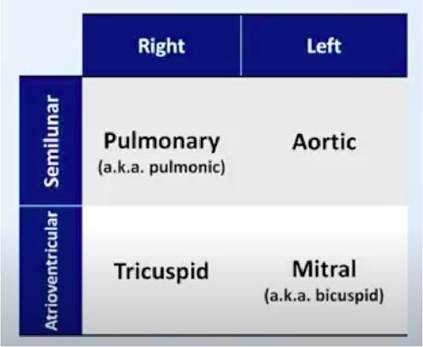
FIGURE 4.1 Pulmonary and Tricuspid valve
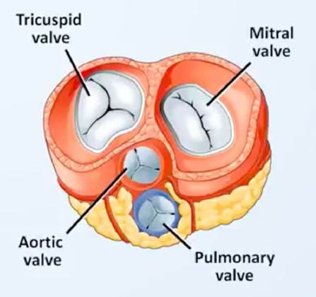
FIGURE 4.2 Coronary circulation
TABLE 4.1
Blood Vessels and their Function
Arteries All blood from the heart leaves into the aorta. From the aorta, they move into the large and medium sized arteries which are thick walled containing a significant amount of elastic tissues and smooth muscles. This allows them to handle the high pressure.
Arterioles Arteries divide into smaller arterioles which are the sight of greatest resistance to the blood flow through the circulation.
Capillaries Arterioles further divide into the further microscopic network of vessels called the capillaries. Which are sometimes as small as lines by a single layer of endothelial cells. Capillaries are the place where the actual exchange of fas between the blood vessels and the target peripheral tissue occurs. Although the capillaries are tiny, their numbers cross in billions in the body. Such thattheor net cross sectional area is much larger than any other level of the blood vessels. Capillaries are the place where the blood flows very slowly through the lumen. Hence allowing sufficient time for the gas, nutrients and waste exchange.
Venules From the capillaries, the blood travels into the venules Veins From venules the blood flows into the veins. There is
comparatively less elastic tissue in the veins than in arteries. Hence the veins have a larger capacity of distensibility. This allows the veins to act as the reservoir for the blood volume.
Lymphatics In addition to the blood vessels, there are also conduits. These are responsible to carry the interstitial fluids back to the main circulatory system. Interstitial fluids are the extravascular fluid which surrounds the cells. They sit in the connective tissue. These lymphatic vessels merge together and feed into a conduit called the thoracic duct. Which further joins the large veins of the thoracic cavity.
• They form extensive networks throughout the body, allowing for efficient exchange of substances.
4.3
More on the topic ANATOMY OF THE CARDIOVASCULAR SYSTEM:
- CLINICAL ANATOMY AND PHYSIOLOGY OF PAEDIATRIC PATIENTS
- CHAPTER MENU
- Background Information of Clinical Importance
- Diagnosis of Cardiovascular Disease by Presenting Sign
- Congenital Cardiovascular Disease
- Classification of cases: Sheep
- Classification of cases: Cattle
- PINNIPEDS
- Valvular Heart Disease
- Epidemic and Epizootic Expansions