Autoregulation Is a Relative Constancy of Blood Flow in an Organ Despite Changes in Perfusion Pressure
Metabolic control mechanisms also help to account for the phenomenon known as blood flow autoregulation. Autoregulation is evident in denervated organs and organs in which local control of blood flow is predominant over neural and humoral control (e.g., in coronary circulation, brain, and working skeletal muscle).
Figure 24-3 summarizes an experiment that demonstrates autoregulation in the brain. Initially, the perfusion pressure (arterial pressure minus venous pressure) in this animal is IOO mm Hg, and the blood flow to the brain is 100 milliliters per minute (mL/min) (point A). When perfusion pressure is increased suddenly to 140 mm Hg, brain blood flow rises initially to 140 mL/min but returns toward its initial level over the next 20 to 30 seconds. Eventually, blood flow reaches a stable level of about 110 mL/min (point B). Conversely, if the perfusion pressure is decreased suddenly from 100 to 60 mm Hg, blood flow in the brain decreases initially to 60 mL/min but
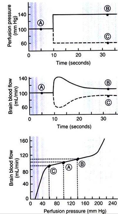
FIGURE 24-3 ■ The experiment summarized here demonstrates autoregulation of blood flow in the brain. Perfusion pressure was artificially set to various levels (top), and the resulting steady-state values of blood flow were measured (middle). The steady-state values of brain blood flow were then plotted against the perfusion pressure (bottom). Circled points A, B, and C are discussed in the text.
returns toward its initial level over the next 20 to 30 seconds (dashed lines in the top and middle graphs of Figure 24-3). Eventually, blood flow reaches a stable level of about 90 mL/min (point C). These stable responses are plotted in the bottom graph. The remainder of the bottom graph is obtained in a similar way; that is, perfusion pressure is set artificially to various levels, ranging from 40 to 220 mm Hg, and the resulting steady-state levels of blood flow are plotted.
Over a considerable range of perfusion pressure (about 60-190 mm Hg), relatively little change occurs in steady-state blood flow to the brain; that is, brain blood flow is autoregulated. The range of perfusion pressures over which flow remains relatively constant is called the autoregulatory range. Autoregulation fails at very high and very low perfusion pressures. Extremely high pressures result in marked increases in blood flow, and extremely low pressures result in marked decreases in blood flow. Nevertheless, over a considerable range of perfusion pressure, autoregulation keeps blood flow in the brain relatively constant.
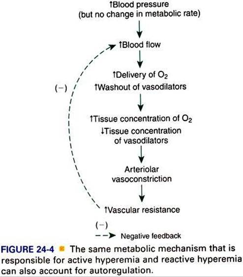
Figure 24-4 shows how the metabolic control mechanisms previously described can account for the phenomenon of autoregulation. If the metabolic rate of an organ does not change but perfusion pressure is increased above normal, the increased pressure forces additional blood flow through the organ. The additional blood flow accelerates the removal of metabolic products from the interstitial fluid and increases the rate of oxygen delivery to the interstitial fluid. Therefore the concentration of vasodilating metabolic products in the interstitial fluid decreases, and the concentration of oxygen in the interstitial fluid increases. These changes cause the arterioles of the tissue to constrict, which increases the resistance to blood flow above normal and decreases blood flow back toward its initial level, despite the continuation of the elevated perfusion pressure.
To summarize, metabolic control mechanisms bring about active hyperemia (the increase in blood flow in an organ in response to an increased metabolic rate, in the absence of any blood pressure change). The same metabolic mechanisms can also account for reactive hyperemia (the increase in blood flow above normal in an organ after a period of flow restriction). In addition, the same metabolic mechanisms can account for autoregulation (the relative constancy of blood flow in an organ when there has been no change in metabolic rate but blood pressure has either increased or decreased). Other mechanisms contribute to autoregulation, and the reader may encounter discussions of these under the terms myogenic hypothesis and tissue pressure hypothesis. However, metabolic control is the most likely explanation for autoregulation of blood flow in the critical tissues of a body (brain, coronary vessels, and exercising skeletal muscle).