Cardiac Action Potentials Are Extremely Long
Two major differences between action potentials in skeletal muscle and cardiac muscle have already been mentioned: First, cardiac muscle cells are electrically connected, whereas skeletal muscle cells are electrically isolated.
Second, the heart has pacemaker cells, which form spontaneous action potentials, whereas skeletal muscle cells only depolarize and form action potentials when “commanded” to do so by motor neurons.A third important difference between skeletal and cardiac action potentials is their duration (Figure 19-4). The entire action potential in a skeletal muscle lasts only 1 to 2 msec. A cardiac action potential lasts about 100 times longer (100-250 msec). Prolongation of the cardiac action potential is brought about by prolonged changes in the permeability of the cardiac muscle membrane to sodium (Na+), potassium (K+), and Ca2+ ions. Cardiac muscle cell membranes have Na* and K+ channels similar to those found in skeletal muscle, but the timing of their opening and closing is different in cardiac muscle. In addition, cardiac cell membranes also have special Ca2+ channels that are not present in skeletal muscle. The movement Ofextracellular Ca2t through cardiac Ca2+ channels has an especially important role in prolonging the cardiac action potential. The presence of Ca2 4 channels and the important role of extracellular Ca2' in the action potential is the fourth major difference between cardiac and skeletal muscle.
To understand the special significance of the membrane Ca2+ channels in cardiac muscle, it is useful to review the roles of K4 and Na+ channels in skeletal muscle and to emphasize some ways in which cardiac K* and Na' channels are similar to those in skeletal muscle. As explained in Chapter 4, many of the Kt channels in a neuron or skeletal muscle cell membrane
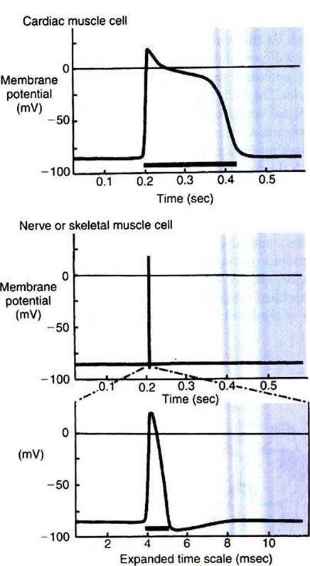
FIGURE 19-4 Action potentials in cardiac muscle cells (top) last 100 times longer than action potentials in nerve or skeletal muscle cells (middle).
Bottom,The nerve Orskeletal muscle action potential is shown on a greatly expanded time scale. The prolonged phase of depolarization in cardiac muscle cells is called the plateau of the action potential.The dark bars under each action potential indicate the length of the absolute refractory period.are open when the cell is at rest, and most of the Na1 channels are closed. As a result, the resting cell is much more permeable to K’ than to Na'. The tendency of the positively charged Kf to leave the cell creates a resting membrane potential (polarization) in which the inside of the cell membrane is negative in comparison with the outside. The resting membrane potential is typically between -70 and -85 mV (see Figure 19-4, bottom). An action potential is created when something depolarizes the cell (makes it less negative inside). Depolarization to the threshold voltage for opening the voltage-gated Na1 channels allows an influx of extracellular Na4 into the cell. This rapid entry of positive ions causes the cell membrane to become positively charged on its inside surface. This positive membrane potential persists for only a moment, however, because the Nar channels become inactivated very quickly, and the cell rapidly repolarizes toward its resting membrane potential. Repolarization is also promoted by the opening of additional K+ channels. In fact, this opening of extra K+ channels may cause neurons and skeletal muscles to become hyperpolarized (even more negative than normal) for a few moments at the end of each action potential (see Figure 19-4, bottom).
In a resting skeletal muscle cell, calcium ions are stored within the intracellular organelle called the sarcoplasmic reticulum. The occurrence of an action potential in the skeletal muscle cell causes Ca2* to be released from the sarcoplasmic reticulum into the free intracellular fluid, which is called the cytosol. The increase in cytosolic Ca2' concentration initiates muscle contraction (see Figure 1-5). The contraction initiated by a single action potential is very brief in skeletal muscle, because the cytosolic Ca2+ is rapidly pumped back into the sarcoplasmic reticulum by active transport, and the muscle relaxes. Note that the Ca2i responsible for initiating skeletal muscle contraction comes entirely from the intracellular storage site, the sarcoplasmic reticulum. No extracellular Ca2* enters the cell during the action potential, because skeletal muscle cells do not have membrane Ca2+ channels. In cardiac muscle, in contrast, membrane Ca2" channels and the entry of extracellular Ca2+ into the cells play key roles in both action potentials and contractions.