SUPERFICIAL STRUCTURES
Other organs that are visible or palpable in life may be identified with the assistance of Figure 25-2. Relatively little of the skull lies directly below the skin, but large areas have thin coverings of fascia and cutaneous muscle that offer little obstacle to palpation.
In addition to the broad forehead and dorsum of the nose, the temporal line, zygomatic arch, facial tuberosity, nasoin- cisive notch, and ventral border of the mandible are all easily palpated. The supraorbital, infraorbital, and mental foramina can also be identified (Figures 25-1, 25-2, and 25-8).Few specific features of the mimetic musculature are important. It is supplied by the facial nerve (VII), which divides into its principal terminal branches under cover of the parotid gland. The auriculopalpebral nerve supplies muscles of the external ear and eyelids. It reaches these by crossing the zygomatic arch directly in front of the temporomandibular joint, where its superficial position makes it vulnerable (Figure 25-6/5). Damage to the nerve may be evidenced by drooping of the ear and
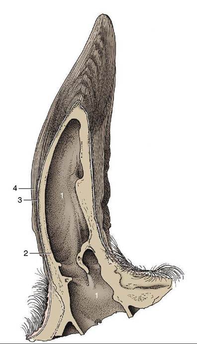
Figure 25-4 Longitudinal section of a bovine horn. 1, Cornual diverticulum of frontal sinus; 2, cornual process; 3, periosteum, dermis, and epidermis; 4, horn tubules.
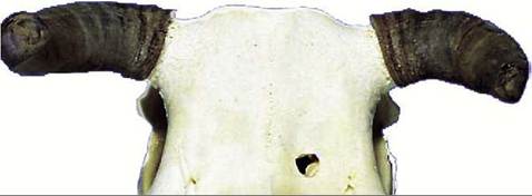
Figure 25-5 Horn rings resulting from variation in horn production and wear in cattle.
sagging of the eyelids, particularly the lower one. Paralysis of the orbicularis makes it impossible to close the eye. It is therefore clear that it may be advantageous to block the nerve to eliminate the blink reflex when examining the eye. It is most easily palpated where it passes over the zygomatic arch.
The dorsal buccal branch continues the parent trunk, crossing the masseter muscle in an exposed position that carries considerable risk of injury.
The effects of such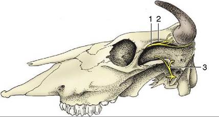
Figure 25-6 Bovine skull with cornual nerve (1) and auricu- lopalpebral nerve (3). The cornual nerve follows the temporal line (2) to the base of the horn. The auriculopalpebral nerve is palpable where it crosses the zygomatic arch.
injury include loss of innervation to the muscles of the nose and upper lip and to the buccinator. The first loss leads to slight distortion of the face, which is drawn toward the unaffected side; the second allows food to collect in a wad within the oral vestibule. The ventral buccal branch takes a more protected course caudome- dial to the ramus of the mandible and reaches the face in company with the facial artery and vein. It has a limited distribution, and the visible effects of injury are minimal (Figure 25-2/5,6).
The distribution of the cutaneous nerves is shown in Figure 25-8. Specific “blocks” of certain of these nerves are occasionally attempted. The large infraorbital nerve can be palpated where it leaves the infraorbital foramen, about 3 cm dorsal to the first cheek tooth. The mental nerve is found where it leaves the mental foramen of the mandible, about 3 to 4 cm caudal to the lateral incisor tooth.
The facial artery and vein are the most important superficial vessels. They cross the ventral margin of the mandible in front of the masseter muscle and are distributed to the lips, cheeks, muzzle, and periocular structures. The pulse may be examined where the artery lies on the side of the bone; it is less easily located in the notch of the ventral border.
The position of the frontal vein should also be noted because this fair-sized vessel is at some risk in trepanation of the caudal frontal sinus. The vein takes a caudo- rostral course in a palpable groove over the frontal bone to enter the supraorbital foramen; it then traverses a canal in the lateral part of the sinus. The foramen is located about 2 cm medial to the temporal line and about 2 cm caudal to the lateral angle of the eye (see Figure 25-12∕√).
A system of veins on the external surface of the pinna becomes engorged and prominent when a tourniquet is applied around the base of the ear. The central member of the set is sometimes used as an alternative to the jugular vein for the placement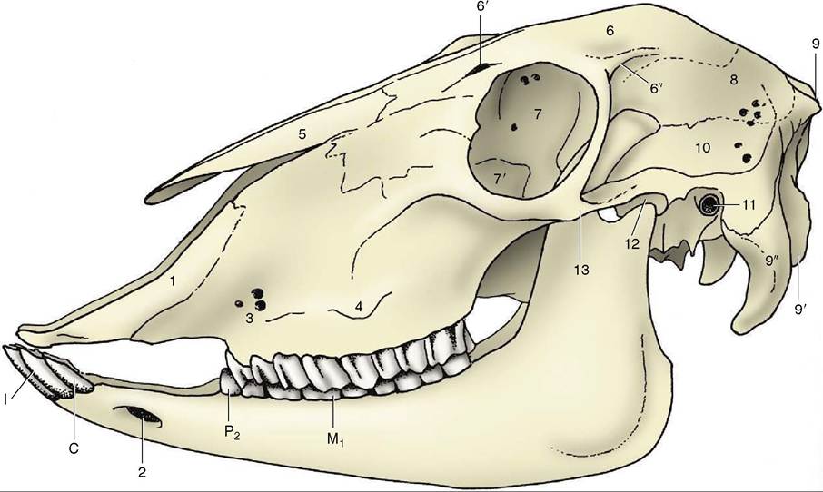
Figure 25-7 Lateral view of the skull of a sheep. 1, Incisive bone; 2, mental foramen; 3, infraorbital foramina; 4, facial tuberosity; 5, nasal bone; 6, frontal bone; 6', supraorbital foramen and groove; 6", temporal line; 7, orbit; 7, lacrimal bulla; 8, parietal bone; 9, external occipital protuberance; 9', occipital condyle; 9", paracondylar process; 10, temporal fossa; 11, external acoustic meatus; 12, temporomandibular joint; 13, zygomatic arch.
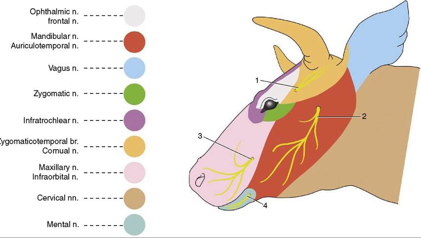
Figure 25-8 Skin innervation of the head. 1, Cornual n.; 2, auriculotemporal n.; 3, infraorbital n.; 4, mental n.
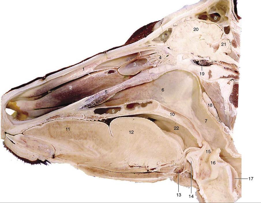
Figure 25-9 Paramedian section of the head. 1, Dorsal nasal concha; 2, ventral nasal concha; 3, middle nasal concha; 4, ethmoidal conchae; 5, vomer; 6, choana; 7, nasopharynx; 8, rostral frontal sinus; 8’, caudal frontal sinus; 9, palatine sinus; 10, soft palate; 11, apex of tongue; 12, torus linguae; 13, basihyoid; 14, thyroid cartilage; 15, epiglottis; 16, arytenoid cartilage; 17, cricoid cartilage; 18, medial retropharyngeal lymph node; 19, venous plexus surrounding hypophysis; 20, cerebrum; 21, cerebellum; 22, entrance to tonsillar sinus.
of an indwelling catheter. Neither site is free from problems.
The ventral end of the mandibular gland forms a conspicuous swelling in the intermandibular space. When palpated, this gland is often mistaken for the adjacent mandibular lymph node (Figure 25—2/20); its larger size, softer consistency, and more medial and more rostral extent make confusion unnecessary. The lymph node can be separately identified on the medial aspect of the sternomandibularis tendon. Normally the parotid lymph node is also palpable rostroventral to the temporomandibular joint.
In the last part of its course along the rostral margin of the masseter, the parotid duct accompanies the facial vessels and ventral buccal nerve. The duct penetrates the cheek opposite the fifth upper cheek tooth.