Eye
Camelids have eyes almost as large as horses and cattle, despite having a proportionately smaller head, and eye injuries are common because of the prominent and protruding eyes. Very little of the sclera is visible in a normal camelid, and what can be seen is often highly pigmented.
Camelids have long eyelashes, and they lack meibomian glands within the eyelids. They do, however, have sebaceous glands on the nictitating membrane and the caruncle of the eye. The lacrimal caruncle normally has hair. Fig. 38.16 illustrates features of the normal llama eye.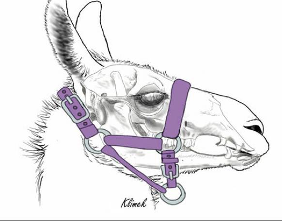
FIG. 38.15 Semi-schematic illustration of llama head with properly fitted halter. The noseband should fit very close to the eye to avoid compressing the nose.
FIG. 38.16
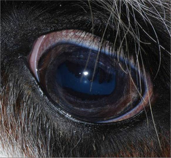
The normal appearance of the globe and adnexa of an alpaca eye. The cornea and pupil are
oblong in a horizontal plane, and the pupillary margins contain prominent iridal folds that are larger on the upper margin. The bulbar conjunctiva is usually pigmented, as in this individual. (From Cebra C, Anderson DE,
Tibary A, et al: Llama and alpaca care: medicine, surgery, reproduction, nutrition, and herd health, St. Louis, 2014, Elsevier, Fig. 38.11.)
The lacrimal glands are in the typical location and are about 31 mm ? 0.9 mm ? 0.5 mm in size. The superficial gland of the third eyelid is also in a typical position. It surrounds the crossbar portion of the cartilage of the third eyelid, and it is 25 mm ? 1.6 mm ? 0.8 mm in size, on the ventral aspect of the orbit. The lacrimal glands and superficial glands of the third eyelid are seromucous, and there are numerous lymphoid nodules on the bulbar surface of the third eyelid.
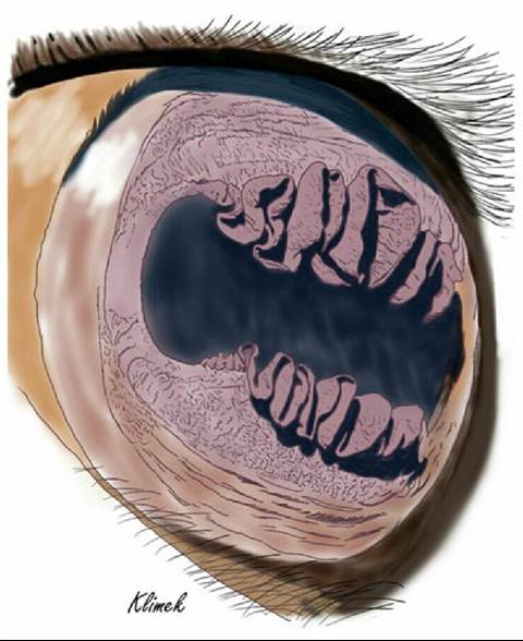
FIG.
38.17 Illustration of the iris of a llama showing the folded corpora nigra.Camelid eyes have structures analogous to the corpora nigra on the pupillary margin of cattle and horses. In llamas and alpacas, these proliferations of the iridic pigmented epithelium are elaborately folded vertically and are called pupillary ruffs, iridic granules, or corpora nigra. They are larger and very prominent on the upper pupillary margin, and the folds interdigitate with each other when the pupil is constricted. Fig. 38.17 is a drawing illustrating these folds on the pupillary margin.
The pigmentation of the iris varies with coat color. Animals with dark coats tend to have a brown iris, and those with light coats have combinations of gray, blue, and brown iris pigmentation. There is no tapetum lucidum or fovea, but the fundus is reflective, and the pigmentation of the fundus also varies with coat color, from nonpigmented to red-brown to brown, with dark-coated animals having heavier pigmentation. The vascular pattern normally has three to five pairs of prominent vessels originating at the optic disk.
Characteristics of the Alpaca Eyes: There are several characteristics of the eyes with which practitioners should be familiar, based on a study of alpaca eyes. The eyes of the neonate may have visible hyaloid arteries coming from the area of the optic disk and coursing toward the lens, and remnants of these are sometimes visible in older animals. As in other species, there is an association among white coat, blue eyes, and deafness, although deafness does not occur in all animals with a white coat and blue eyes. Finally, out of the 50 animals in the study, only one had a clear lens with no opacities of any kind; the most common finding was a ring or rings of opacity in the lens. Three animals with large focal dense opacities had no indication of visual deficits; thus lens opacities may be an incidental finding in the alpaca. However, this frequency of lens opacities was not noted in another study of 29 alpacas; in this study 2 animals had focal incipient primary cataracts. The eyelids may exhibit a slight ectropion when the animal is excited.
The nasolacrimal duct system follows the usual pattern. The lacrimal puncta are easily visible about 5 to 7 mm from the medial canthus and accessible to cannulate. The nasal opening of the nasolacrimal duct is laterally placed in the ventrocaudal aspect of the vestibule, about 1.5 to 2 cm proximal to the wing of the nostril, near the mucocutaneous junction.