» Neck and Trunk
Fig. 38.18 shows the skeleton of a llama. The vertebral formula of the llama and alpaca (cervical, thoracic, lumbar, sacral, and caudal) is C7T12L7S5Cd1^15.
The neck is long and very flexible in all directions and has sparse musculature and prominent cervical vertebrae. The normal neck carriage is vertical in the llama and about a 70-degree angle in the alpaca.The length and flexibility of the neck, along with the relative lack of supporting musculature, make cervical subluxations and fractures more common in camelids. In fact, bone tissue in general in camelids is relatively thin and easily fractured.
No description of the nuchal ligament is available for the llama or alpaca. The nuchal ligament is illustrated in one source as only consisting of the funicular part, similar to the nuchal ligament of the dog; however, the camel has both funicular and laminar parts of the nuchal ligament.
Fig. 38.19 shows the skull and cervical vertebrae of a llama. In the cervical region the bodies of the vertebrae are relatively long with the exception of C1 and C7, and the dorsal spinous processes are much reduced compared to other species. Cervical vertebrae 3 through 7 have downwardprojecting transverse processes that protect structures in the visceral space of the neck. These are very well developed cranially and caudally on the sixth cervical vertebra and are easily identified on a radiograph.
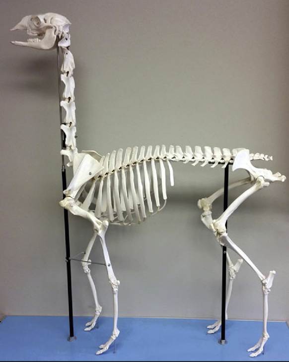
FIG. 38.18 Llama skeleton.
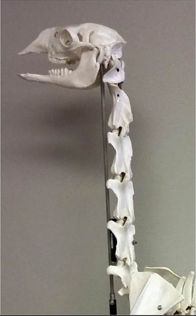
FIG. 38.19 Llama skull and cervical vertebrae. Llamas carry their head and neck in a nearly vertical orientation.
The cervical vertebrae of camelids lack transverse foramina.
The vertebral artery in the llama, and presumably in the alpaca as well, travels mostly within the vertebral canal, passing through osseous canals within the cranial portion of the vertebral canals of the second through the sixth cervical vertebrae. According to one source, the seventh cervical vertebra may or may not have a transverse foramen. The authors could find no information on the vertebral nerve in llamas and alpacas, but in the camel this nerve is inside this osseous channel; presumably it is in the same location in llamas and alpacas. This could have implications for the location of the communicating branches that normally leave the vertebral nerve to join the cervical spinal nerves.There are some interesting differences from the usual mammalian pattern with some of the nerves of the neck in llamas and alpacas. Both the recurrent laryngeal nerve and the external branches of the accessory nerve have been reported to be lacking in the alpaca. In one study the recurrent laryngeal nerve was absent, with both branches of the vagus to the extrinsic muscles of the larynx arising from the vagus nerve just outside the jugular foramen. In addition, the trapezius was innervated by branches of cervical nerves in the absence of the cervical branch of the accessory nerve. The external branch of the accessory nerve has also been found to be absent in the camel and the llama. The dromedary camel has cranial and caudal laryngeal nerves that originate from the vagus near its origin but also a small recurrent laryngeal nerve that follows the conventional route and travels cranially to the larynx to anastomose with the caudal laryngeal nerve.
The paired thyroid glands are on the dorsolateral aspect of the trachea. They are 4 cm long and 2 cm wide and extend from the cricoid cartilage to the third or fourth tracheal ring.
Jugular Venipuncture: Several features of cervical anatomy combine to make jugular venipuncture a particular challenge. There is only one jugular vein present in the llama, the internal jugular vein, and it is therefore relatively deep in the neck.
The skin on the neck is up to 1 cm thick, which is a protective mechanism against fighting but makes venipuncture more challenging and, in the case of catheterization, necessitates making a small stab incision through the skin over the vein to avoid damaging the tip of the catheter. There is no jugular groove in camelids because of the sparse musculature, and the jugular vein and carotid artery are in close proximity for much of their course through the neck. The jugular vein is relatively superficial only in the cranial part of the neck near the mandible and is separated from the carotid artery by a small omohyoideus muscle for only a short distance.The general rule for jugular venipuncture is to stay in either the rostral or the caudal third of the neck. Both the high and low locations have advantages and disadvantages. In either location, valves in the jugular vein may interfere with venipuncture. It is not always necessary to clip the fleece to locate the vein, and owners may resent the veterinarian clipping fleece because it can take 12 to 18 months to regrow. The fibers can be parted and taped out of the way to access the site for venipuncture or catheterization. The right side of the neck is preferred because a hematoma on the left side, from either jugular or carotid puncture, may compress the esophagus or cause choking.
The high jugular venipuncture location is near the ramus of the mandible, where the jugular vein is most superficial and there is more separation between it and the common carotid artery, and the skin is thinner, but landmarks are harder to palpate. To estimate the correct location for jugular venipuncture in this area, the needle is inserted dorsal to the intersection of a line drawn at the ventral border of the mandible and the tendon of the sternomandibularis muscle. The jugular vein in this location is lateral to the tendon as it inserts on the mandible but then moves dorsal, and then medial, to the tendon as it moves down the neck.
Also in this location, the omohyoideus, much smaller than that in cattle or horses, is positioned deep to the jugular vein, between it and the common carotid artery. The omohyoideus only extends for a distance of about 14 cm caudal to the ramus of the mandible. There is one set of valves approximately 1 cm caudal to the origin of the jugular vein and angle of the mandible and another set 5 cm caudal to that.As the jugular vein moves caudally in the neck, it becomes surrounded by the same fascial sheath as the common carotid artery and the vagosympathetic trunk. These structures are located between the trachea medially and the ventral projections of the transverse processes of cervical vertebrae laterally. The low position for venipuncture is medial to the ventral projections of the transverse processes of the fifth or sixth cervical vertebrae, with the advantage that here the vein is larger and landmarks are easy to palpate. The disadvantage of this location is a heavier fiber coat here and closer proximity of the carotid artery to the vein in this position. To find the jugular vein in this location, the ventral projection of the sixth cervical vertebra is located, and the thumb or fingers of the occluding hand are wrapped medial to this projection, between it and the trachea. The jugular vein should be medial to this projection. The carotid artery should be close, and pulsations may be felt. The needle is advanced slightly medial to the projection and toward the center of the neck. There may be a set of valves in this location as well.