Upper Airway
The soft palate is relatively long in the camelid. Llamas, alpacas, and Bactrian camels lack the dulaa (the diverticulum of the soft palate) present in dromedary camels. Camelids are obligate (or nearly obligate) nasal breathers because of the relatively long length of the soft palate and the arrangement of the intrapharyngeal ostium to the glottis in a manner similar to horses.
The caudal end of the soft palate is usually ventral to the epiglottis. The nasal cavity also has less rigid support than most other species, because the nasal septum is mainly cartilaginous and the nasal bones do not extend as far rostrally.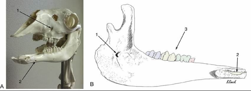
FIG. 38.13 (A) Rostrolateral view of a llama skull. 1, Infraorbital foramen; 2, mental foramen. (B) Medial view of a llama mandible. 1, Mandibular foramen; 2, incisor tooth; 3, one premolar and three molars (color added for contrast of individual teeth).
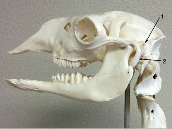
FIG. 38.14 Llama skull. 1, External acoustic meatus; 2, tympanic bulla.
Fitting a Halter: The less rigid support of the nasal cavity is important to remember when restraining the animal and when fitting a halter because pressure on the top of the nose can easily collapse and block the nasal passage, causing discomfort and distress to the animal and possibly making handling more difficult. For this reason, the nosepiece of the halter must fit close to the eyes and must not be allowed to slip forward. This also needs to be balanced against the need to allow movement of the jaws for mastication, so there must be slack in the nosepiece. Fig. 38.15 illustrates an appropriately fitted halter.
About 8 cm caudal to the nares, the nasal septum ends, and a common meatus is formed.
This allows observation of the caudal portion of the entire nasal cavity via an endoscope passed through the ventral nasal meatus on either side. The anatomy of the larynx is typical, although the larynx is relatively narrow.Endotracheal, Orotracheal, and Nasotracheal Intubation: The anatomy of the oral cavity and oropharynx complicates endotracheal intubation for inhalation anesthesia. Visualization of the glottis for orotracheal intubation is difficult or impossible without the use of a laryngoscope. Nasotracheal intubation is possible, especially when access to the oral cavity is necessary during anesthesia, but llamas and alpacas have a pharyngeal diverticulum that must be avoided if this method is chosen. The opening is approximately 1 cm in diameter, and the diverticulum extends caudally for approximately 2 cm between the longus capitis muscles. In adult llamas, when the tube is passed in the ventral meatus, the ethmoid conchae are about 10 cm from the nares and may be contacted by the tube. The pharyngeal diverticulum is about 25 cm from the nares and can also prevent further passage if it is entered. Camelids are at risk of dorsal displacement of the soft palate following extubation from the orotracheal route because of relaxation of the pharynx after the tube is removed.