Fascia and Fat
The connective tissue that separates and surrounds the more obviously important structures is generically known as fascia; many of its larger accumulations, particularly those of a sheetlike nature, have specific names.
The fascia has important functions as it is encountered in surgery.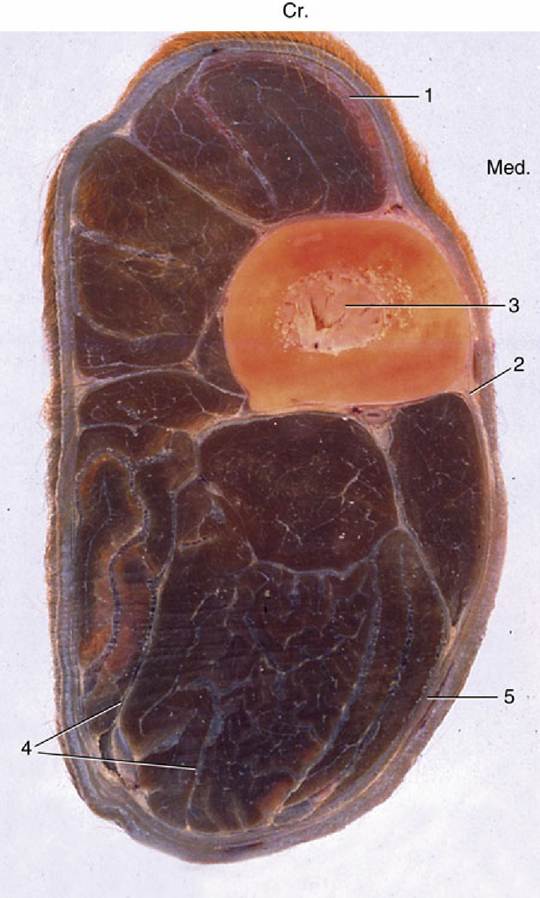
FIG. 1.8 Osteofascial compartments in the forearm of a horse. 1, Superficial fascia; 2, cephalic vein; 3, radius; 4, septa of deep fascia enclosing individual muscles or groups of muscles; 5, deep fascia. (In transverse sections of the limbs, cranial [Cr.] and medial [Med.] are identified.)
The superficial fascia (subcutis) is a loose (areolar) tissue extensively spread below the skin of animals that possess a hairy coat. A similar tissue surrounds many deeper organs, and in both situations, the loose fascia allows neighboring structures to change in shape and to move easily against each other. Its looseness varies with the amount of fluid it contains and may provide an indication of ill health. The superficial fascia is one of the principal sites for the storage of fat. In naked species, the fat forms a continuous layer, the panniculus adiposus.
The deep fascia is generally organized into much tougher fibrous sheets. A layer beneath the superficial fascia extends over most of the body and fuses to bony prominences. In many places it detaches septa that penetrate between the muscles, enclosing them individually or in groups (Fig. 1.8); sometimes the periosteum, the fibrous covering of the bones, participates in outlining the enclosures. This division into fascial or osteofascial compartments is very prominent in the forearm and leg and plays a part in the circulation, assisting the return of blood and lymph to the heart. The contraction of muscles presses the structures such as valved veins contained within their unyielding walls to return the blood toward the heart.
For this reason, muscular paralysis or prolonged inactivity may lead to stasis of blood or lymph flow. Arteries and nerves whose functions would not be assisted by compression often travel in small tunnels within the septa.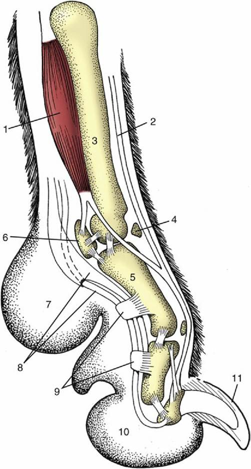
FIG. 1.9 Axial section of a dog's paw; the metacarpal pad (7) is in contact with the ground during standing. 1, Interosseous muscle; 2, extensor tendon; 3, metacarpal bone; 4, dorsal sesamoid bone; 5, proximal phalanx; 6, proximal sesamoid bone; 8, flexor tendons; 9, retinacula; 10, digital pad; 11, claw.
More specific functions can be assigned to localized thickenings (e.g., retinacula, tethers) of deep fascia, which hold tendons in place and sometimes provide pulleys around which the tendons wind to change direction. Good examples are provided by the retinacula on the dorsal aspect of the hock and the palmar aspect of the digits (Fig. 1.9/9).
Because dense fascia is relatively impermeable, it determines the direction taken by spreading fluids such as pus, which sometimes tracks below a fascial sheet before breaking through far from its source. Surgeons exploit both the toughness of the deep fascia to hold sutures securely and its cleavage planes to gain relatively bloodless access to deeper parts during surgery.
Most deposits of fat (adipose tissue) may be regarded primarily as food reserves. Small amounts of fat are widely distributed, but the bulk is contained in three or four places: in the superficial fascia (Fig. 1.10/2); between and within muscles; below the peritoneum (the delicate membrane lining the abdominal cavity); and in the marrow cavities of long bones. Subcutaneous fat deposits help mold the body contours and often show specific and gender differences in localization and development. Animals that are adapted to hot habitats often develop localized fat depots (e.g., humped zebu cattle and camels, fat-tailed sheep), leaving the rest of the body surface to release heat to the environment.
The onset of puberty leads to fat deposition in the breasts and over the hips in women. In many male animals, much fat is deposited in the tissues of the dorsal part of the neck: the thickened crest of stallions is a good example.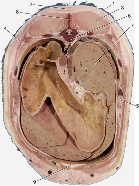
FIG. 1.10 Transverse section of the back of a pig. 1, Skin; 2, fat (panniculus adiposus) associated with the superficial fascia; 3, back muscles; 4, cutaneous muscle enclosed within superficial fascia; 5, rib; 6, thoracic vertebra; 7, liver; 8, spinous process of vertebra; 9, additional fat deposited between muscles.
Some fat deposits, like that enclosed within a fibrous lattice in the footpad of the dog, function as mechanical buffers (see Fig. 1.9/7 and 10). Fat with a mechanical function is usually resistant to mobilization during starvation.
The chemical and physical differences in fat may reflect diet as much as specific genetic factors. The fat of horses and of Channel Island breeds of cattle is yellow, that of sheep hard and white, and that of pigs soft and grayish. It should also be remembered that fat at body temperature is softer (semifluid) than that exposed in a colder environment. Certain procedures employed by the cosmetic surgeon—liposuction and lipofixation—depend on this fortunate circumstance.
All these remarks refer to the common sort of fat. A second variety, brown fat, is of much more restricted distribution in time and place. Brown fat differs in structure (Fig. 1.11) and function as well as in color. In domestic species, it is found especially during the fetal and neonatal periods; in wild species, it is especially prominent in those that hibernate (Fig. 1.12). The brown adipocyte contains numerous smaller droplets and a much higher number of mitochondria. It is richly vascularized. It provides both groups in which it is especially found with a readily available source of heat, which is equally useful in newborn animals with imperfect thermoregulation and in hibernators required to awaken rapidly from a deep winter sleep.
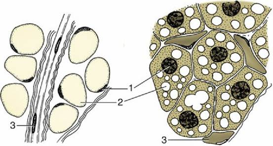
FIG. 1.11 Fat cells of white (left) and brown (right) fat. In white fat a single large fat vacuole displaces the cytoplasm and the nucleus to the periphery of the cell. The small fat vacuoles are evenly distributed in the cells of brown fat. 1, Nuclei; 2, fat vacuoles; 3, capillaries.
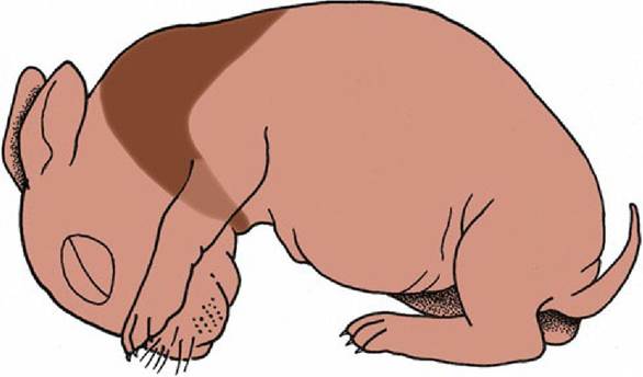
FIG. 1.12 The distribution of brown fat in the newborn rabbit, concentrated around the neck and between the shoulder- [Per Dorland's and other chapters.] blades.