INNERVATION AND VASCULARIZATION
The principal gastric nerves, parasympathetic efferent and afferent, run in the trunks formed along the esophagus by the regrouping of vagal fibers (see Figure 27-3/79,20). The sympathetic nerves that reach the stomach through periarterial plexuses have a subordinate role.
Section of both vagal trunks abolishes all motor activity of the forechambers. Section of the dorsal trunk alone results in almost complete but not necessarily permanent paralysis of the rumen, while the effect on the reticulum is generally less marked. The effects of division of the ventral trunk are unpredictable and range from little or no discernible change to almost complete paralysis of the forechambers. It is presumed
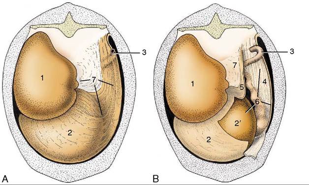
Figure 28-24 Attachment of the greater omentum to the stomach and the dorsal body wall. A, Caudal view of intact greater omentum. B, Caudal view of greater omentum fenestrated to permit a view into omental bursa. 1, Dorsal sac of rumen; 2, ventral sac of rumen, covered by superficial wall of greater omentum; 2’, ventral sac of rumen projecting into omental bursa; 3, caudal flexure of duodenum; 4, superficial wall of greater omentum; 5, deep wall of greater omentum; 6, omental bursa; 7, supraomental recess.
that these inconstant results can be explained by differences in the regrouping of fibers where the vagus nerves combine to form the dorsal and ventral trunks and by the later assumption of part of these functions by association neurons in the stomach wall.
Abomasal contractions are greatly reduced after bilateral vagal section but are not wholly interrupted, possibly because some intrinsic control is vested in a submucosal nerve plexus present in the wall of this chamber alone. Division of the splanchnic nerves brings only slight alteration to the gastric movements.
Clinically, disturbances of stomach function may follow involvement of the vagus nerves at any point along their courses from the brainstem; the most common causes are mediastinal infections and traumatic reticulitis.The stomach is supplied with blood through several branches of the celiac artery. The large right ruminal artery runs caudally in the right longitudinal groove and continues into the left groove by passing between the dorsal and ventral blind sacs. It supplies most of the rumen wall and ends in anastomosis with the left ruminal artery, which follows the cranial groove (between atrium and ventral sac) to supply adjoining parts of the rumen and reticulum. The omasum and abomasum are supplied by the left gastric and left gastroepiploic arteries that follow their curvatures.
The veins are mainly satellite to the arteries. The left ruminal vein joins veins draining other chambers of the stomach; the right one, the veins leading from the spleen; their union produces a major radicle (splenic vein) of the portal vein.
Many small lymph nodes are scattered over the stomach, particularly in the ruminal grooves and over the omasal and abomasal curvatures. Lymph from the forechambers leads, after serial passage through these peripheral nodes, to a number of large atrial nodes situated between the cardia and omasum and thence to the visceral root of the cisterna chyli. The nodes placed along the abomasal curvatures direct their efferent vessels to the hepatic lymph nodes.