Introductory Survey
The brain* and spinal cordt are continuous without any clear demarcation. The brain is a very irregular organ whose shape conforms very approximately to the cranial cavity in which it is lodged, whereas the slender elongated cord has a more regular and uniform appearance.
The size of the brain does not bear a linear relationship to that of the animal from which it came but is relatively smaller in large species and is proportionately larger in more advanced mammals. The ratio of brain mass to body mass is of the order of 1:50, 1:200, and 1:800 in the human, dog, and horse, respectively. As a general rule, domestication of a species leads to a smaller brain to body mass ratio than in nondomesticated species of the same kind; the process is not reversed by putting domesticated animals back into the wild. Perhaps more important is the relative development of particular parts of the brain; mammals possess a relative preponderance of phylogenetically newer parts, particularly in the cerebrum generally and in "higher" mammals specifically, in comparison with other forms. The great size and complexity of the human cerebral hemispheres provide the extreme example of this evolutionary trend (Fig. 8.8).
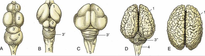
FIG. 8.8 Vertebrate brains illustrating the phylogenetic development. The increase in volume and complexity of the telencephalon and cerebellum is most striking. (A) Fish (carp); (B) reptile (python); (C) bird (duck); (D) mammal (cattle); (E) mammal (human). 1, Telencephalon; 2, mesencephalon; 3' and 3”, metencephalon (3' archicerebellum in A-C; 3” neocerebellum in D); myelencephalon; 5, spinal cord.
An initial survey of the brain as a whole is given here, followed by an account of its development. Detailed descriptions of the parts of the central nervous system follow in this chapter.
Repeated consultation should be made to the figures indicated so that the structures named can be located and identified.Dorsal views show the cerebral hemispheres and cerebellum as the dominant features of the mammalian and avian brain; only a small part of the medulla oblongata is visible in continuity with the spinal cord (Fig. 8.8 C, D). The semiovoid cerebral hemispheres are divided from each other by a deep longitudinal fissure and from the cerebellum by a transverse fissure; when the brain is in situ, both fissures are occupied by folds of the tough dural membrane that lines the cranial cavity. In many domestic mammals, each hemisphere is molded to display ridges (gyri) and grooves (sulci) in patterns that differ significantly among the various species. The cerebellum has an even more pronounced surface folding, the purpose of which is to increase the area available for neural processing.
The ventral aspect of the brain is flatter overall and reveals the subdivisions of the brain more clearly (see Fig. 8.19). The caudalmost part is the medulla oblongata, which widens cranially until it terminates at a prominent transverse ridge. This ridge represents the ventral surface of the next part of the brain, the pons. The ridge is formed by the transverse fibers of the pons, which can be followed dorsally over the lateral aspect to join the cerebellum (Fig. 8.19). The midbrain is located cranial to the pons and is represented on the ventral aspect of the brain by two divergent columns, the crura cerebri or cerebral peduncles (Fig. 8.19/12). They continue rostrally to disappear into the depths of the hemispheres. They are separated by the interpeduncular fossa, a shallow depression on the ventral midline of the midbrain (Fig. 8.19/13). The forebrain lies rostral to this depression; its most prominent ventral median features of the forebrain are the hypothalamus (to which the hypophysis [pituitary gland] is attached by a stalk) and the crossing or chiasm formed by the optic nerves. The larger part of the forebrain is provided by the paired cerebral hemispheres, which have as their most prominent ventral features the rounded piriform lobes (Fig. 8.19/3), flanking the crura cerebri, and the rostrally located olfactory tracts (Fig. 8.19/2), which originate in the olfactory bulbs that project at the rostral extremity. The superficial origins of the cranial nerves, all except the trochlear (IV) pair, are also visible on the ventral surface.
The cerebral hemispheres and cerebellum dominate the dorsal aspect of the brain, and when they are removed, all that remains is referred to as the brainstem, which is directly continuous with the spinal cord (see Fig. 8.22).