Joints
Bones form joints or articulations, of which some unite the bones firmly and others allow free movement. The many variations in joint shapes and structures do not permit an easy system of classification.
Periodic revisions of terminology have seen new categories defined and former categories merged or renamed so that some confusion now exists and many superfluous terms circulate. The current official system recognizes three major categories—namely, the fibrous joint, in which the bones are united by dense connective tissues; the cartilaginous joint, in which the bones are united by cartilage; and the synovial joint, in which a fluid-filled cavity intervenes between the bones. It is obvious that most joints of the first and second categories must be relatively immovable or even rigid; these classes were formerly together known as synarthroses. In contrast, most joints of the third category are freely movable; they were formerly termed diarthroses. Both of these terms are obsolete.Fibrous Joints
Most fibrous joints occur in the skull and are known as sutures (Fig. 1.18). The narrow strips of fibrous tissue that outline and unite the margins of the bones represent the surviving part of the originally continuous membrane in which the separate ossification centers appeared. Sutures play an important role in the young animal, allowing for the growth of the skull through the extension of individual bones at their margins while proliferation of the membrane continues. Sutures are gradually eliminated when ossification extends across the membrane after it has ceased to grow. This is a slow and uneven process that is not complete even in the aged. The gradual modification of the suture pattern is used in anthropology and forensic medicine as a not very reliable guide to the age of the individual. In comparison with sutures of the adult skull, the wider sutures of the fetal skull allow some useful passive deformation during birth in some species, including primates.
FIG.
1.18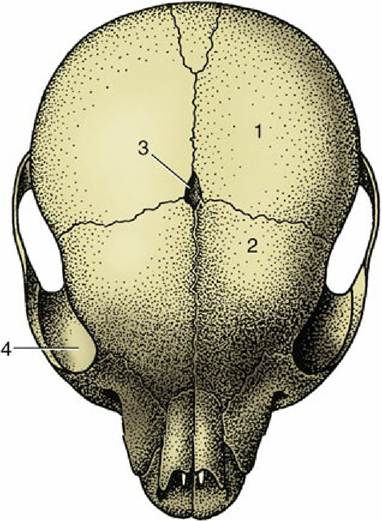
Sutures between the bones of a puppy's skull. 1, Parietal bone; 2, frontal bone; 3, fontanelle
(fonticulus); 4, orbit.
The syndesmoses are fibrous joints in which two bones are joined by connective tissue ligaments. In some syndesmoses, relatively broad areas of bone are united by short ligaments, allowing very restricted movement; examples are the joints between the major and minor bones of the horse's metacarpus. In others the ligaments are longer and their attachments narrower so that more appreciable movement is possible; an example is the joint between the shafts of the radius and ulna in the forearm of the dog.
The attachment of a tooth to the bone of its socket, or gomphosis, may be included among the fibrous joints.
Cartilaginous Joints
Most cartilaginous joints are known as synchondroses. They include the joints between the epiphyses and diaphyses of juvenile long bones and the corresponding joints of the base of the skull. These temporary joints disappear with ossification of the cartilage. The few permanent synchondroses include the joint between the skull and hyoid apparatus (p. 57), which allows appreciable movement in some species.
In the more complicated symphysis the articulating bones are divided by a succession of tissues; usually cartilage covers the bones, with fibrocartilage or fibrous tissue in the middle. The category includes the joints between the symmetrical halves of the mandible (in species such as the dog, cat, and ruminants, in which fusion is not complete) and of the pelvic girdle and the joints between the bodies of successive vertebrae (Fig. 1.19). Each of these joints presents its own, sometimes specifically variable, features that are best considered later.
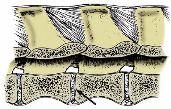
FIG. 1.19 Intervertebral disk (arrow) joining bodies of adjacent vertebrae.
Synovial Joints
Structure
In synovial joints the articulating bones are separated by a fluid-filled space, the joint cavity (Fig. 1.20). The boundaries of the space are completed by a sleeve of delicate connective tissue, the synovial membrane. The membrane is attached around the periphery of the articular surfaces. However, in most synovial joints the synovial membrane is strengthened externally by a fibrous capsule, and additional fibrous bands (ligaments) are strategically placed to join the bones and to restrict movement to the required directions and extents. Synovial joint injuries are highly prevalent in domestic animals.
The articular surface is covered with articular cartilage that is generally of the hyaline variety, although fibrocartilage or even dense fibrous tissue is found in a few locations. The thickness of the cartilage ranges from about a millimeter in the joints of the dog to several millimeters in the larger joints of horses and cattle. It accentuates the curvature of the underlying bone, being thickest in the center of convex surfaces and about the periphery of concave ones. The cartilage is a pliant material that is translucent and glassy in appearance, and although generally white with a blue or pink tinge in young animals, it becomes yellowish and less elastic with age. The surface is smooth to the touch and to the naked eye but quite irregular when seen at low magnification.
The cartilage has a complex structure in which fine fibers within its matrix pass from the underlying bone to the surface, where they bend to lie close together. Because splitting of the cartilage, common in joint disease, tends to follow the fiber course, superficial lesions lead to tangential flaking, whereas lesions that extend more deeply create more or less vertical cracks.
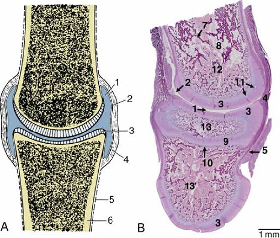
FIG. 1.20 (A) A synovial joint in section. (B) Sagittal section through the decalcified digit of a newborn lamb stained with Masson's trichrome.
1, Joint cavity; 2, synovial membrane; 3, articular hyaline cartilage;4, fibrous layer of joint capsule; 4,, joint capsule; 5, periosteum; 6, compact bone; 7, trabeculae; 8, hemopoietic tissue; 9, shaft; 10, growth plate; 11, vascular channels in hyalline cartilage; 12, proximal phalanx; 13, middle phalanx.
Articular cartilage is insensitive and avascular. The insensitivity explains why joint lesions may progress far before the patient becomes aware of their existence. The oxygen and nutritive requirements are met by diffusion from three sources: fluid within the joint cavity, vessels in the tissues at the periphery of the cartilage, and vessels in the subjacent marrow spaces. Diffusion is assisted by the porosity of the cartilage matrix, which soaks up and releases fluid as the cartilage is alternately unloaded and compressed during movements of the joint.
Certain large articular cartilages are interrupted by depressed areas that may indent the periphery or appear as islands. These naked areas (synovial fossae) are clothed by a thin connective tissue resting on the underlying bone. Their significance is disputed but not the constancy of their occurrence nor their frequent coincidence in opposing bones in certain positions of the joint.*
The synovial membrane, which completes the lining of the joint, is a glistening pink connective tissue sheet. It may be left entirely unsupported, may rest directly on a tough outer fibrous capsule, or may be separated from it by the interposition of pads of fat; all three arrangements may occur in different regions of the same joint. The membrane may pouch where it is unsupported, and these diverticula may extend quite far, a point of potential significance because it explains how joints may be entered by apparently remote wounds. The inner surface of the membrane carries many projections of various sizes and degrees of permanency that greatly increase its surface area (see Fig. 1.20B). Unlike mucous membranes, the synovial membrane has no continuous covering of cells.
The more cellular parts, limited to relatively protected situations, are responsible for the production of the lubricant component (aminoglycans) of the synovial fluid. The other components are derived from the blood plasma. The membrane is both vascular and sensitive.Synovia, the fluid within the cavity, obtains its name from its resemblance to egg white. It is a viscous, fluid whose color ranges from pale straw to medium brown. It is usually said to be present in very small amounts but is, in fact, quite copious in the larger joints; as much as 20 to 40 mL can sometimes be aspirated from limb joints of horses and cattle. The quantity is greatest in animals permitted free exercise.
Synovia has both lubricant and nutritive functions. The ways in which it acts as a lubricant are disputed, but it is certainly very efficient, the friction being such that virtually no wear occurs in healthy joints. The fluid helps nourish the articular cartilage, any intraarticular structures, and, possibly, the surface layer of the synovial membrane itself.
An outer fibrous layer usually completes the capsule. It attaches around the margins of the articular surfaces and contains local thickenings, which are named individually as ligaments when well developed and discrete. Some, of which the cruciate ligaments of the stifle are good examples, appear to run within the joint cavity from bone to bone. Such ligaments are sometimes designated intracapsular to distinguish them from the majority in peripheral and clearly extracapsular positions; however, they are actually excluded from the cavity by a covering of synovial membrane (Fig. 1.21). The fibrous layer and ligaments are supplied with proprioceptive nerve endings that register the position and the rate of change in position of the joint; other receptors register pain.
A few joints possess disks or menisci that are truly intracapsular (Fig. 1.22). A disk, such as occurs in the temporomandibular joint formed between the mandible and the skull, fuses with the synovial membrane around its periphery and so divides the cavity into upper and lower compartments.
Paired menisci, which are semilunar as the name suggests, are found within the stifle joint. They are attached only around their convex borders and therefore divide the cavity incompletely. Both of these structures are composed of hyaline cartilage, fibrocartilage, and fibrous tissue in proportions that vary with the part, the species, and the age. Menisci and disks provide congruence of incompatible articulating surfaces, but this feature can hardly explain their presence because congruence is achieved at other joints more simply. The most probable alternative explanation is that they are a means of resolving complicated movements into simpler components that are assigned to different levels of the articulation. Thus, in the temporomandibular joint the hinge movement involved in opening the mouth occurs at the lower level (between the disk and the mandible), and the translatory movements that protrude, retract, or slide the lower jaw sideways occur at the upper level (between the disk and the skull).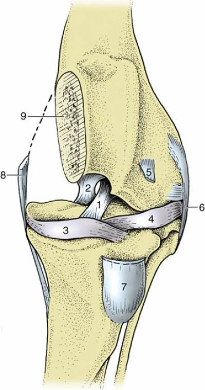
FIG. 1.21 Cranial view of left stifle joint of the dog, resected to show intracapsular (1, 2) and extracapsular (6, 8) ligaments. 1, Cranial cruciate ligament; 2, caudal cruciate ligament; 3, medial meniscus; 4, lateral meniscus; 5, tendon of origin of long digital extensor; 6, lateral collateral ligament; 7,
patellar ligament; 8, medial collateral ligament; 9, medial condyle, partly removed.
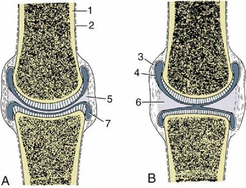
FIG. 1.22 (A) Synovial joint with articular disk. (B) Synovial joint with meniscus. 1, Compact bone; 2, periosteum; 3, fibrous layer of joint capsule; 4, synovial membrane; 5, articular disk; 6, meniscus; 7, joint cavity.
An articular labrum is a fibrocartilaginous lip or rim placed around the circumference of certain concave articular surfaces, including the acetabulum (the deep socket at the hip). A labrum serves to extend and deepen the articular surface, increasing the load-bearing area and helping to spread the synovial fluid. Because a labrum is deformable, it allows the surface to adapt to disparities in the curvature of the bone with which it comes in contact.
Synovial pads or cushions are formed where fat masses are included between the synovial and fibrous layers of the joint capsule. They are sometimes interpreted as swabs that spread the synovia over the surface, but their main purpose is to allow the synovial membrane to accommodate its shape to the part of the bone with which it is momentarily in contact.
Movements
Although many joint movements appear to be complicated, they can always be resolved into simple components. Moreover, many activities are the result of coordinated movement at several neighboring joints; the sum of changes can be considerable even when the movement at each individual joint is modest.
The simplest type of movement is translation. In its pure form, translation consists of one flat surface sliding over another while the bodies to which the surfaces belong maintain their original orientation. True translatory movements probably never occur because the prerequisites are perfectly flat surfaces and the absence of spin. Nonetheless, a category of joint (plane joint) is defined in which movement is supposed to be of this kind. These joints have small articular surfaces that appear flat at first scrutiny; in reality, articular surfaces are always curved.
All other movements involve angular change. In some, the moving bone turns (spins) about an axis perpendicular to its articular surface, a movement called rotation. Rotation can always be reversed, so its direction must always be specified. According to convention, an internal rotation of a limb carries the cranial surface medially (Fig. 1.23/4); an external rotation carries this surface laterally (see Fig. 1.23/5).
Other movements involve the moving bone turning about an axis parallel to its articular surface in a pendular or rolling movement (Fig. 1.24/3); this is a slide between curved surfaces and may be described as a swing. Most swings are accompanied by some rotation, although it often goes undetected.
Pendular movements in sagittal planes predominate in the joints of the limbs and are known as flexion and extension. Flexion reduces the angle between the two segments of the limb. The opposite movement, extension, opens the angle and brings the two segments more closely into alignment (see Fig. 1.24). However, the movement at some joints ranges from one flexed position through full extension (180 degrees) to a second flexed position at the other limit. The fetlock joint of the horse is a good example of a joint with such a wide range of movement. In such cases the two terminal positions may be distinguished as overextension (or dorsal flexion), the posture of the animal standing at rest, and (palmar) flexion, the posture when the foot is passively raised. Fig. 1.24 may make this rather confusing distinction plain.
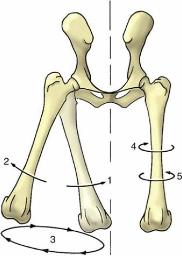
FIG. 1.23 Limb movements illustrated by the femurs of the dog, cranial view. 1, Adduction; 2, abduction;
3, circumduction; 4, inward rotation; 5, outward rotation.
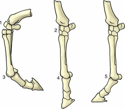
FIG. 1.24 Flexion, extension, and overextension illustrated by the distal part of the horse's forelimb. 1, Flexed carpal joint; 2, extended carpal joint; 3, flexed fetlock joint; 4, extended fetlock joint; 5, overextended fetlock joint.
Adduction and abduction are pendular movements in transverse planes (see Fig. 1.23/1 and 2). Adduction carries the moving part toward the median plane, and abduction carries it away from this plane. When applied to the digits, adduction and abduction describe movement with reference to the axis of the limb and indicate the convergence and the spread of the digits, respectively.
The combination of flexion and extension and adduction and abduction allows the extremity of the limb to describe a circle or ellipse, a movement known as circumduction.
Several limitations are placed on the movements of all joints. The shape of the articular surfaces is obviously relevant. A degree of incongruence is required to maintain a wedge of the lubricant synovia between the surfaces. This wedge is reduced when the radius of curvature of the convex surface increases toward its margin to approximate to the radius of curvature of the opposing concave surface. The surfaces thus become congruent in the closely packed terminal position, and further movement is checked by their being squeezed together.
Tension in extracapsular ligaments can certainly arrest movement, although it is uncertain whether this method of braking is required in normal circumstances. Some ligaments appear to be moderately taut throughout the normal range of movement, whereas others are generally slack and become taut only when movement threatens to go beyond the normal limit.
In some situations, contact between extra-articular structures may be of importance; the olecranon obviously prevents forceful overextension of the elbow, and apposition of the caudal muscles of the thigh and calf prevents overflexion of the human knee. Tension in muscles and other soft structures in the neighborhood of a joint may first decelerate and then arrest movement; inability of the muscles of the caudal aspect of the human thigh to stretch beyond a certain limit— passive insufficiency—prohibits many people from touching their toes. The contraction of muscles that oppose a given movement may be the most important factor; its significance is discussed in the following section.
Classification
Synovial joints may be classified according to numerical and geometrical criteria. The numerical system distinguishes simple joints, with one pair of articular surfaces, and composite joints, in which more than two opposing surfaces are involved and movement occurs at more than one level within a shared capsule. The shoulder joint illustrates the first and the carpal joint the second variety.
There are seven categories in the current version of the geometrical system. One, the plane joint (Fig. 1.25A), has already been mentioned.
The hinge joint (ginglymus; Fig. 1.25B) has one articular surface shaped like a segment of a cylinder and the other excavated to receive it. Pendular movement is possible in one plane only. The other movements are prohibited by stout collateral (one to each side) ligaments and possibly by the development of matching ridges and grooves on the articular surfaces. The elbow joint between the humerus and bones of the forearm is an example.
The pivot joint (articulatio trochoidea; Fig. 1.25C) comprises a peg fitted within a ring. Movement takes place about the long axis of the peg. In some joints (e.g., the proximal radioulnar joint) the peg rotates within the fixed ring; in others (e.g., the atlantoaxial joint between the first two vertebrae) the ring rotates about the fixed peg.
The condylar joint (Fig. 1.25D) is formed by two knuckle-shaped condyles that engage with corresponding concave surfaces. The two complexes may be close together, as in the femorotibial joint, or widely separate and provided with independent joint capsules, as are the twin articulations of the mandible. In each case the whole arrangement is regarded as constituting a single condylar joint. Movement is primarily uniaxial, about a transverse axis common to the two condyles; certain amounts of rotation and slide are also permitted.
The ellipsoidal joint (Fig. 1.25E) consists of an ovoid convex surface that fits into a corresponding concavity. Movements occur principally in two planes at right angles to each other (flexionextension; adduction-abduction), but a small amount of rotation may be possible. The radiocarpal joint of the dog is ellipsoidal.
The saddle joint (articulatio sellaris; Fig. 1.25F) combines two surfaces, each maximally convex in one direction and maximally concave in a second direction at right angles to the first. These are also biaxial joints, allowing flexion-extension and adduction-abduction but with a certain amount of rotation permitted or imposed by the geometry of the surfaces. An example is the distal interphalangeal joint of the dog.
The ball-and-socket or spheroidal joint (Fig. 1.25G) consists of a portion of a sphere received within a corresponding cup. This multiaxial joint enjoys the greatest versatility of movement. The hip joint is the best example; the human shoulder joint also conforms closely to the pattern, but the shoulder of domestic species largely restricts its movement to flexion and extension.
It must be emphasized that anatomic joints correspond very imperfectly to the theoretical models. Sometimes, the departure from the ideal can be sufficiently large to make it a matter of controversy as to which category best accommodates a particular articulation.