» Palpation
Abdominal palpation is an important diagnostic tool in the examination of companion animals as it allows identification and assessment of a number of abdominal organs.
Abdominal palpation is an important skill and competency for the veterinarian.
Abdominal palpation can be reliably used to identify the masses in 20%-50% of dogs with intestinal tumors.The system of reference to abdominal regions that is preferred by clinicians divides the abdomen into 18 compartments. Epigastrium, mesogastrium, and hypogastrium are visualized as being defined by two transverse planes:
1. The cranial plane is situated just caudal to the last rib.
2. The caudal plane is situated just cranial to the thigh musculature.
The depth of the abdomen, between the lumbar muscles and the abdominal floor, is then visualized as divided into three, more or less equal parts — dorsal, middle, and ventral parts — yielding nine compartments to each side of the median plan. Palpation of these compartments is performed in a systematic way, generally commencing with the dorsal epigastrium, continuing ventrally, and proceeding from superficial (muscle tension, overfilled intestines) to deep. Palpation is usually performed with the subject standing and with the converging extended fingers of the examiner's hands placed over the flanks. For some purposes it is helpful to have the cranial part of the body raised, allowing the intrathoracic abdominal organs to slide caudally, and for other purposes to have the subject laterally recumbent or supine. A one-handed approach, with the converged fingers opposed to the thumb, is useful with cats and small dogs. Whatever the technique, it is important to allay anxiety so that the animal relaxes its abdominal muscles. The procedure is most rewarding in cats and small dogs and least rewarding in large, well-muscled, or obese dogs.
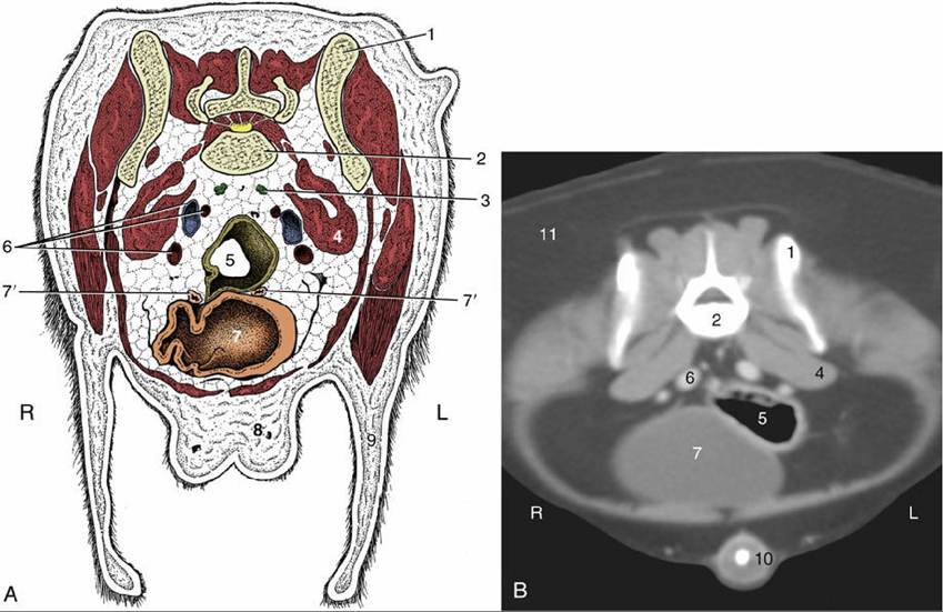
FIG. 14.32 (A) Transverse section of the canine abdomen at the level of the seventh lumbar vertebra. (B) Corresponding computed tomography image at about the same level. 1, Wing of ilium; 2, seventh lumbar vertebra; 3, sacral lymph nodes; 4, iliopsoas; 5, descending colon; 6, internal iliac artery (most dorsal), external iliac vein, and external iliac artery; 7, bladder; 7', uterine horns; 8, mammary gland; 9, flank fold; 10, penis with os penis; 11, fat; L, left; R, right.
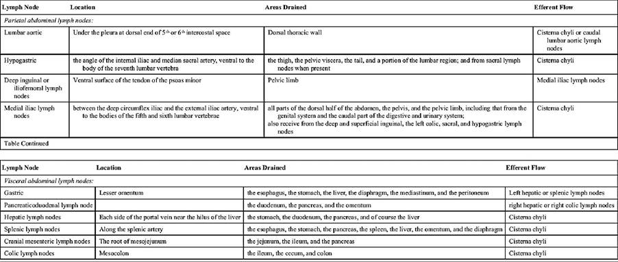
» TABLE 14.2
Abdominal lymph structures
The normal liver projects only slightly, and variably, behind the costal arches and is difficult if not impossible to recognize when the bilateral approach is used. Greater success may be obtained if the fingertips are insinuated deep to the costal arch, a maneuver possible only during full relaxation of the flank muscles. It may then be possible to identify the sharp free margin and narrow adjoining strip of the liver. Homogenous enlargement of the liver can first be palpated in the ventral epigastrium (on superficial palpation) and, following further enlargement, in the medial epigastrium, especially when one places the fingers within the costal arch. The liver can be more easily reached at the left than at the right side.
The empty stomach is tucked under the ribs, out of reach on the left side, but when full of ingesta or distended with gas, it projects behind the costal cartilages. It is more easily found in narrow, deep-chested dogs than in barrel-chested breeds. The stomach, when empty, does not contact the abdominal wall, but when moderately filled, it lies against the wall ventrally and to the left. The completely filled stomach, especially in pups, lies largely in contact with the ventral body wall, toward a transverse plane just caudal to the umbilicus.
The spleen occupies the same region against the left flank, but its soft and deformable consistency does not make it easy to palpate unless the organ is considerably enlarged and firmed. Normally the spleen is located to the left in the epigastrium, close to the major curvature of the stomach (completely within the costal arch in the dog). In the case of enlargement the spleen moves ventrally and caudally, and can be felt in the ventral and medial mesogastrium.Success in locating the kidneys is rather unpredictable in the dog. Most often, only the caudal pole of the left kidney is within reach, and it may be identified by its firm, rounded contours. The right kidney is commonly inaccessible. In some dogs, generally of the larger breeds, the left kidney is pendulous and "floats" at a more ventral level than usual; this is the normal condition for both kidneys in the cat, and both may be steadied through the abdominal wall for biopsy puncture. The entire surface of a "floating" kidney, including the depression at the dorsally facing hilus, may be examined. The left kidney contacts the dorsal part of the left lateral abdominal wall.
The fluctuating intestinal mass occupies a large part of the abdomen, extending from the roof to the floor and from one flank to the other. Identification of most individual parts is problematical. The descending duodenum may sometimes be identified on the right side if the fingers are first pressed against the abdominal roof and then drawn laterally. There is no difficulty in finding the jejunum, whose coils may be made to slip between the hands. In the dog the only part of the large intestine that may be sought with confidence is the descending colon on the left side. It is most readily identified when occupied by a column of hard or granular feces. The ascending colon and cecum may sometimes be identified, most readily when gas-distended, but the transverse colon is too deeply tucked under the ribs to be within reach. All parts of the large intestine are more readily found in cats, in which a useful guide to the positions of the cecum and ascending colon is provided by the firmness at the ileocecocolic junction. The lymph nodes associated with the intestine evade detection unless enlarged.
Comprehension Check
1. Using cadavers and available imaging modalities (radiographs or CT scans), develop a thorough understanding of the topography of the abdominal organs.
2. Explore the inguinal canal and its contents, and gain an understanding of the process of inguinal hernia.
>------------------------- <
* Where we give an indication of weight or measure, we have in mind a subject of the size of a beagle, an animal weighing about 15 to 20 kg. Cats or course vary less if we exclude such atypical breeds as the Maine Coon.