NEUROGLIA
8.6.1 Astrocytes
Astrocytes, the most abundant glial cells, are star-shaped and found in the brain and spinal cord. They provide structural support to neurons and help maintain the integrity of the blood-brain barrier, which regulates the passage of substances between the bloodstream and the brain.
They also regulate the extracellular environment by taking up and releasing ions and neurotransmitters. Moreover, they provide energy substrates such as glucose to neurons and help regulate synaptic activity. Further, they help clear neurotransmitters from the synaptic cleft, thereby terminating synaptic transmission and preventing excessive neuronal excitation. Neurotrophically, astrocytes are involved in the secretion of growth factors and other molecules that promote neuronal survival and growth during development and in response to injury. They also play a major role in the modulation of synaptic function as they can influence synaptic transmission and plasticity through the release of signaling molecules such as glutamate, ATP, and D-serine. Additionally, they are involved in regulating the pH, ion concentration, and water balance of the extracellular space, contributing to the overall homeostasis of the CNS.8.6.2 Ependymal Cells
Glia, also known as glial cells or neuroglia, are non-neuro- nal cells that provide support and protection for neurons in the nervous system. They outnumber neurons and include various types such as astrocytes, oligodendrocytes, microglia, Schwann cells, ependymal cells, and satellite cells (Figure 8.4), each with specific functions.
Glia are crucial for maintaining the structural integrity of the nervous system, regulating the extracellular environment, modulating synaptic transmission, and participating in immune responses and repair processes. They play
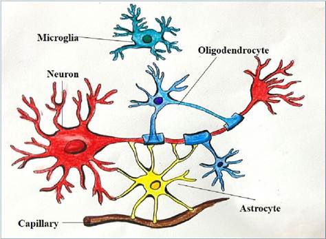
FIGURE 8.4 Various types of glial cells
Ependymal cells are a type of glial cell that lines the ventricles of the brain and the central canal of the spinal cord.
These cells are cuboidal to columnar in shape and have cilia for the movement of CSF and microvilli on their surfaces. They are involved in the production and circulation of cerebrospinal fluid (CSF), which cushions the brain and spinal cord, provides nutrients, removes waste, and helps regulate the extracellular environment. They also form a barrier between the CSF-filled ventricles and the surrounding brain tissue. This barrier helps regulate the exchange of substances between the CSF and the brain, contributing to the overall homeostasis and protection of the CNS. Additionally, in some regions of the CNS, such as the lining of the lateral ventricles, ependymal cells possess stem celllike properties and can give rise to new neurons and glial cells in response to injury or disease.8.6.3 Microglia
Microglia are the highly dynamic and primary immune cells of the brain and spinal cord, constantly surveying their surroundings for signs of damage, infection, or inflammation. They can rapidly change their morphology and function in response to various signals. When activated by injury, infection, or inflammation, microglia can proliferate, migrate to the site of injury, and release cytokines and other signaling molecules to initiate an immune response. They thereby engulfing and digesting the cellular debris, pathogens, and dying neurons, helping to maintain tissue integrity and support neuronal survival. In addition to their role in immune defense, they also play roles in synaptic pruning, neurogenesis, and modulating neuroinflammation, thereby contribute to the maintenance of neuronal networks. Since the microglia play a crucial role in immune surveillance and neuronal homeostasis, dysfunction of microglia has been implicated in various neurological disorders, highlighting their importance in brain health and disease.
8.6.4 Oligodendrocytes
Oligodendrocytes have a relatively small cell body with a round or oval shape. The processes from the cell body wrap around the axons of neurons, forming a segment of the myelin sheath around multiple axons, creating a layered, insulating structure.
The primary function of oligodendrocytes is to produce myelin, a fatty substance that insulates axons, allowing for efficient and rapid transmission of electrical signals along the neurons. They also provide structural support to neurons and contribute to the overall stability of the CNS. In the event of injury, oligodendrocytes can contribute to the repair and regeneration of damaged myelin.8.6.5 Schwann Cells
Schwann cells are elongated glial cells that play a crucial role in the maintenance and function of peripheral nerves. Schwann cells wrap around the axons of neurons in the PNS to form the myelin sheath. Unlike oligodendrocytes, each Schwann cell typically myelinates only one segment of a single axon. Gaps between adjacent Schwann cells along the axon where the myelin sheath is absent are called Nodes of Ranvier. These gaps are essential for rapid signal transmission through saltatory conduction. The primary function of Schwann cells is to myelinate axons in the PNS, which speeds up the transmission of electrical impulses. Following a nerve injury, these cells can proliferate and prompt regrowth of axons, aiding in the repair of damaged nerves.
8.6.6 Satellite Cells
Satellite cells have a small and flattened cell body. They are found in the peripheral nervous system, specifically within ganglia. Within sensory, sympathetic, and parasympathetic ganglia, they surround the cell bodies and form a continuous layer around the neurons, providing a physical barrier between the neurons and the surrounding connective tissue. Satellite cells provide structural support to the neurons in the ganglia. They help regulate the exchange of nutrients and waste products between neurons and their surrounding environment. They play a role in modulating the microenvironment of the ganglia, including ion balance and neurotransmitter levels, which can affect neuronal excitability and function. In response to nerve injury, the activated satellite cells proliferate, produce growth factors and other molecules that support neuronal survival and regeneration. They can respond to changes in the neuronal environment, potentially playing a role in chronic pain conditions and neuropathies.
8.7