Oral Cavity
The mouth is long and narrow, and it does not open widely. The tongue is not as mobile as other large animals and usually does not extend beyond the lips, so it is not used for prehension of food.
The rostral two-thirds of the tongue is about 2 cm thick in the llama. The tongue has a prominent dome-shaped projection in the root that is 5 cm in depth, similar to the torus linguae of ruminants. There are several kinds of mechanical and gustatory papillae on the tongue and buccal mucosa. Foliate papillae are lacking, and the vallate papillae are large and oval in shape.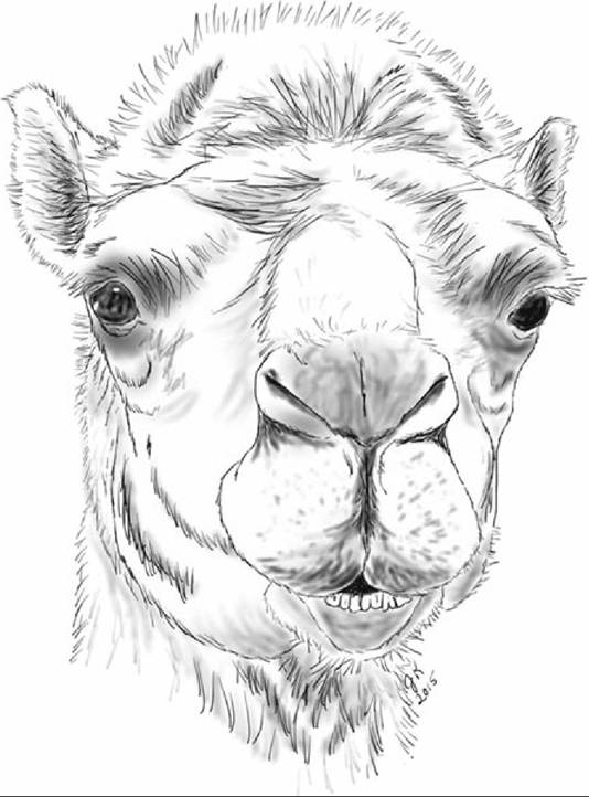
FIG. 38.9 Camels have the ability to completely close the nostril via muscular depression of the alae of the nostril, whereas llamas and alpacas lack this ability.
The dentition of camelids is complex compared to ruminants and horses, and dental disease is common in llamas and alpacas. Similar to ruminants, camelids have a dental pad, shown in Fig. 38.10, and a reduced number of upper incisors. The deciduous dental formula of llamas and alpacas (incisors, canine, premolars) is:

Note that one source indicates there may be 1 or 2 upper deciduous incisors. The permanent dental formula (incisors, canine, premolars, molars) is:
1-1-1(2)-3
3-1-1(2)-3
Fig. 38.7A is an illustration of an individual with two upper premolars and one lower premolar. Eruption dates are discussed later.
Deciduous incisors of both llamas and alpacas are spatula shaped. They are slightly smaller than permanent incisors and have a chalky color; permanent incisors are more translucent.
Fig. 38.11 is a schematic illustration of the radiographic appearance of llama and alpaca permanent incisors, demonstrating some differences in their shape.
Llama permanent incisors are spatula shaped with tapered roots, do not grow continuously, and have enamel over the entire crown. Alpaca permanent incisors are long and narrow and rectangular in cross section and erupt throughout life. The occlusal surface of the alpaca incisor is chisel shaped. Although alpacas have been reported to lack enamel on the lingual surface of the permanent incisors, similar to the vicuna, one histologic study demonstrated that there is enamel on both surfaces of the alpaca incisor. The tips of the incisors should meet the dental pad; incisors are oriented somewhat vertically early in life and become more horizontally oriented as the animal ages.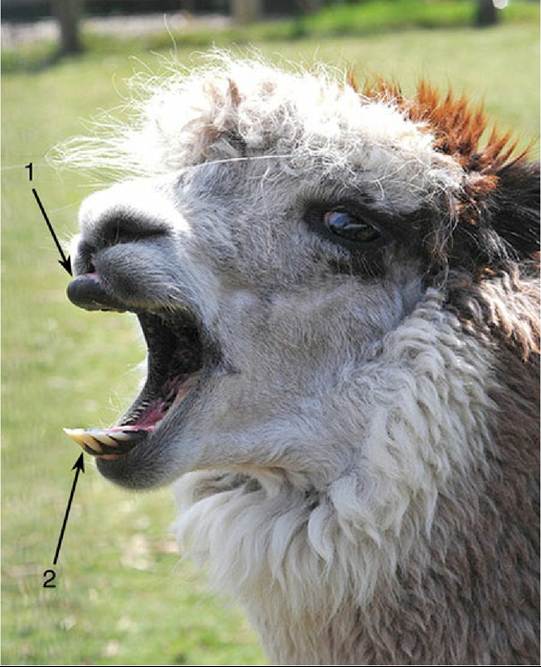
FIG. 38.10 Alpaca. 1, Dental pad; 2, lower incisors. (Image by Arbutus Photography. Available at:
https://www.flickr.eom/photos/arbutusridge/8672528601/in/pool-1087584@n20. This work is licensed under the Creative Commons Attribution-Share Alike 2.0 Generic license.)
Although camelids have a dental pad opposing the lower incisors, they do not have a total absence of upper incisors. They have one upper incisor and one upper canine on each side, and the upper incisor is morphologically identical to the upper canine. The upper incisor and canine and the lower canine together are referred to as the "fighting teeth," because male camelids use them as weapons. They are much larger in intact males than in females or geldings. These teeth are located in the diastema between the lower incisors or dental pad and the premolars, and care should be taken to avoid lacerations from them when examining the mouth. The fighting teeth of a llama are illustrated in Fig. 38.7A.
There is some gender and individual variability in the deciduous and permanent premolars, and premolars can be difficult to tell apart from molars, but premolars are much smaller than molars when present. The premolars on the upper arcade are rostral to the infraorbital foramen.
The deciduous upper premolars 3 and 4 are usually present, but deciduous upper premolar 2 is present in only 65% of males and 45% of females. Deciduous lower premolar 4 is consistent; lower premolar 3 is present in 91% of males and 82% of females.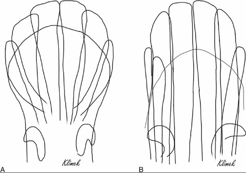
FIG. 38.11 Schematic illustration of the permanent lower incisors of the llama (A) and alpaca (B), as viewed on ventrodorsal intraoral radiographs. Alpaca incisors have straight, parallel outlines, whereas llama incisors taper at the roots.
The lower permanent premolar 3 is absent in the male and sometimes present in the female (15%). The upper permanent premolar 3 is consistently present in males and usually present in females. There are consistently three upper and three lower permanent molars in all individuals. Fig. 38.7A shows permanent premolars and molars in a llama. Molars increase in size from rostral to caudal.
The form of the cheek teeth is similar to that of cattle and horses. Although camelid cheek teeth do not erupt throughout life, they are subject to wear, and new bone formation below the teeth does force them out gradually to compensate for this wear.
Cheek Teeth of Camelids: The upper arcade is wider than the lower arcade, and the upper cheek teeth themselves are wider than the lower cheek teeth. Thus, similar to the cheek teeth of horses, the premolars and molars of camelids develop normal points on the labial side of the upper teeth and the lingual side of the lower teeth. These should not be floated in the camelid, however, in the absence of any evidence of trauma resulting from the points or in the absence of abnormal mastication.
Upper premolars have three roots, and upper molars have four roots. The lower cheek teeth all have two roots, but in the last lower molar the caudal root is a fusion of two roots.
It is challenging to observe the teeth of camelids because of the restriction to opening the mouth widely and the long and narrow oral cavity.
Cheek teeth can be observed with the use of a speculum or mouth gag. The cheek teeth may also be palpated through the cheek tissue. In one study of tooth eruption in 235 alpacas, alpaca-vicuna crosses, and llamas of known ages, the investigator found very little variation between llamas and alpacas, so alpacas and llamas can be assumed to follow the same pattern of eruption.At birth in full-term crias (the term describing neonatal camelids), all of the deciduous mandibular incisors are erupted. The deciduous fighting teeth are present in all animals but often not erupted. It is possible to estimate the age of the animal by eruption of the incisors up to about age 5; beyond that it is difficult. Permanent incisors erupt caudal to the deciduous incisors, at ages 2 to 2.5 years for incisor 1, 3 to 3.25 years for incisor 2, and 3 and 6 years for incisor 3. Retained deciduous incisors can occur and may need to be removed.
The first two permanent molars can be a reliable estimate of age; the lower molar 1 erupts at age 6 to 9 months; the lower molar 2 erupts between 17 and 24 months of age. The lower molar 3 can erupt anywhere between 2 years 9 months and 3 years 8 months.
Trimming Camelid Teeth: Some of the teeth may require trimming. Lower incisors should meet the tip of the dental pad. If these teeth do not meet the dental pad properly they can become overgrown and will need to be trimmed to restore appropriate function; Fig. 38.12 shows an alpaca with incisor teeth that are overgrown. There are various methods to accomplish trimming, but care should be taken with power tools to avoid overheating or cracking the tooth. The fighting teeth, if present, need to be trimmed to prevent injury to herd mates and handlers. The teeth should be trimmed annually during their active growth period, generally between ages 3 and 8. They can be trimmed to 2 to 4 mm from the gum line to avoid damage to the gums or formation of a recess where food can accumulate. The use of cutters for these teeth is not advised because of the possibility of fracture of the tooth.
In addition, the mandibular canine is immediately adjacent to the mental foramen and to the root of the third incisor, and care must be taken to protect these structures.Camelids have a high incidence of tooth root abscesses, especially on the lower arcade, and especially between the ages of 4 and 8 years. When examining the head, the jaws and the lymph nodes should be examined for swellings. The upper cheek teeth roots can be accessed, and the teeth repulsed, if necessary, through the maxillary sinus, dorsal to the facial crest, and ventral to a line drawn from the medial canthus to the infraorbital foramen. One should be aware that the infraorbital canal is in this area and lies between the medial and lateral roots of the molars. Because its roots are ventral to the orbit, the last upper molar is not accessible unless approached through the zygomatic bone, after reflecting the masseter muscle from the facial crest.
Nerve blocks for the teeth similar to those used for ruminants can be used for anesthesia of teeth that can be removed orally under sedation. The infraorbital nerve can be blocked through the infraorbital foramen, which is palpable dorsal to the premolars. The mental nerve can be blocked at its termination by inserting the needle into the mental foramen, located just caudal to the lower canine tooth (or 2-3 cm caudal to the incisors). Alternately, the inferior alveolar nerve can be blocked as it enters the mandibular foramen on the medial side of the ramus of the mandible. This foramen is located rostral and dorsal to the ventral curvature of the ramus of the mandible.
The locations of foramina associated with nerve blocks for the teeth are illustrated in Fig. 38.13.

FIG. 38.12 Mucho the alpaca, demonstrating teeth that are overgrown as a result of
malocclusion. (Unmodified from original. Available at: https://www.flickr.com/photos/justinlindsay/91878991. This work is licensed
under the Creative Commons Attribution-Share Alike 2.0 Generic license.)